

1 Introduction
Cyanide is an extremely lethal anion that can affect several functions of human health, involving the visual, vascular, cardiac, central nervous, endocrine, and metabolic systems [1–10]. Cyanide could be absorbed through the skin, gastrointestinal tract and lungs, causing convulsion, vomiting, loss of consciousness, and eventual death [1,9]. Its toxicity arises from its ability to bind to the iron in cytochrome c oxidase, interfering with electron transport and resulting in hypoxia [10–15]. Despite its toxicity, cyanide is generally employed in various chemical processes such as raw material for synthetic fibers, herbicides, resins, tanning, metallurgy and the gold extraction process [16–19]. Due to its serious toxicity and general utility, developments of reliable and efficient probes are quite necessary to monitor the presence of cyanide from contaminant sources. The most difficult fact in the anion sensing in aqueous environment is that water is a highly competitive solvent and interferes with the detection process of anions by intervening in the interaction between associates [20,21]. One of the greatest challenges for detecting cyanide arises from the interference of other anions, most especially, fluoride and acetate, working only in organic media [22,25]. The higher solvation energy of cyanide ions in aqueous media is known to negatively affect the hydrogen-bonded adduct formation between the receptor unit and the CN− [26,27]. With these considerations in mind, it is evident that the development of highly selective and operative sensors, capable of detecting cyanide in aqueous media, is an important target for real-life applications. So far, most chemo-dosimeters for cyanide are based on hydrogen bonding [20,28,29], complexation to Lewis acids [30–32], time-gated fluorescence [33], quantum dots [34], Au nano-particle [35], sensing on Al2O3-based thin-film matrix [36] dyes equipped with a metal binding unit [21] or covalent bond formation [37–43]. Hence, receptors with chromogenic subunits are interesting as naked eye detection systems, which have been actively studied over the past 10 years because of their high sensitivity, ease of operation, rapid response rates and relatively low costs [14,43,44]. Based on our previous works [45–47], in this study, we present a chromogenic chemosensor L based on azo-azomethine receptors, for the rapid detection and colorimetric sensing of cyanide anions. Above all, it is worthy of note that sensor L could clearly detect cyanide from other interfering anions by color change from yellow to reddish orange through naked-eye detection in aqueous media. To the best of our knowledge, this is the first unsymmetrical azo-azomethine receptor derived from 2-amino-1-propanol for the qualitative and quantitative detection of toxic cyanide anions in water and other samples. Colorimetric receptor L possessing two phenolic and alcoholic OH binds to cyanide ions by involving azomethine cyanide reaction sites and phenolic OH via hydrogen bonding interaction. To gain a deeper insight into the mechanism and structure in addition to the spectral aspects of the complex, quantum-chemical calculations were performed using Density Functional Theory (DFT) to supplement the experimental results.
2 Experimental
2.1 Materials and equipment
All chemicals were purchased from Sigma–Aldrich and Merck and were used without further purification. Electronic spectral measurements were performed utilizing an Optizen 3220 UV spectrophotometer in the range of 200–900 nm at room temperature. 1H NMR spectra were recorded on a Bruker AV 300 MHz spectrometer and chemical shifts were recorded in ppm. The sensor L was synthesized based on our previous reported procedure [48]. Fourier transform infrared (FT-IR) spectra were recorded as pressed KBr discs utilizing a Unicom Galaxy Series FT-IR 5000 spectrophotometer within the region of 400–4000 cm−1.
2.2 Results and discussion
2.2.1 Colorimetric and spectral response of L toward CN−
With seldom exceptions [20,49,50], the sensing of cyanide was limited to only tetrabutylammonium (TBA) as the counter ion within pure organic media since the solvation of KCN/NaCN in aqueous/mixed aqueous systems generally leads to sensing silence as a result of competitive and interfering problems of solubility in water. From this point, at first, the sensing behavior of the receptor L toward various anions such as F−, Cl−, Br−, AcO−, H2PO4−, HSO4−, ClO4−, N3−, NO2−, SCN− and CN−, as their inorganic sodium salts, was probed qualitatively by naked-eye colorimetric examination and then, absorption spectral studies were conducted on sensor L (5 × 10⁻⁵ mol L−1) in DMSO/H2O (95:5, v/v) upon the addition of 10 equivalent of typical anions in water in order to survey the selectivity of receptor L as a colorimetric sensor. As illustrated in Fig. 1, sensor L was yellow in color and displayed a broad band with a maximum absorption wavelength, centered at about 400 nm in the UV−vis spectrum, corresponding to π → π* transition of azo and azo-methane chromophores and intra-molecular charge transfer (ICT) transition [51–53]. Upon the addition of different anions to the solution of L, only CN− caused a remarkable color change from yellow to reddish orange instantly (Fig. 1a) and demonstrated a bathochromic shift (from 400 to 505 nm) in absorption spectra (Fig. 1b), while other anions demonstrated almost no change in color and UV–vis spectra under identical conditions.

Color changes (a) and UV–vis absorption spectra (b) of receptor L (5 × 10⁻⁵ mol L−1) in DMSO/H2O (95:5) in the presence of 10 equiv. of different anions.
In order to inspect the further detailed response of the designed receptor L toward CN− ions, we conducted UV–vis spectroscopic titrations by adding 10 equivalents of cyanide anions in water into the diluted solution of L (2 × 10⁻⁵ mol L−1) in DMSO/H2O (95:5). As shown in Fig. 2, with incremental addition of cyanide ions, the absorption band at 400 nm was gradually reduced and a new band centered at 505 nm progressively appeared with an obvious isosbestic point at 460 nm, indicating the formation of new species. The extinction coefficient of the chemosensor L at 400 nm is 24,250 M−1 cm−1 and 10,338 M−1 cm−1 at 505 nm for the L–CN− adduct. Consequentially, these distinct changes can be attributed to the charge transfer interactions due to the complexation between cyanide ions and reactant sites of L at high concentrations. The conclusive stoichiometric ratio between L and CN− was inferred to be 1:2 with the aid of Job's plot [54] experiments (Fig. 3a). Furthermore, the binding constant of complexation was calculated to be 2.71 × 10⁴ M−1 utilizing the Benesi–Hildebrand method [55] as shown in Fig. 3b. In addition, based on UV–vis measurements, the detection limit of the sensor towards cyanide was found to be 1.03 × 10⁻⁶ mol L−1, which was lower than the maximum contaminant level (MCL) for cyanide in drinking water (1.9 × 10⁻⁶ mol L−1) as adjusted by the World Health Organization (WHO) [56]. Therefore, the present sensor should be applicable as a practical system for the monitoring of cyanide concentrations in aqueous samples.

Absorption spectral changes of receptor L (2 × 10⁻⁵ mol L−1) in DMSO/H2O (95:5) with the incremental addition of CN− ions (0–10 equiv.) at room temperature. Inset: absorbance at selected wavelengths versus different concentrations of CN− added.

(a) Job's plot and (b) Benesi–Hildebrand plot of sensor L binding with CN− anions associated with absorbance change at 505 nm in DMSO/H2O (95:5, v/v).
By close examination, the calibration curve at 505 nm, plotting of absorbance against concentration of the complex formed with L and CN−, underwent three different steps (Fig. 4). With the dissociation of these steps, a linear relationship appeared for each step with different slopes, confirming that L interacts with CN− in a stoichiometric ratio of 1:2. In the first step, the addition of 1 equivalent of cyanide ions into solution of the receptor leads to no significant UV–vis spectral changes and a light yellow color was observed, confirming that nucleophiles such as cyanide could attack the azo-methine group of receptor L via the Michael addition reaction [57]. As a result, the extent of conjugation in the receptor molecule should be broken, which would affect the electronic structure and slightly optical properties because the sensor L was yellow in color; thus, the nucleophilic addition of CN− to the azo-methine group was expected to produce the light yellow color [38]. In the second step, more addition of CN− (from 1 to 2 equivalents) to the indicator solution led to the emergence of a absorption band with absorption maxima at 505 nm with a concurrent reduction in the absorbance at 400 nm. Simultaneously, the light yellow color of the sensor solution instantly turned to reddish orange. These findings propose that the OH of the phenolic receptor L interacts with CN− via hydrogen bonding. With the addition of cyanide ions, deprotonation occurs and leads to a drastic color change in the solution. In the third step, the calibration curve becomes a horizontal line with a near zero slope and the color of the solution remains unchanged.

Plotting of absorbance vs. concentration of sensor L in DMSO/H2O (95:5, v/v) with the incremental addition of CN− ions at 505 nm.
The recognition mechanism of the sensor L for CN− was also verified by FT-IR spectroscopy (Fig. S1). A comparison of L and L/CN− adducts showed two new peaks at 2254 and 2171 cm−1, when the cyanide ion was added. Furthermore, a peak at 1645 cm−1 corresponding to the ν(CN) band completely disappeared in the infrared spectrum of the L–CN− complex; this also verified that the cyanide anion was indeed added to the carbon of the imine group of chemosensor L via nucleophilic addition [58].
To further check the practical applicability of receptor L as a CN−-selective colorimetric receptor, we conducted a competitive experiment with different anions in an aqueous solution, which plotted an orange bar graph as illustrated in Fig. 5b. The selectivity of receptor L for CN− was plotted evidently as a yellow bar graph at 505 nm in Fig. 5b. It was found that the presence of other anions did not cause any obvious disturbance to the detection of CN− by L. These results further confirmed that only the addition of cyanide could induce the ostensible color changes from yellow to orange as depicted in Fig. 5a.

(a) Color changes and (b) bar graph for the behavior of receptor L toward CN− and other anions as measured by UV–vis in DMSO/H2O (95:5) at 505 nm.
2.2.2 1H NMR titration of sensor L with cyanide
To examine the sensing mechanism of receptor L for CN−, the reaction mixture of L with excess CN− was assayed by 1H NMR spectrometry as illustrated in Fig. 6. During the 1H NMR titration of receptor L in DMSO-d6 with 1.0 equivalent of CN− (as its sodium salt) in D2O, the sharp singlet bond at δ = 8.68 ppm corresponding to the azo-methine proton (Ha) completely disappeared. Simultaneously, two new peaks emerged at δ = 3.25 ppm and δ = 2.89 ppm, which is attributed to the formation of Hb and NH protons. Meanwhile, most of the aromatic and olefinic protons exhibited an upfield shift and simultaneous appearance of the new Hb and NH protons clearly indicates that the cyanide anion was added to the azo-methine group and the breaking of the conjugation has occurred. In the next stage, with the addition of 2.0 equivalents of CN− into the solution of the sensor, all signals for the 1:1 adduct in the aliphatic section remain unchanged, but the single resonance at 10.22 ppm corresponding to the phenolic proton disappeared. This shows that the interaction of cyanide ions with the phenolic OH with a vivid light red color change has occurred. Furthermore, the aromatic protons shifted upfield, which suggests that the negative charges developed from the deprotonation of L by CN− are delocalized through the whole receptor molecule [59]. Evidently, 1H NMR titration data powerfully uphold our supposition of stepwise cyanide addition. Expansion of aliphatic and aromatic sections of 1H NMR spectra is shown in Figs. S2 and S3 (Supplementary data). The proposed reaction mechanism of sensor L with cyanide anions according to the results of UV–vis and 1H NMR spectroscopy is shown in Scheme 1.

1H NMR titration of (a) L (in DMSO), (b) L + CN− (1:1) and (c) L + CN− (1:2) complexes.

Proposed reaction mechanism of sensor L with cyanide anions.
3 Computational studies
To have a better understanding of the photo physical properties of sensor L and its cyanide complex, they were carefully studied by density functional theory (DFT) calculations at the B3LYP2 level and 6-31+G(d) basis set of the Gaussian03 program package [60]. Molecular geometry optimizations of the chemosensor L and its cyanide complex are shown in Fig. 7, which demonstrates that the N atom of the azo-methine group interacts with phenolic OH through hydrogen bonding in free L. After the nucleophilic addition reaction between sensor L and CN− (1:1), the double bond of the azo-methine group (CN) with a distance of 1.284 Å changed to a single bond with 1.461 Å. With the addition of anions (1:2) in the next step, second CN− interacts with phenolic OH through hydrogen bonding and intermolecular distances found for O⋯H⋯N are 1.565 Å and 1.067 Å. The outcome of this reaction is that the conjugation in the chemosensor L would be broken, which would cause structural and spectral alterations.

(a) optimized structure of L, (b) L + CN− (1:1) and (c) L + CN− (1:2) complexes by B3LYP/6-31+G(d).
To gain a deeper insight into the nature of the response mechanism of sensor L, the typical transition energy diagram for the highest occupied molecular Orbital (HOMO) and the lowest unoccupied molecular orbital (LUMO) of the receptor L along with the resultant complex was obtained, as shown in Fig. 8. The complexation of L with cyanide anions distorted the electron density distribution of the sensor because of the nucleophilic addition and formation of hydrogen bonds. The total energy of the 1:1 and 1:2 optimized complexes (−1947.2457 a.u.) and (−2040.1798 a.u.), respectively, in DFT were lower compared to the energy of the free receptor (−1853.8150 a.u.) protecting the greater stability of the complexes. The difference in the energy gap of the HOMO and LUMO for the 1:1 complex (ΔE = 3.5638 (eV)) is a little more when compared to this value for the free receptor (ΔE = 3.4577 (eV)). This can be attributed to the lack of π-bond conjugation in the L + CN− complex. Theoretical calculations also disclosed the reduction in the energy gap of the HOMO and LUMO, on complexation with 2 equivalents of CN− leading to red shift in the UV–vis absorption spectra [61].

Energy level diagrams of the HOMO and LUMO of (a) L, (b) L + CN− (1:1) and (c) L + CN− (1:2) complexes (using the DFT/B3LYP/6-31+G(d) method).
4 Study as a function of time
As a probe for the assigned anion, the response time is of importance to the practical detection of analytes [62]. The effect of the reaction time on the binding process of CN− to receptor L was investigated. As shown in Fig. 9, UV–vis spectroscopic measurements were carried out by adding 10 equivalents of cyanide anions in water into the solution of L (4 × 10⁻⁵ mol L−1) in DMSO/H2O (95:5, v/v) at different times. After 72 h, there was a slight or no significant change in the absorption spectrum, which suggests that the sensor L is a very suitable probe for practical purposes over a long time.

Time evolution of receptor L in DMSO/H2O in the presence of 10 equiv. of CN− ions.
5 Effect of pH
To check the effect of pH on the host–guest binding affinity, the absorption band of receptor L at 505 nm attributed to the formation of the L–cyanide complex under different pH conditions was examined. As shown in Fig. 10, at an acidic pH of 3, the color of the L–CN− complex changed to pale yellow while no band appeared at 505 nm; suggesting that the L–CN− complex does not exist at acidic pH. However, at a basic pH of 10, the absorption band at 505 nm heightened rapidly and the color of the solution turned to reddish orange. The result showed that receptor L is a sensitive sensor with respect to pH and could be applied in physiological systems. As shown in Fig. 10, the absorption of the L–CN− complex is same as the absorption of the complex at basic pH; this indicates that the cyanide ions act as a base in the receptor (L) solution.
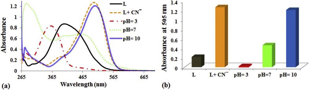
Effect of pH on the UV–vis spectra of sensor L (4 × 10⁻⁵ mol L−1) containing cyanide anions at 505 nm.
To explore more potential and analytical applications of chemosensor L for CN− ions, we prepared test strips by immersing filter papers (1 × 5 cm2) into the DMSO/H2O solution of the sensor (2 × 10⁻³ mol L−1) and then dried in air. To detect the cyanide anion in water, test strips coated with L were put into the aqueous solution of cyanide for a few seconds. An immediate obvious color change from yellow to red was observed with different cyanide concentrations (Fig. 11). Consequently, the development of such “easy-to-prepare” test kits was extremely attractive to approximately and quantitatively discern and estimate the concentration of cyanide ions that do not require any additional equipment, similar to that generally utilized as a pH-meter. Fig. 11

Photographs showing (a) solution and (b) the test strips of sensor L (2 × 10⁻³ mol L−1) in DMSO/H2O at various CN− concentrations.
6 Conclusion
In summary, we have studied a highly selective and sensitive azo-azomethine based anion chemosensor for the facile detection of cyanide present in aqueous media without any spectroscopic instrumentation. The detailed anion binding characteristics were also investigated by UV–vis and 1H NMR spectroscopic techniques. The non-interfering absorption band with considerable wavelength shift (from 400 to 505 nm), dramatic color change from yellow to reddish orange and the possibility to probe the binding of CN− in aqueous media through naked-eye detection make the receptor L a unique colorimetric chemosensor for the qualitative detection of cyanide in real samples. Significantly, the designed sensor can be used for the rapid detection of cyanide anions in the basic pH range and also under physiological conditions in a long time. Notably, the DFT calculation supported the chemosensor selectivity by the reduction in the energy gap between the HOMO and LUMO.
Acknowledgements
The authors would like to thank the Research Council of Arak University for financial support of this research.