1 Introduction
Lignin is present in plant cell walls as a three-dimensional (3D) macromolecule associated with cellulose and hemicelluloses via both covalent bonds and non-covalent interactions [1]. Determining the 3D structure of macromolecular lignin is essential for a proper understanding of physical, chemical and biological properties of lignin in the cell walls. For this purpose, the following information with respect to lignin macromolecule in different morphological regions of the cell wall is necessary: (i) distribution and frequencies of different kind of monomer units, (ii) distribution and frequencies of inter-unit bonds; (iii) type and distribution of bonds between lignin and polysaccharides; (iv) higher-order structure and size of lignin macromolecule; (v) 3D assembly of lignin, hemicelluloses and cellulose. Ginkgo (Ginkgo biloba Linn.) is a particularly suitable tree species for investigation of structure of softwood lignin. The anatomical features of the vascular tissue and xylem tissue of ginkgo are similar to those of conifers [2]. A major part of ginkgo lignin is composed of guaiacylpropanoid units [3], and the structure of ginkgo lignin is very close to that of conifer lignin. In applying an isotope tracer technique for structural analysis of lignin, selective labeling of hydrogen or carbon with radio- or stable isotopes in guaiacyl lignin can be conveniently achieved by feeding specifically labeled coniferin to the growing stem of ginkgo wood [4,5]. Non-destructive analysis of the labeled isotopes by microautoradiography or by differential 13C-nuclear magnetic resonance spectroscopy can afford the necessary information (i), (ii), (iii) and a part of (v) [3–5]. Direct observation of the lignin deposition process through a powerful electron microscope will be a promising approach to obtain information (iv) and (v) with respect to the different morphological regions of the cell wall.
This paper deals with observations of lignifying ginkgo cell walls by a field emission scanning electron microscope (FESEM).
2 Materials and methods
Small blocks containing differentiating xylem were cut out from a growing stem of a ginkgo tree (Ginkgo biloba Linn.), and fixed with 3% glutaraldehyde in 0.1 M phosphate buffer (pH 7.1). Radial and cross sections of 60–100-μm thickness were prepared using a freezing/sliding microtome. Sections were postfixed with 1% osmium tetroxide for 2 h at room temperature. The sections were dehydrated through a graded ethanol series and isoamyl acetate, and dried using a critical point dryer (HCP-2, Hitachi) using liquid carbon dioxide. Some sections were subjected to delignification according to the procedure applied for pinewood by Hafrén et al. [6] with slight modification. Wood sections were heated in acetate buffer (pH 3.5, 4 ml) containing sodium chlorite (40 mg) and acetic acid (12 mg) at 75 °C with addition of the same amount of sodium chlorite and acetic acid seven times over 2 h. Removal of the hemicelluloses was carried out by extraction of the delignified section with 24% potassium hydroxide overnight followed by extraction with 18% sodium hydroxide containing 4% boric acid at room temperature [7]. These sections were washed with water, acetate buffer (pH 3.5), and phosphate buffer (pH 7.0), then stained with 0.1% ruthenium tetroxide in phosphate buffer in the dark under cooling with ice water for 10 min. After washing with buffer and water followed by drying with graded ethanol series, the sections were subjected to freeze-drying using t-butanol. The sections were coated with approximately 1-nm-thick platinum/carbon by rotary shadowing at an angle of 60°. Some sections were coated with approximately 1-nm-thick platinum and palladium using ion sputter (Hitachi E-1030). The sections were examined using a FESEM (S-4500, Hitachi) at an accelerating voltage of 1.5 kV and 3–5 mm of working distance.
3 Results and discussion
Fig. 1 shows the sectional view of a cell wall in the formation stage of the middle layer of the secondary wall (S2). A higher magnification view (Fig. 2) shows that in the outer part of S2 (S2o), lignin deposited on the CMFs, while in the inner part of S2 (S2i) lignin deposition is not completed yet and CMFs are clearly visible. In the micrograph taken at higher magnification (Fig. 3), deposited lignin covers cellulose microfibrils (CMF), making the image of CMFs obscure. Fig. 4 shows the higher magnification view of S2o. Macromolecular lignins of almost the same size are observed as bead-like modules on a CMF. For determination of the size of the module, careful consideration should be given on the shrinkage of the module during preparation of a dry wood section for observation by FESEM. The thickness of the coating (about 1 nm) should be also taken into consideration. However, because the shrinkage will be negligible in the direction along the crystalline CMF, the observed average distance between the modules along the CMF in S2 should indicate the distance between initiation sites of polylignol formation regardless of the coating thickness. In the outer layer of the secondary wall (S1) (Fig. 5), the distance between the modules (about ) along the CMF is larger than that in S2, while the CMFs in S1 seem to be thinner than those in S2 (Fig. 6). In the middle lamella and cell corner regions, larger modules of are distributed randomly (Fig. 7). Similar large irregular structures have been observed in the true middle lamella of developing xylem of pine by rapid-freeze deep-etching (RFDE) technique combined with transmission electron microscopy (TEM) [6]. Donaldson observed lignifying cell wall of pine by TEM and permanganate staining, and he found that growing lignin particles in the middle lamella and primary wall form roughly spherical structures within the randomly arranged matrix, while in the S2, lignin lamellae form greatly elongated structures, following the orientation of CMFs [8]. Fig. 6 shows the innermost surface of the developing cell wall at its S2 formation stage. We tentatively interpret this image that several fine elementary fibrils (generally considered wood cellulose of 3–5 nm in width) form a thick bundle of about including associated hemicelluloses on/inside of a bundle, and the bundles are laid down slightly changing orientation with respect to each other. Combined with the cross sectional view of S2 (Fig. 8), the 3D assembly of the CMFs in S2 of ginkgo is best understood as a kind of twisted honeycomb structure [1]. Similar assembly of CMFs in twisted honeycomb structure in S2 has been observed also in pine by RFDE-TEM [9]. It is assumed that glucomannan associates with CMFs on their surface to prevent further thickening [1]. Simultaneous deposition of glucomannan with CMFs was confirmed by an immuno-gold labeling technique on the innermost surface of a developing secondary wall of Cryptomeria japonica tracheids [10]. Though a careful consideration of shrinkage must be also given in determination of the distance between the CMFs, a tentatively estimated distance between CMFs in S2 of the dried cross section (Fig. 8) is around . Delignification does not have much effect on this estimated distance between CMFs as observed in Fig. 9. In fiber S2 of beech (Fagus crenata), delignification also did not cause significant changes in diameter or appearance of the CMFs [11]. However, after hemicelluloses are removed, the CMFs form thicker bundles with smooth surfaces, though some thin crossing CMFs make bridges to keep bundles of CMFs separate (Figs. 10 and 11). This suggests that the twisted honeycomb assembly of CMFs would be maintained mainly by cross-linking hemicelluloses and partly by thin cross-linking CMFs to keep the distance of around 25 nm between CMFs even after lignin is removed. So, hemicellulose and lignin are assumed not to be in definitely separate layers surrounding CMFs in the secondary wall, and lignin may be present as an intimate complex with the hemicellulose skeleton. However, lignin and hemicelluloses may not be distributed uniformly in the module, but the ratio of lignin to hemicelluloses may be high in outer part of the module.

A view of developing cell wall cut parallel to CMFs in middle layer of secondary wall (S2) at a stage of lignin formation.

Higher magnification view of Fig. 1. Compound middle lamella (CML), outer layer of secondary wall (S1) and outer part of S2 (S2o) are lignified, while inner part of S2 (S2i) is not yet lignified.

Higher magnification view of Fig. 2. CMFs are covered with deposited lignin.

Higher magnification view of Fig. 3. Lignin modules (arrows) are growing along CMFs at an almost regular distance.

S1 layer at a stage of formation of lignin modules (arrows).

Innermost surface of a tracheid at a S2 formation stage.

Cell corner region. Large globular modules form aggregates at random.
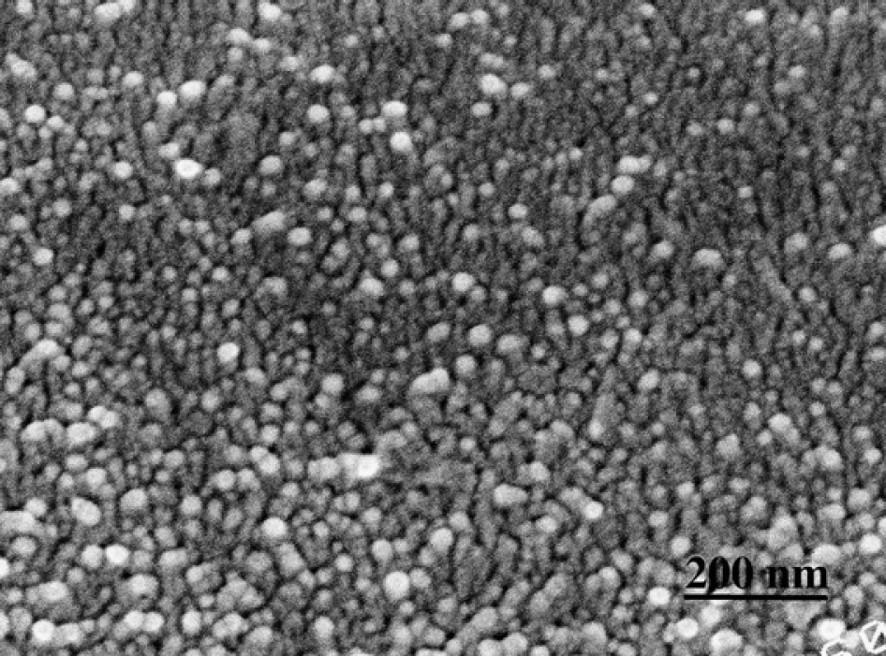
S2 layer cut perpendicular to CMFs. Lignifying wood section.
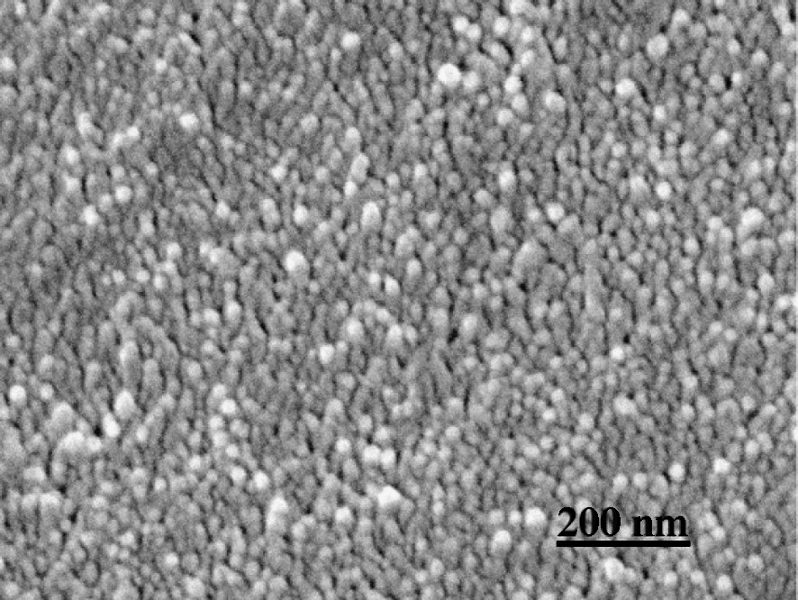
S2 layer cut perpendicular to CMFs. Delignified wood section.
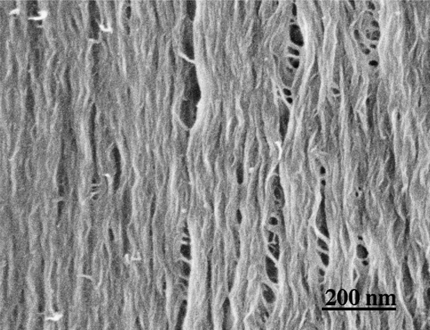
S2 layer cut parallel to CMFs. A wood section from which lignin and hemicelluloses are removed.
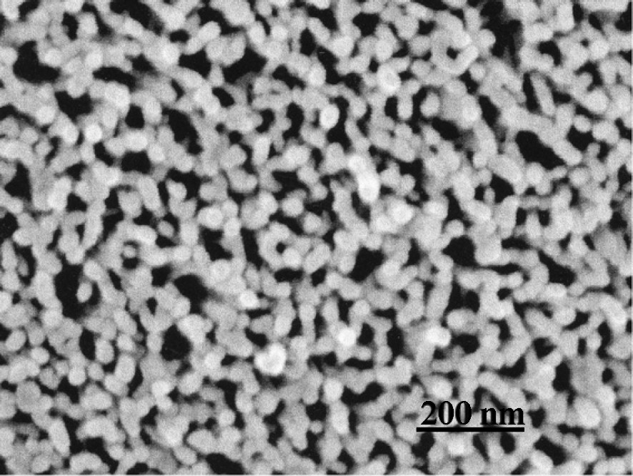
S2 layer cut perpendicular to CMFs. A wood section from which lignin and hemicelluloses are removed.
Though the detailed mechanism of polylignol formation in wood cell walls is not yet fully elucidated, Ralph et al. showed that ferulate polysaccharide esters or diferulate polysaccharide esters appear to be acting as initiation or nucleation sites for lignification in grasses [12,13]. Carnachan and Harris reported that ferulic acid is bound to the primary cell walls of all gymnosperms including ginkgo [14]. If this kind of ferulate-carrying hemicellulose would associate at regular intervals along the CMF, lignin modules could be formed at regular intervals. Fig. 12 shows thin bead-like hemicelluloses strung with the CMFs in ginkgo S2 that remained after lignin is removed. The interval of the remaining hemicellulose is around , that is comparable to the interval of lignin-module formation. These substances disappeared by treatment for removal of hemicelluloses (Fig. 10). In delignified fiber secondary wall of beech, globular hemicellulose has been observed also on the CMFs, and the globular material was removed by treatment with xylanase to result in a smooth CMFs surface [11]. Model experiments on the association of hemicellulose and CMF have been carried out by formation of bacterial cellulose in the presence of glucuronoxylan or glucomannan, as observed by immuno-gold labeling [15]. Glucomannan associates with CMF covering a wide area of CMF, while glucuronoxylan associates locally with some intervals [15]. All these observations suggest that the formation of bead-like lignin modules at regular intervals on the CMFs could be initiated by a special kind of polysaccharide (e.g. feruloylated hemicelluloses), which associates with CMFs, providing periodic initiation sites for polymerization of monolignols. Structural periodicity of the hemicellulose or its regulated association with the CMF would provide controlled formation of modules, as observed in the ginkgo cell walls. It is noted that polysaccharide–polysaccharide cross-linking is effected by radical dimerization of the ferulates–polysaccharide esters [13]. Regulation or limitation of the distance between polysaccharides by the cross-linking through diferulate could control the space for growing modules between the polysaccharide-associated CMFs. This could be one of the factors controlling the dimension of the modules.
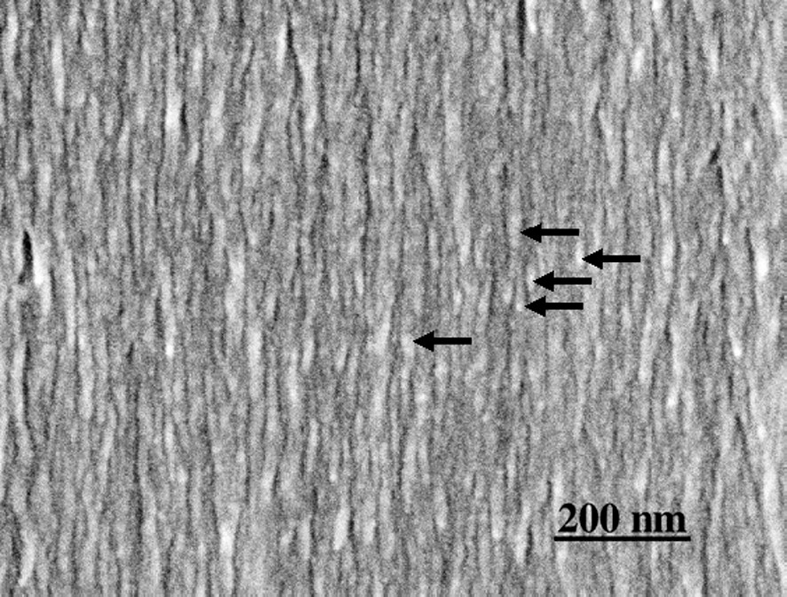
S2 layer cut parallel to CMFs. Delignified wood section. Thin modules composed mainly of hemicelluloses (arrows) remain along the CMFs.
The lignin modules start growing at a regulated distance surrounding the CMFs in the differentiating secondary walls, and the tubular bead-like modules physically and chemically bound to hemicelluloses grow finally to fill up the space between the CMFs. Well-defined modular structures are not observed in completely lignified cell walls. Because the growing end of the module will be mainly 4-O-β bonded guaiacylglycerol units, frequent formation of dibenzodioxocin structures [16] may occur at the boundaries of fusing modules as the final step of lignification. However, the frequency of the bonds formed at the fusion boundary may not be so high compared with the bond frequency inside of the module. A modular structure of lignin has been proposed based on molecular weight distribution of chemically degraded lignin [17].
The size of one tubular-bead of lignin-hemicellulose module in the S2 of dry ginkgo tracheids is tentatively estimated to be in length along the direction of CMF. The final outer diameter of the tube just before fusion should correspond to distance between CMFs, about . The thickness of the tube is tentatively estimated to around from the width of CMFs (about ) and the width of the module (). In S1, larger (about in length) and thicker tubular modules are formed surrounding thinner CMFs. The composition and amount of polysaccharides in different cell wall layers are different from those in S2 [18,19]. It is noted that the size of the module depends on the morphological region, decreasing in the order: cell corner, compound middle lamella, S1, and S2; both the concentration of lignin and the content of condensed substructures are in the same decreasing order [1,3]. The structure of non-cellulosic polysaccharides providing the initiation sites and their assembly with CMFs, and available space for polylignol formation, seem to be important factors controlling the structure and concentration of lignin macromolecules in the cell wall.
4 Conclusion and future prospects
Observation of lignifying secondary cell walls of ginkgo tracheids by FESEM showed that lignin–hemicellulose complexes are formed as tubular bead-like modules surrounding the CMFs with regular spacing. The modules grew finally to fill up the spaces between CMFs. The size of one tubular module in S2 was tentatively estimated to be about in length, about in outer diameter, with a thickness of , and the module in S1 was larger and thicker than that in S2. Aggregates of more large globular modules were formed in the middle lamella and cell corner regions. More accurate size determination of the modules in growing tree cell walls should be made in the future by the application of suitable techniques such as RFDE, avoiding possible shrinkage of the specimen during drying. It was suggested that the size of the module, structure, and concentration of lignin in different morphological regions of the cell wall might be controlled by the structure of non-cellulosic polysaccharides and the mode of their association with CMFs. For detailed elucidation of 3D structure of macromolecular lignin in the cell walls, it is necessary to obtain information on the frequency and distribution of chemical and physical bonds between lignin and polysaccharides in the modules with respect to different morphological regions of the cell wall.
Acknowledgments
The authors are grateful to Prof. M. Fujita and Dr T. Higasa (Kyoto University), and Prof. T. Okuyama, Prof. K. Fukushima, Mr Y. Hosoo (Nagoya University) for providing useful information and conveniences for experiments by FESEM. Thanks are due to Prof. J. Ralph (US Dairy Forage Research Center) for reading the manuscript and helpful discussions.


