Plan
Comptes Rendus

Article de revue
Impact of R-loops on oncogene-induced replication stress in cancer cells
[Impact des R-loops sur le stress réplicatif induit par les oncogènes dans les cellules cancéreuses]
Comptes Rendus. Biologies, Volume 346 (2023), pp. 95-105.

Résumés

Replication stress is an alteration in the progression of replication forks caused by a variety of events of endogenous or exogenous origin. In precancerous lesions, this stress is exacerbated by the deregulation of oncogenic pathways, which notably disrupts the coordination between replication and transcription, and leads to genetic instability and cancer development. It is now well established that transcription can interfere with genome replication in different ways, such as head-on collisions between polymerases, accumulation of positive DNA supercoils or formation of R-loops. These structures form during transcription when nascent RNA reanneals with DNA behind the RNA polymerase, forming a stable DNA:RNA hybrid. In this review, we discuss how these different cotranscriptional processes disrupt the progression of replication forks and how they contribute to genetic instability in cancer cells.

Le stress réplicatif correspond à une altération de la progression des fourches de réplication causé par une variété d’événements d’origine endogène ou exogène. Dans les lésions précancéreuses, ce stress est aggravé par la dérégulation de voies oncogéniques, qui perturbe notamment la coordination entre la réplication et la transcription du génome et entraine une instabilité génétique contribuant au développement du cancer. Il est maintenant bien établi que la transcription peut interférer avec la réplication du génome de différentes façons, telles que des collisions frontales entre polymérases, l’accumulation de supertours positifs de l’ADN ou la formation de R-loops. Ces structures se forment au cours de la transcription lorsque l’ARN naissant se réassocie avec l’ADN derrière l’ARN polymérase, formant un hybride ADN :ARN stable. Dans cette revue, nous discutons comment ces différents processus cotranscriptionnels perturbent la progression des fourches de réplication et comment ils contribuent à l’instabilité génétique des cellules cancéreuses.

Métadonnées
Reçu le :
Révisé le :
Accepté le :
Publié le :
DOI : 10.5802/crbiol.123
Keywords: Replication stress, Transcription–replication conflicts, R-loops, Genomic instability, Oncogenes, Cancer
Mot clés : Stress réplicatif, Conflits réplication–transcription, R-loops, Instabilité génomique, Oncogènes, Cancer
Jonathan Heuzé 1 ; Yea-Lih Lin 2 ; Armelle Lengronne 2 ; Jérôme Poli 2 ; Philippe Pasero 2

1 Institut de Génétique Humaine, CNRS UMR 9002, Université de Montpellier, 141 rue de la Cardonille, 34396 Montpellier Cedex 5, France
2 Institut de Génétique Humaine, CNRS UMR 9002, Université de Montpellier, 141 rue de la Cardonille, 34396 Montpellier Cedex 5, France
Licence : CC-BY 4.0
Droits d'auteur : Les auteurs conservent leurs droits
@article{CRBIOL_2023__346_G2_95_0,
     author = {Jonathan Heuz\'e and Yea-Lih Lin and Armelle Lengronne and J\'er\^ome Poli and Philippe Pasero},
     title = {Impact of {R-loops} on oncogene-induced replication stress in cancer cells},
     journal = {Comptes Rendus. Biologies},
     pages = {95--105},
     publisher = {Acad\'emie des sciences, Paris},
     volume = {346},
     year = {2023},
     doi = {10.5802/crbiol.123},
     language = {en},
}
TY  - JOUR
AU  - Jonathan Heuzé
AU  - Yea-Lih Lin
AU  - Armelle Lengronne
AU  - Jérôme Poli
AU  - Philippe Pasero
TI  - Impact of R-loops on oncogene-induced replication stress in cancer cells
JO  - Comptes Rendus. Biologies
PY  - 2023
SP  - 95
EP  - 105
VL  - 346
PB  - Académie des sciences, Paris
DO  - 10.5802/crbiol.123
LA  - en
ID  - CRBIOL_2023__346_G2_95_0
ER  - 
%0 Journal Article
%A Jonathan Heuzé
%A Yea-Lih Lin
%A Armelle Lengronne
%A Jérôme Poli
%A Philippe Pasero
%T Impact of R-loops on oncogene-induced replication stress in cancer cells
%J Comptes Rendus. Biologies
%D 2023
%P 95-105
%V 346
%I Académie des sciences, Paris
%R 10.5802/crbiol.123
%G en
%F CRBIOL_2023__346_G2_95_0
Jonathan Heuzé; Yea-Lih Lin; Armelle Lengronne; Jérôme Poli; Philippe Pasero. Impact of R-loops on oncogene-induced replication stress in cancer cells. Comptes Rendus. Biologies, Volume 346 (2023), pp. 95-105. doi : 10.5802/crbiol.123. https://comptes-rendus.academie-sciences.fr/biologies/articles/10.5802/crbiol.123/

Version originale du texte intégral (Proposez une traduction )

1. Introduction

Our genome is particularly vulnerable during the S phase of the cell cycle. During this process, the two strands of the double helix are separated and the complementary strands are faithfully synthesized so that the mother cell can pass two identical copies of the genome to the daughter cells. This task is performed by thousands of molecular micromachines called replisomes, which polymerize DNA at structures called replication forks [1, 2]. This process is initiated at well-defined genetic elements called replication origins [3]. It depends on the assembly of pre-replication complexes on origins during the G1 phase of the cell cycle and their activation in S phase under the control of CDK and DDK kinases [4, 5, 6]. Origin activation allows the recruitment of the CMG helicase complex, consisting of the CDC45 protein, the MCM2-7 hexamer and the GINS tetrameric complex. The CMG helicase interacts with the Fork Protection Complex (composed of Claspin, Timeless, Tipin), as well as DNA polymerases, and various accessory factors of the replisome [1, 7, 8]. Replication origins are activated sequentially throughout the S phase, following a precise spatiotemporal program [9, 10]. The replication forks progress at a rate of 1–2 kb per minute along the chromosomes until they encounter forks progressing in the opposite direction or reach the end of the chromosomes [11]. The two replisomes are then disassembled in a process called replication termination [12]. Correct execution of the replication program allows each portion of the genome to be replicated once and only once during the S phase and ensures faithful transmission of genetic material to daughter cells. However, the replication forks encounter various obstacles during their progression along the chromosomes, causing what is commonly referred to as replication stress [13].

2. Mechanisms of response to replication stress

Blocking replication forks can induce chromosomal breaks and rearrangements. In fact, replication defects represent the major source of endogenous genomic instability at the origin of cancers. Stalled forks are unstable structures that must be rapidly stabilized and restarted to avoid the formation of toxic recombination intermediates and the induction of chromosomal rearrangements [11]. During evolution, complex surveillance mechanisms have emerged in eukaryotic cells, the main one being the S phase checkpoint mediated by the proteins ATR and CHK1 [14].

The ATR kinase is recruited to the forks via its ATRIP subunit, which interacts with RPA, a complex that protects single-stranded DNA. ATR is activated by TopBP1, a factor recruited to junctions between single- and double-stranded DNA [15]. Once activated, ATR phosphorylates the effector kinase CHK1, which allows the amplification and propagation of the stress signal. CHK1 activation is mediated by the Claspin protein, which is part of the replication fork protection complex [16, 17, 18]. The ATR-CHK1 pathway acts at multiple levels to coordinate fork repair processes, prevent premature entry into mitosis, and enable completion of DNA replication [14].

Fork restart depends on the concerted action of numerous enzymes including homologous recombination factors, helicases, nucleases, translocases and chromatin remodelers [19]. Under replication stress conditions, these factors ensure extensive remodeling of the fork structure. Specifically, this involves a remodeling of nascent DNA strands to form a four-way junction called a reversed fork [20, 21]. Fork reversal allows for the controlled degradation of nascent DNA strands by the nucleases MRE11, DNA2, and EXO1 [22] and the recruitment of the recombinase RAD51 to initiate fork repair by homologous recombination [23]. Excessive nascent strand resection is prevented by BRCA1 and BRCA2, two repair proteins frequently mutated in breast and ovarian cancers [24, 25]. The inability of cells to respond effectively to replication stress results in chronic genomic instability and contributes to tumorigenesis.

3. Main sources of replication stress

Cellular responses to replication stress have been particularly studied in the presence of drugs such as hydroxyurea (HU) or aphidicolin, which block replication by inducing nucleotide pool depletion or inhibiting DNA polymerase activity, respectively. Replication stress is also induced by most drugs commonly used in chemotherapy, such as alkylating agents, antimetabolites and topoisomerase inhibitors [26]. These drugs induce uncoupling between helicase and polymerase activities and increase single-stranded DNA, which is recognized as a universal stress signal by the ATR kinase [13, 27]. Interestingly, recent evidence indicates that HU can also slow down fork progression independently of dNTP pools by inducing oxidative stress and displacing Timeless from the replisome [28, 29].

Under normal growth conditions, cells are also exposed to endogenous sources of replication stress. This stress can originate from protein complexes strongly associated with DNA, secondary structures of DNA (G-quadruplexes, hairpins, etc.), or lesions induced by cellular metabolism byproducts [13, 19]. These obstacles are generally well managed by the cell and their impact on replication remains limited. A more complex problem concerns the availability of deoxyribonucleotides (dNTPs) during S phase. Indeed, the production of dNTPs must be closely coordinated with DNA synthesis to ensure optimal progression of replication forks [11]. Thus, it has been shown that a deficit of dNTPs slows down the forks and an excess increases their speed [30]. A too large or unbalanced pool of dNTPs can also increase the frequency of mutations. Maintaining a balanced pool of dNTPs throughout S phase requires a tight coupling between the cell cycle and cell metabolism [31]. Thus, cells must be able to detect ongoing replication in order to produce dNTPs at the right time. Recent evidence indicates that this is an essential function of the ATR-CHK1 pathway [32]. In addition, ribonucleotides (rNTPs) can be mistakenly incorporated during DNA synthesis. These ribonucleotides are normally removed by RNase H2 [33, 34, 35]. In the absence of RNase H2, excision of ribonucleotides by topoisomerase I (Top1) generates non-repairable DNA lesions leading to deletions and double-strand breaks [36].

Another major source of endogenous replication stress results from conflicts between replication and transcription. This is due to the fact that DNA and RNA polymerases must share the same DNA substrate as they move along the chromosomes, making conflicts inevitable [37]. These conflicts can manifest in different ways, such as head-on collisions between the transcription and replication machineries, accumulation of topological stress along the DNA or the formation of R-loops [38]. R-loops are three-stranded nucleic acid structures that form during transcription when nascent RNA reanneals with the template DNA strand, leaving the non-coding strand unpaired [39, 40]. Recent evidence indicates that R-loops represent barriers to replication, but the molecular mechanisms involved in this interference are still poorly understood [38].

In the absence of exogenous sources of replication stress (Figure 1A), the low level of endogenous replication stress is properly managed by the cell. Indeed, it enters S phase only when its metabolic state allows it, regulates dNTPs pools and coordinates transcription and replication programs to limit conflicts [11]. In contrast, aberrant activation of oncogenes in pre-tumor cells disrupts this coordination (Figure 1B). Endogenous replication stress increases, which induces genetic instability and contributes to cancer development [41].

Figure 1.

Major sources of endogenous replication stress in normal and cancer cells. (A) Replication fork progression depends on a constant supply of deoxyribonucleotide triphosphate (dNTPs) to fuel the activity of DNA polymerases α, δ and 𝜀. The cell must also avoid collisions between replication forks and transcription complexes (RNAPII). Under normal growth conditions, this coordination is achieved by tight coupling between replication and transcription programs, cell cycle progression, and cell metabolism. (B) Deregulation of oncogenic pathways increases transcription and R-loop formation, alters the G1/S transition, and disrupts cell metabolism. This leads to deregulation of dNTP pools, increased replication–transcription conflicts and accumulation of chromosomal rearrangements.

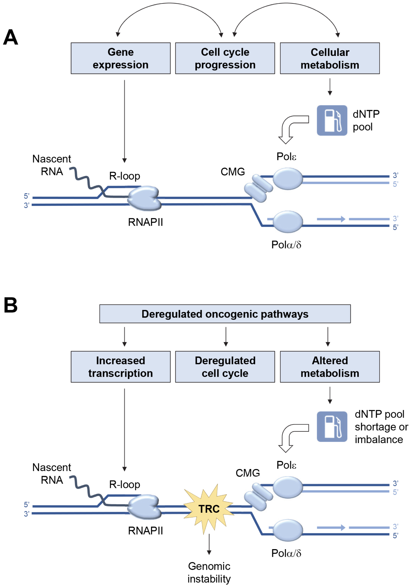
4. Oncogene-induced replication stress

It has been shown that aberrant expression of oncogenes such as Ras, Myc, or CycE can disrupt DNA replication in a variety of ways, for example by deregulating dNTP pools, increasing replication–transcription conflicts, or causing premature entry into S phase [39, 41, 42, 43]. This oncogene-induced replication stress represents a double-edged sword for cancer cells. While it contributes to cancer development by promoting genetic instability, it also slows tumor cell proliferation and activates anticancer barriers leading to apoptosis or senescence [44, 45, 46, 47]. To continue to proliferate, cancer cells must circumvent these barriers, while limiting the deleterious effects of replication stress on DNA replication. To do so, cancer cells become very dependent on the ATR-CHK1 pathway [48, 49, 50]. They also adapt to oncogene-induced replication stress by overexpressing components of the replication fork protection complex such as Claspin and Timeless, independently of ATR signaling [51]. This overexpression is observed in primary colon, breast and lung carcinomas and is generally associated with poor prognosis. Surprisingly, overexpression of Claspin and Timeless appears spontaneously in immortalized human fibroblasts expressing the H-RasV12 oncogene and allows them to escape replication stress and senescence [51].

5. Replication–transcription conflicts and cancer

Recent evidence indicates that the replication stress detected in cancer cells is largely due to replication–transcription conflicts [43]. Since transcription and replication complexes progress at roughly the same rate, the most severe conflicts result from head-on collisions [52, 53]. In order to limit the deleterious effects of head-on conflicts, the genome of bacterial species such as Bacillus subtilis has evolved such that most genes are codirectionally oriented with respect to replication forks [54]. In contrast, genes associated with pathogenesis and stress resistance are oriented in the opposite direction to replication, which locally increases DNA damage and promotes targeted evolution of these genes [55, 56]. This codirectional organization of replication and transcription is not found in eukaryotes because unlike bacterial chromosomes, which have only one origin of replication, eukaryotic chromosomes can carry several thousand. Moreover, the diversity of gene expression programs in metazoans does not allow to define a unique organization that would be adapted to all cell types. However, eukaryotes are not helpless against transcription–replication conflicts. Indeed, in the dynamic context of cell differentiation, human cells use a trick of promoting replication initiation upstream of highly transcribed genes [57, 58, 59], allowing these genes to be mostly replicated codirectionally. Thus, the average fork direction profile is positive (codirectional) between the transcriptional start (TSS) and termination (TTS) site in HeLa cells (Figure 2A). Interestingly, the direction of fork progression reverses at the end of the genes, indicating that replication forks initiated downstream of the genes converge with transcription at the TTS [60].

Figure 2.

Functional organization of the human genome and replication–transcription conflicts. (A) Distribution of replication fork direction (blue), R-loops (red) and a replication stress marker (p-RPA32-S33, black) at a metagene (16,000 human genes) aligned between transcription start (TSS) and termination (TTS) sites. A positive replication fork direction indicates that the fork is progressing in the same direction as transcription. (B) Schematic representation of the distribution of replication origins and R-loops (1), as well as RNA polymerases prior to replication (2), when replication is initiated upstream of the TSS (3), and when replication and transcription converge at the TTS, inducing replication stress (yellow spark) and accumulation of phospho-RPA32-S33 (4).

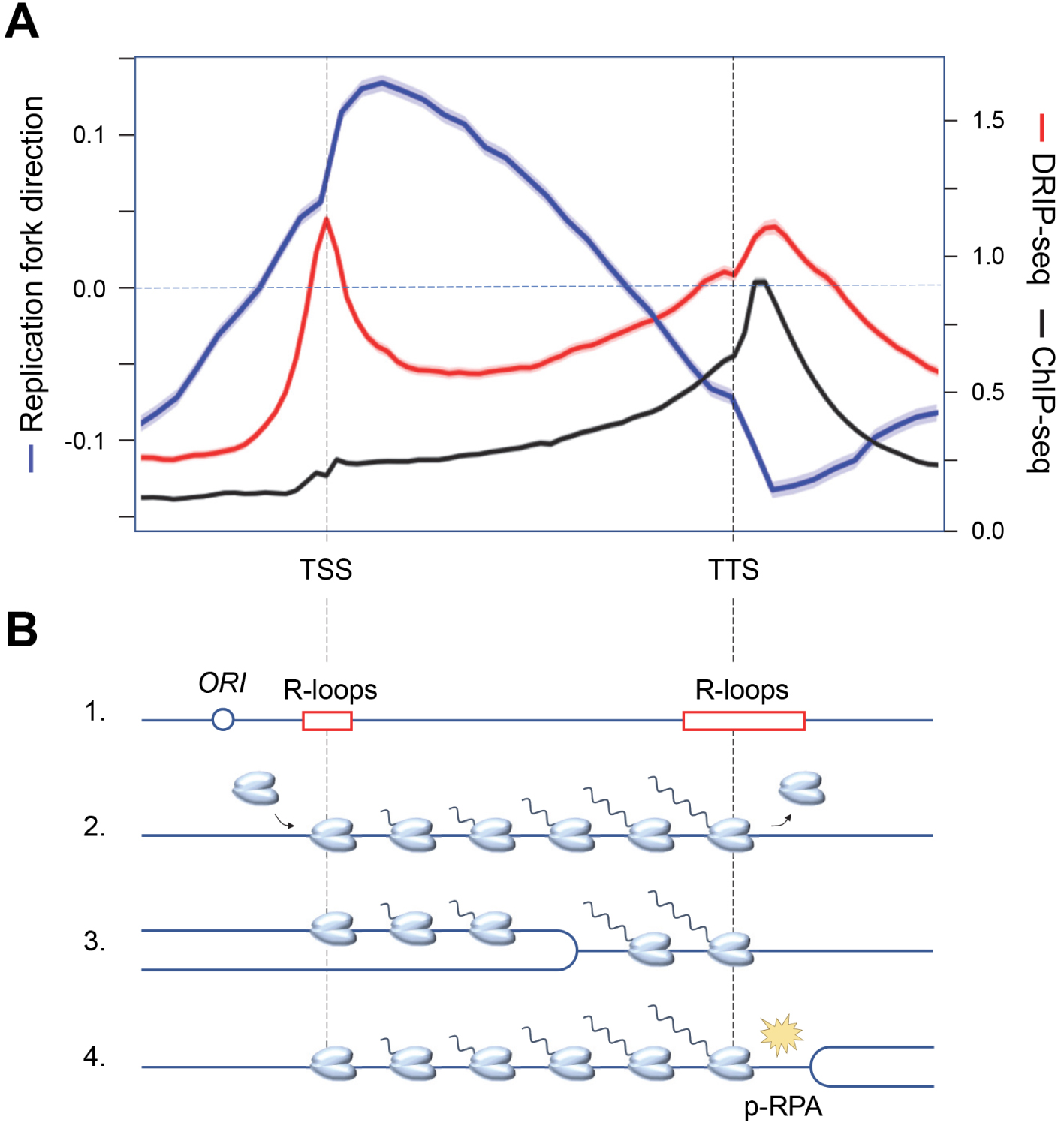
Another important question concerns the impact of R-loops on replication–transcription conflicts. Using methods such as DRIP-seq, which involve immunoprecipitating R-loops with an antibody specifically recognizing DNA:RNA hybrids and sequencing the associated nucleic acids, it has been shown that R-loops are highly abundant and can occupy up to 5% of the human genome [61, 62]. These structures play important physiological roles, such as control of gene expression, recombination of immunoglobulin genes, and mitochondrial DNA replication [39]. The question then arises as to whether all R-loops are inherently difficult to replicate, or whether a particular class of R-loops is particularly toxic to replication forks.

Analysis of the distribution of replication stress markers such as the ATR substrate phospho-RPA32-S33 showed that most R-loops are not associated with replication stress in the human genome [60]. Indeed, cotranscriptional R-loops are mainly located at the transcription initiation (TSS) and termination (TTS) sites, whereas an enrichment of phospho-RPA32-S33 is only detected at TTS (Figure 2A). Since these regions are on average replicated in the opposite direction to the direction of transcription, this means that replication–transcription conflicts occur preferentially at the TTS of highly transcribed genes enriched in R-loops, when the replication and transcription machineries converge (Figure 2B). Importantly, these conflicts do not automatically induce chromosomal breakage. Indeed, these events are only detected in Top1-depleted cells, which fail to manage the topological problems induced by replication and transcription convergence (Figure 3A). Furthermore, the replication stress observed in the absence of Top1 is suppressed by overexpression of RNase H1, an enzyme involved in the degradation of DNA:RNA hybrids, attesting to the fact that R-loops contribute to replication–transcription conflicts [60, 63].

Figure 3.

Model of replication–transcription conflict involving the formation of post-replicative RNA:DNA hybrids. (A) Convergence of the replisome and RNA polymerases (RNAPII) at transcription termination sites (TTS) induces accumulation of positive DNA supercoiling, which causes replication fork arrest and activation of the ATR kinase. The release of this topological stress is mediated by Top1. (B) ATR promotes the removal of fork-blocking RNA polymerases. At the same time, the DNA:RNA hybrid is degraded by RNase H to remove the R-loop. (C) In the absence of RNase H, the replicative helicase progresses beyond the position of the R-loop, transferring the DNA:RNA hybrid behind the fork and stops at the next RNAPII. (D) RNA:DNA hybrids located behind the fork interfere with post-replicative mechanisms such as resection of nascent DNA and prevent fork restart.

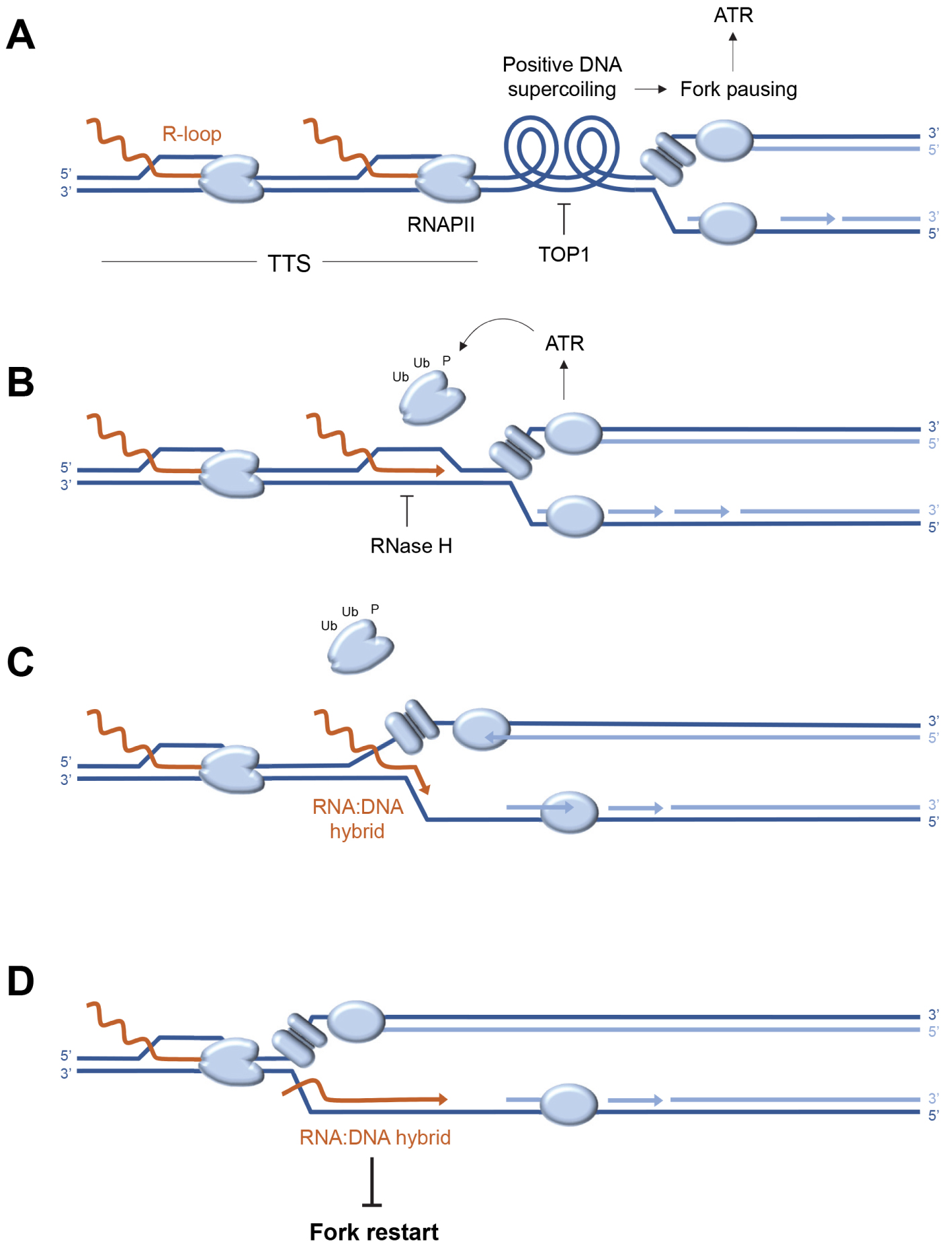
In conclusion, the functional organization of the human genome reduces the genetic instability associated with replication–transcription conflicts by limiting these conflicts to well-defined areas downstream of the genes. However, this regulation cannot operate if the initiation of replication takes place inside the genes, which occurs only very rarely under normal physiological conditions. Interestingly, a new class of replication origins has been identified within genes under oncogenic stress [64]. This intragenic initiation is associated with chromosomal rearrangements in cancers, possibly because it prevents cells from handling replication–transcription conflicts in an optimal manner.

6. R-loops and post-replicative DNA:RNA hybrids

The cell is well equipped to manage conflicts between replication and transcription. On the one hand, the replication fork is able to displace transcription complexes that block its passage (Figure 3B) by mobilizing various factors coordinated by the ATR kinase [65, 66, 67, 68]. On the other hand, the cell has a whole array of enzymes capable of degrading or displacing DNA:RNA hybrids [40]. These include RNases H1 and H2, which specifically degrade the RNA portion of DNA:RNA hybrids [69, 70, 71]. RNase H2 is frequently mutated in a severe interferonopathy called Aicardi–Goutières syndrome [72, 73]. Other key players include helicases such as Senataxin [74, 75] and FANCM [76]. Yet, despite this redundancy of repair mechanisms, some R-loops interfere with replication, following a mechanism that remains to be elucidated.

It is generally accepted that DNA:RNA hybrids are inherently difficult to replicate and block fork progression. However, when replication and transcription converge, the DNA:RNA hybrid is positioned on the strand opposite the replicative helicase (CMG), which should not block its progression (Figure 3C). Recent data derived from an in vitro replication system indicate that DNA:RNA hybrids do not block forks in this configuration, unless the displaced DNA strand is capable of forming a G4-like secondary structure [77]. Alternatively, DNA:RNA hybrids could interfere with post-replicative mechanisms acting behind the forks to promote their restart [78, 79]. This model is supported by unpublished data from our team, showing that in the absence of RNase H2, the resection of nascent DNA strands is inhibited by the persistence of undegraded DNA:RNA hybrids. Since fork resection contributes to fork restart [23, 80], these post-replicative DNA:RNA hybrids could induce replication stress not only by blocking forks, but also by preventing their restart (Figure 3D). This model is in agreement with recent data from the team of Massimo Lopes [81], demonstrating by electron microscopy the presence of DNA:RNA hybrids behind the replication forks.

These recent findings led us to propose a model in which R-loops and transcriptional complexes would not only represent an obstacle to the replication fork, but that the limited progression of replicative helicase at the site of conflict could convert the R-loop into a toxic DNA:RNA hybrid, interfering with fork restart if not rapidly degraded by RNase H2 (Figures 3C,D). This model emphasizes the importance for the cell of coordinated removal of the transcription complex and the R-loop to avoid the formation of a post-replicative DNA:RNA hybrid. It is worth noting that cells could be particularly sensitive to transcription–replication conflicts when replication forks are slowed by HU or oxidative stress [29] and that different mechanisms may operate when fork progression is blocked. Indeed, it has been reported that fork recovery at lesions caused by the Top1 poison camptothecin (CPT) involves the MUS81-dependent cleavage of stalled forks in human cells [82]. In fission yeast, fork restart at CPT lesions and at the RTS1 replication fork barrier depends on the degradation by RNase H of RNA primers on Okazaki fragments [83].

7. Conclusion and perspectives

In conclusion, the data gathered by different groups in the last few years shed new light on the molecular mechanisms at the origin of genetic instability in cancers. It now appears that despite optimal functional organization of the genome and redundancy of repair mechanisms, replication–transcription conflicts represent a major source of genetic instability, especially when oncogenic pathways are deregulated. R-loops contribute significantly to these conflicts, but the molecular mechanisms involved are more complex than initially thought. Indeed, these structures would not only act as replication barriers, but also act as postreplicative RNA:DNA hybrids to interfere with the restart of stalled forks. Such postreplicative RNA:DNA hybrids have recently been imaged by electron microscopy [81], but the mechanism of their formation remains unclear. New technologies such as nanopore sequencing could help determine the relative position of nascent DNA and RNA:DNA hybrids at the single molecule level. Another important question that remains to be elucidated is whether these post-replicative hybrids pre-exist before fork passage, as shown in Figure 3, or whether they are synthesized de novo by RNA polymerases acting behind the stalled fork. From this point of view, it is interesting to note that DNA:RNA hybrids are also detected at DNA double-strand breaks and that these structures interfere with DNA end resection, thus affecting homologous recombination repair mechanisms [84]. Finally, it is now well established that the repair mechanisms of forks and chromosomal breaks generate small DNA fragments that accumulate in the cytoplasm, activate the cGAS-STING pathway, and induce an inflammatory response [85, 86]. A very recent study also shows that DNA:RNA hybrids from R-loops after cleavage by repair enzymes can also activate the cGAS-STING pathway [87]. These data indicate that replication stress generated by replication–transcription conflicts and R-loops induces cellular responses acting beyond the boundaries of the cell. This endogenous stress could thus stimulate the elimination of abnormal cells by the immune system, opening promising prospects for the development of new cancer therapies.

Declaration of interests

The authors do not work for, advise, own shares in, or receive funds from any organization that could benefit from this article, and have declared no affiliations other than their research organizations.

Acknowledgements

The author thanks the members of the team “Maintenance of genome integrity during replication” for their contribution to the work mentioned in this review. Our work is supported by the Agence Nationale de la Recherche (ANR), the Institut National du Cancer (INCa), the Ligue contre le Cancer (labelled team) and the MSDAvenir Fund.


Bibliographie

[1] K. Labib Building a double hexamer of DNA helicase at eukaryotic replication origins, EMBO J., Volume 30 (2011), pp. 4853-4855 | DOI

[2] N. Y. Yao; M. O’Donnell Replisome structure and conformational dynamics underlie fork progression past obstacles, Curr. Opin. Cell Biol., Volume 21 (2009), pp. 336-343 | DOI

[3] M. Fragkos; O. Ganier; P. Coulombe; M. Mechali DNA replication origin activation in space and time, Nat. Rev. Mol. Cell Biol., Volume 16 (2015), pp. 360-374 | DOI

[4] P. Pasero; E. Schwob Think global, act local—how to regulate S phase from individual replication origins, Curr. Opin. Genet. Dev., Volume 10 (2000), pp. 178-186 | DOI

[5] S. P. Bell; A. Dutta DNA replication in eukaryotic cells, Annu. Rev. Biochem., Volume 71 (2002), pp. 333-374 | DOI

[6] J. F. Diffley Regulation of early events in chromosome replication, Curr. Biol., Volume 14 (2004), p. R778-R786 | DOI

[7] A. Johnson; M. O’Donnell Cellular DNA replicases: components and dynamics at the replication fork, Annu. Rev. Biochem., Volume 74 (2005), pp. 283-315 | DOI

[8] A. Gambus; R. C. Jones; A. Sanchez-Diaz; M. Kanemaki; F. van Deursen; R. D. Edmondson; K. Labib GINS maintains association of Cdc45 with MCM in replisome progression complexes at eukaryotic DNA replication forks, Nat. Cell Biol., Volume 8 (2006), pp. 358-366 | DOI

[9] D. Boos; P. Ferreira Origin firing regulations to control genome replication timing, Genes (Basel), Volume 10 (2019), 199 | DOI

[10] A. E. Vouzas; D. M. Gilbert Mammalian DNA replication timing, Cold Spring Harb. Perspect. Biol., Volume 13 (2021), a040162 | DOI

[11] H. Técher; P. Pasero The replication stress response on a narrow path between genomic instability and inflammation, Front. Cell Dev. Biol., Volume 9 (2021), 702584 | DOI

[12] Y. Xia The fate of two unstoppable trains after arriving destination: replisome disassembly during DNA replication termination, Front. Cell Dev. Biol., Volume 9 (2021), 658003

[13] M. K. Zeman; K. A. Cimprich Causes and consequences of replication stress, Nat. Cell Biol., Volume 16 (2014), pp. 2-9 | DOI

[14] J. C. Saldivar; D. Cortez; K. A. Cimprich The essential kinase ATR: ensuring faithful duplication of a challenging genome, Nat. Rev. Mol. Cell Biol., Volume 18 (2017), pp. 622-636 | DOI

[15] L. Zou Four pillars of the S-phase checkpoint, Genes Dev., Volume 27 (2013), pp. 227-233 | DOI

[16] A. Kumagai; W. G. Dunphy Claspin, a novel protein required for the activation of Chk1 during a DNA replication checkpoint response in Xenopus egg extracts, Mol. Cell, Volume 6 (2000), pp. 839-849 | DOI

[17] C. C. S. Chini; J. Chen Human Claspin is required for replication checkpoint control, J. Biol. Chem., Volume 278 (2003), pp. 30057-30062 | DOI

[18] K. Unsal-Kacmaz; P. D. Chastain; P.-P. Qu; P. Minoo; M. Cordeiro-Stone; A. Sancar; W. K. Kaufmann The human Tim/Tipin complex coordinates an intra-S checkpoint response to UV that slows replication fork displacement, Mol. Cell. Biol., Volume 27 (2007), pp. 3131-3142 | DOI

[19] S. Saxena; L. Zou Hallmarks of DNA replication stress, Mol. Cell, Volume 82 (2022), pp. 2298-2314 | DOI

[20] K. J. Neelsen; M. Lopes Replication fork reversal in eukaryotes: from dead end to dynamic response, Nat. Rev. Mol. Cell Biol., Volume 16 (2015), pp. 207-220 | DOI

[21] A. Quinet; D. Lemaçon; A. Vindigni Replication fork reversal: players and guardians, Mol. Cell, Volume 68 (2017), pp. 830-833 | DOI

[22] A. Delamarre; A. Barthe; C. de la Roche Saint-André; P. Luciano; R. Forey; I. Padioleau; M. Skrzypczak; K. Ginalski; V. Géli; P. Pasero et al. MRX increases chromatin accessibility at stalled replication forks to promote nascent DNA resection and cohesin loading, Mol. Cell, Volume 77 (2020), pp. 395-410 (e393) | DOI

[23] P. Pasero; A. Vindigni Nucleases acting at stalled forks: how to reboot the replication program with a few shortcuts, Annu. Rev. Genet., Volume 51 (2017), pp. 477-499 | DOI

[24] K. Schlacher; N. Christ; N. Siaud; A. Egashira; H. Wu; M. Jasin Double-strand break repair-independent role for BRCA2 in blocking stalled replication fork degradation by MRE11, Cell, Volume 145 (2011), pp. 529-542 | DOI

[25] S. Tye; G. E. Ronson; J. R. Morris A fork in the road: where homologous recombination and stalled replication fork protection part ways, Semin. Cell. Dev. Biol., Volume 113 (2020), pp. 14-26 | DOI

[26] T. Ubhi; G. W. Brown Exploiting DNA replication stress for cancer treatment, Cancer Res., Volume 79 (2019), pp. 1730-1739 | DOI

[27] B. Pardo; L. Crabbé; P. Pasero Signaling pathways of replication stress in yeast, FEMS Yeast Res., Volume 17 (2017), fow101

[28] K. Somyajit; R. Gupta; H. Sedlackova; K. J. Neelsen; F. Ochs; M. B. Rask; C. Choudhary; J. Lukas Redox-sensitive alteration of replisome architecture safeguards genome integrity, Science, Volume 358 (2017), pp. 797-802 | DOI

[29] M. Andrs; H. Stoy; B. Boleslavska; N. Chappidi; R. Kanagaraj; Z. Nascakova; S. Menon; S. Rao; A. Oravetzova; J. Dobrovolna et al. Excessive reactive oxygen species induce transcription-dependent replication stress, Nat. Commun., Volume 14 (2023), 1791 | DOI

[30] J. Poli; O. Tsaponina; L. Crabbe; A. Keszthelyi; V. Pantesco; A. Chabes; A. Lengronne; P. Pasero dNTP pools determine fork progression and origin usage under replication stress, EMBO J., Volume 31 (2012), pp. 883-894 | DOI

[31] A. Chabes; L. Thelander DNA building blocks at the foundation of better survival, Cell Cycle, Volume 2 (2003), pp. 171-173 | DOI

[32] R. Forey; A. Poveda; S. Sharma; A. Barthe; I. Padioleau; C. Renard; R. Lambert; M. Skrzypczak; K. Ginalski; A. Lengronne et al. Mec1 is activated at the onset of normal S phase by low-dNTP pools impeding DNA replication, Mol. Cell, Volume 78 (2020), pp. 396-410 | DOI

[33] F. Lazzaro; D. Novarina; F. Amara; L. Watt Danielle; E. Stone Jana; V. Costanzo; M. Burgers Peter; A. Kunkel Thomas; P. Plevani; M. Muzi-Falconi RNase H and postreplication repair protect cells from ribonucleotides incorporated in DNA, Mol. Cell, Volume 45 (2012), pp. 99-110 | DOI

[34] M. A. Reijns; B. Rabe; R. E. Rigby; P. Mill; K. R. Astell; L. A. Lettice; S. Boyle; A. Leitch; M. Keighren; F. Kilanowski et al. Enzymatic removal of ribonucleotides from DNA is essential for mammalian genome integrity and development, Cell, Volume 149 (2012), pp. 1008-1022 | DOI

[35] J. S. Williams; S. A. Lujan; T. A. Kunkel Processing ribonucleotides incorporated during eukaryotic DNA replication, Nat. Rev. Mol. Cell Biol., Volume 17 (2016), pp. 350-363 | DOI

[36] S. M. Cerritelli; J. Iranzo; S. Sharma; A. Chabes; R. J. Crouch; D. Tollervey; A. El Hage High density of unrepaired genomic ribonucleotides leads to Topoisomerase 1-mediated severe growth defects in absence of ribonucleotide reductase, Nucleic Acids Res., Volume 48 (2020), pp. 4274-4297 | DOI

[37] B. Gomez-Gonzalez; A. Aguilera Transcription-mediated replication hindrance: a major driver of genome instability, Genes Dev., Volume 33 (2019), pp. 1008-1026 | DOI

[38] S. Kemiha; J. Poli; Y. L. Lin; A. Lengronne; P. Pasero Toxic R-loops: cause or consequence of replication stress?, DNA Repair (Amst.), Volume 107 (2021), 103199 | DOI

[39] T. Garcia-Muse; A. Aguilera R loops: from physiological to pathological roles, Cell, Volume 179 (2019), pp. 604-618 | DOI

[40] J. R. Brickner; J. L. Garzon; K. A. Cimprich Walking a tightrope: the complex balancing act of R-loops in genome stability, Mol. Cell, Volume 82 (2022), pp. 2267-2297 | DOI

[41] M. Macheret; T. D. Halazonetis DNA replication stress as a hallmark of cancer, Annu. Rev. Pathol.: Mech. Dis., Volume 10 (2015), pp. 425-448 | DOI

[42] C. Bester Assaf; M. Roniger; S. Oren Yifat; M. Im Michael; D. Sarni; M. Chaoat; A. Bensimon; G. Zamir; S. Shewach Donna; B. Kerem Nucleotide deficiency promotes genomic instability in early stages of cancer development, Cell, Volume 145 (2011), pp. 435-446 | DOI

[43] E. Petermann; L. Lan; L. Zou Sources, resolution and physiological relevance of R-loops and RNA–DNA hybrids, Nat. Rev. Mol. Cell Biol., Volume 23 (2022), pp. 521-540 | DOI

[44] R. Di Micco; M. Fumagalli; A. Cicalese; S. Piccinin; P. Gasparini; C. Luise; C. Schurra; M. Garre; P. G. Nuciforo; A. Bensimon et al. Oncogene-induced senescence is a DNA damage response triggered by DNA hyper-replication, Nature, Volume 444 (2006), pp. 638-642 | DOI

[45] J. Bartkova; N. Rezaei; M. Liontos; P. Karakaidos; D. Kletsas; N. Issaeva; L. V. Vassiliou; E. Kolettas; K. Niforou; V. C. Zoumpourlis et al. Oncogene-induced senescence is part of the tumorigenesis barrier imposed by DNA damage checkpoints, Nature, Volume 444 (2006), pp. 633-637 | DOI

[46] L. I. Toledo; M. Murga; O. Fernandez-Capetillo Targeting ATR and Chk1 kinases for cancer treatment: a new model for new (and old) drugs, Mol. Oncol., Volume 5 (2011), pp. 368-373 | DOI

[47] L. I. Toledo; M. Murga; P. Gutierrez-Martinez; R. Soria; O. Fernandez-Capetillo ATR signaling can drive cells into senescence in the absence of DNA breaks, Genes Dev., Volume 22 (2008), pp. 297-302 | DOI

[48] J. Bartek; M. Mistrik; J. Bartkova Thresholds of replication stress signaling in cancer development and treatment, Nat. Struct. Mol. Biol., Volume 19 (2012), pp. 5-7 | DOI

[49] M. Murga; S. Campaner; A. J. Lopez-Contreras; L. I. Toledo; R. Soria; M. F. Montaña; L. D’Artista; T. Schleker; C. Guerra; E. Garcia et al. Exploiting oncogene-induced replicative stress for the selective killing of Myc-driven tumors, Nat. Struct. Mol. Biol., Volume 18 (2011), pp. 1331-1335 | DOI

[50] A. J. Lopez-Contreras; O. Fernandez-Capetillo The ATR barrier to replication-born DNA damage, DNA Repair, Volume 9 (2010), pp. 1249-1255 | DOI

[51] J. N. Bianco; V. Bergoglio; Y. L. Lin; M. J. Pillaire; A. L. Schmitz; J. Gilhodes; A. Lusque; J. Mazieres; M. Lacroix-Triki; T. I. Roumeliotis et al. Overexpression of Claspin and Timeless protects cancer cells from replication stress in a checkpoint-independent manner, Nat. Commun., Volume 10 (2019), 910 | DOI

[52] S. Hamperl; M. J. Bocek; J. C. Saldivar; T. Swigut; K. A. Cimprich Transcription–replication conflict orientation modulates R-loop levels and activates distinct DNA damage responses, Cell, Volume 170 (2017), pp. 774-786 (e719) | DOI

[53] M. Lalonde; M. Trauner; M. Werner; S. Hamperl Consequences and resolution of transcription–replication conflicts, Life (Basel), Volume 11 (2021), 637

[54] E. P. C. Rocha The organization of the bacterial genome, Annu. Rev. Genet., Volume 42 (2008), pp. 211-233 | DOI

[55] H. Merrikh Spatial and temporal control of evolution through replication–transcription conflicts, Trends Microbiol., Volume 25 (2017), pp. 515-521 | DOI

[56] K. S. Lang; A. N. Hall; C. N. Merrikh; M. Ragheb; H. Tabakh; A. J. Pollock; J. J. Woodward; J. E. Dreifus; H. Merrikh Replication–transcription conflicts generate R-loops that orchestrate bacterial stress survival and pathogenesis, Cell, Volume 170 (2017), pp. 787-799 (e718) | DOI

[57] Y. H. Chen; S. Keegan; M. Kahli; P. Tonzi; D. Fenyo; T. T. Huang; D. J. Smith Transcription shapes DNA replication initiation and termination in human cells, Nat. Struct. Mol. Biol., Volume 26 (2019), pp. 67-77 | DOI

[58] N. Petryk; M. Kahli; Y. d’Aubenton-Carafa; Y. Jaszczyszyn; Y. Shen; M. Silvain; C. Thermes; C.-L. Chen; O. Hyrien Replication landscape of the human genome, Nat. Commun., Volume 7 (2016), 10208 | DOI

[59] C. Cayrou; P. Coulombe; A. Vigneron; S. Stanojcic; O. Ganier; I. Peiffer; E. Rivals; A. Puy; S. Laurent-Chabalier; R. Desprat et al. Genome-scale analysis of metazoan replication origins reveals their organization in specific but flexible sites defined by conserved features, Genome Res., Volume 21 (2011), pp. 1438-1449 | DOI

[60] A. Promonet; I. Padioleau; Y. Liu; L. Sanz; A. Biernacka; A. L. Schmitz; M. Skrzypczak; A. Sarrazin; C. Mettling; M. Rowicka et al. Topoisomerase 1 prevents replication stress at R-loop-enriched transcription termination sites, Nat. Commun., Volume 11 (2020), 3940 | DOI

[61] D. Castillo-Guzman; F. Chédin Defining R-loop classes and their contributions to genome instability, DNA Repair (Amst.), Volume 106 (2021), 103182 | DOI

[62] A. Sanz Lionel; R. Hartono Stella; W. Lim Yoong; S. Steyaert; A. Rajpurkar; A. Ginno Paul; X. Xu; F. Chédin Prevalent, dynamic, and conserved R-loop structures associate with specific epigenomic signatures in mammals, Mol. Cell, Volume 63 (2016), pp. 167-178 | DOI

[63] S. Tuduri; L. Crabbé; C. Conti; H. Tourrière; H. Holtgreve-Grez; A. Jauch; V. Pantesco; J. De Vos; A. Thomas; C. Theillet et al. Topoisomerase I suppresses genomic instability by preventing interference between replication and transcription, Nat. Cell Biol., Volume 11 (2009), pp. 1315-1324 | DOI

[64] M. Macheret; T. D. Halazonetis Intragenic origins due to short G1 phases underlie oncogene-induced DNA replication stress, Nature, Volume 555 (2018), pp. 112-116 | DOI

[65] J. S. Im; M. Keaton; K. Y. Lee; P. Kumar; J. Park; A. Dutta ATR checkpoint kinase and CRL1betaTRCP collaborate to degrade ASF1a and thus repress genes overlapping with clusters of stalled replication forks, Genes Dev., Volume 28 (2014), pp. 875-887 | DOI

[66] J. Poli; C. B. Gerhold; A. Tosi; N. Hustedt; A. Seeber; R. Sack; F. Herzog; P. Pasero; K. Shimada; K. P. Hopfner et al. Mec1, INO80, and the PAF1 complex cooperate to limit transcription replication conflicts through RNAPII removal during replication stress, Genes Dev., Volume 30 (2016), pp. 337-354 | DOI

[67] H. B. Landsverk; L. E. Sandquist; L. T. E. Bay; B. Steurer; C. Campsteijn; O. J. B. Landsverk; J. A. Marteijn; E. Petermann; L. Trinkle-Mulcahy; R. G. Syljuåsen WDR82/PNUTS-PP1 prevents transcription–replication conflicts by promoting RNA polymerase II degradation on chromatin, Cell Rep., Volume 33 (2020), 108469 | DOI

[68] V. Hurst; K. Challa; F. Jonas; R. Forey; R. Sack; J. Seebacher; C. D. Schmid; N. Barkai; K. Shimada; S. M. Gasser et al. A regulatory phosphorylation site on Mec1 controls chromatin occupancy of RNA polymerases during replication stress, EMBO J., Volume 40 (2021), e108439 | DOI

[69] M. Hyjek; M. Figiel; M. Nowotny RNases H: structure and mechanism, DNA Rep. (Amst.), Volume 84 (2019), 102672 | DOI

[70] H. D. Nguyen; T. Yadav; S. Giri; B. Saez; T. A. Graubert; L. Zou Functions of replication protein A as a sensor of R loops and a regulator of RNaseH1, Mol. Cell, Volume 65 (2017), pp. 832-847 (e834) | DOI

[71] A. Lockhart; V. B. Pires; F. Bento; V. Kellner; S. Luke-Glaser; G. Yakoub; H. D. Ulrich; B. Luke RNase H1 and H2 are differentially regulated to process RNA–DNA hybrids, Cell Rep., Volume 29 (2019), pp. 2890-2900 (e2895) | DOI

[72] Y. J. Crow; N. Manel Aicardi–Goutieres syndrome and the type I interferonopathies, Nat. Rev. Immunol., Volume 15 (2015), pp. 429-440 | DOI

[73] A. Cristini; M. Tellier; F. Constantinescu; C. Accalai; L. O. Albulescu; R. Heiringhoff; N. Bery; O. Sordet; S. Murphy; N. Gromak RNase H2, mutated in Aicardi–Goutières syndrome, resolves co-transcriptional R-loops to prevent DNA breaks and inflammation, Nat. Commun., Volume 13 (2022), 2961 | DOI

[74] M. Groh; L. O. Albulescu; A. Cristini; N. Gromak Senataxin: genome guardian at the interface of transcription and neurodegeneration, J. Mol. Biol., Volume 429 (2017), pp. 3181-3195 | DOI

[75] U. Aiello; D. Challal; G. Wentzinger; A. Lengronne; R. Appanah; P. Pasero; B. Palancade; D. Libri Sen1 is a key regulator of transcription-driven conflicts, Mol. Cell, Volume 82 (2022), pp. 2952-2966 | DOI

[76] C. Hodson; S. van Twest; M. Dylewska; J. J. O’Rourke; W. Tan; V. J. Murphy; M. Walia; L. Abbouche; J. Nieminuszczy; E. Dunn et al. Branchpoint translocation by fork remodelers as a general mechanism of R-loop removal, Cell Rep., Volume 41 (2022), 111749 | DOI

[77] C. Kumar; S. Batra; J. D. Griffith; D. Remus The interplay of RNA:DNA hybrid structure and G-quadruplexes determines the outcome of R-loop-replisome collisions, Elife, Volume 10 (2021), e72286 | DOI

[78] S. Barroso; E. Herrera-Moyano; S. Muñoz; M. García-Rubio; B. Gómez-González; A. Aguilera The DNA damage response acts as a safeguard against harmful DNA–RNA hybrids of different origins, EMBO Rep., Volume 20 (2019), e47250 | DOI

[79] S. Šviković; A. Crisp; S. M. Tan-Wong; T. A. Guilliam; A. J. Doherty; N. J. Proudfoot; G. Guilbaud; J. E. Sale R-loop formation during S phase is restricted by PrimPol-mediated repriming, EMBO J., Volume 38 (2019), e99793 | DOI

[80] A. Teixeira-Silva; A. Ait Saada; J. Hardy; I. Iraqui; M. C. Nocente; K. Fréon; S. A. E. Lambert The end-joining factor Ku acts in the end-resection of double strand break-free arrested replication forks, Nat. Commun., Volume 8 (2017), 1982 | DOI

[81] H. Stoy; K. Zwicky; D. Kuster; K. S. Lang; J. Krietsch; M. P. Crossley; J. A. Schmid; K. A. Cimprich; H. Merrikh; M. Lopes Direct visualization of transcription–replication conflicts reveals post-replicative DNA:RNA hybrids, Nat. Struct. Mol. Biol., Volume 30 (2023), pp. 348-359 | DOI

[82] N. Chappidi; Z. Nascakova; B. Boleslavska; R. Zellweger; E. Isik; M. Andrs; S. Menon; J. Dobrovolna; C. Balbo Pogliano; J. Matos et al. Fork cleavage-religation cycle and active transcription mediate replication restart after fork stalling at Co-transcriptional R-loops, Mol. Cell, Volume 77 (2020), pp. 528-541 (e528) | DOI

[83] C. Audoynaud; K. Schirmeisen; A. Ait Saada; A. Gesnik; P. Fernández-Varela; V. Boucherit; V. Ropars; A. Chaudhuri; K. Fréon; J. B. Charbonnier et al. RNA:DNA hybrids from Okazaki fragments contribute to establish the Ku-mediated barrier to replication-fork degradation, Mol. Cell, Volume 83 (2023), pp. 1061-1074 | DOI

[84] A. Marnef; G. Legube R-loops as Janus-faced modulators of DNA repair, Nat. Cell Biol., Volume 23 (2021), pp. 305-313 | DOI

[85] F. Coquel; C. Neumayer; Y.-L. Lin; P. Pasero SAMHD1 and the innate immune response to cytosolic DNA during DNA replication, Curr. Opin. Immunol., Volume 56 (2019), pp. 24-30 | DOI

[86] F. Coquel; M. Silva; H. Técher; K. Zadorozhny; S. Sharma; J. Nieminuszczy; C. Mettling; E. Dardillac; A. Barthe; A. Schmitz et al. SAMHD1 acts at stalled replication forks to prevent interferon induction, Nature, Volume 557 (2018), pp. 57-61 | DOI

[87] M. P. Crossley; C. Song; M. J. Bocek; J. H. Choi; J. Kousorous; A. Sathirachinda; C. Lin; J. R. Brickner; G. Bai; H. Lans et al. R-loop-derived cytoplasmic RNA–DNA hybrids activate an immune response, Nature, Volume 613 (2023), pp. 187-194 | DOI


Commentaires - Politique


Ces articles pourraient vous intéresser

SAMHD1 agit sur les fourches de réplication bloquées pour empêcher l’induction d’interféron

F. Coquel; M. J. Silva; H. Técher; ...

C. R. Biol (2020)


A prominent role for segmental duplications in modeling Eukaryotic genomes

Romain Koszul; Gilles Fischer

C. R. Biol (2009)


Gene organization inside replication domains in mammalian genomes

Lamia Zaghloul; Antoine Baker; Benjamin Audit; ...

C. R. Méca (2012)