




Plan

Article de synthèse
Sexual dimorphisms in juvenile development under nutritionnal stress
[Dimorphismes sexuels des adaptations au stress nutritionnel pendant le développement juvénile]
Comptes Rendus. Biologies, Volume 349 (2026), pp. 231-243

Résumés

Nutrition is a fundamental regulator of juvenile development. The persistently high global prevalence of childhood undernutrition and consequent stunting underscores the urgent need to better understand how nutritional status shapes developmental trajectories. Biological sex has recently emerged as a key risk factor for stunting, with boys consistently exhibiting greater vulnerability. This review examines the biological basis of sex differences in physiological adaptation to nutritional stress, focusing specifically on dietary protein deficiency during the juvenile period. Two critical developmental processes display pronounced sexual dimorphism under protein restriction: linear growth and reproductive maturation. Evidence from both preclinical and clinical studies indicates that juvenile protein restriction differentially affects these processes in males and females, highlighting the necessity of considering sex as a biological variable in experimental design and data interpretation. Shared endocrine signals, including Fibroblast Growth Factor 21, may contribute to these sex-specific adaptive responses. Further elucidating the nutritional, endocrine and metabolic mechanisms underlying sexually dimorphic outcomes of juvenile development is essential for enhancing the mechanistic value and translational relevance of preclinical and clinical research.

La nutrition est un déterminant fondamental du développement juvénile. La forte prévalence de la malnutrition infantile à l’échelle mondiale, et du retard de croissance qui en résulte, souligne la nécessité de mieux comprendre l’influence de l’état nutritionnel sur les trajectoires développementales. Le sexe biologique a récemment été identifié comme un facteur déterminant de la susceptibilité au retard de croissance : les garçons présentent une vulnérabilité accrue par rapport aux filles. Cette revue explore les bases biologiques des différences entre les sexes dans l’adaptation physiologique au stress nutritionnel, en portant une attention particulière aux conséquences d’une carence en protéines alimentaires au cours de la période juvénile. Deux processus majeurs du développement présentent un dimorphisme sexuel marqué en situation de restriction protéique : la croissance linéaire et le développement reproducteur. Les données issues d’études précliniques et cliniques montrent que la restriction protéique juvénile affecte différemment ces processus chez les mâles et les femelles, soulignant l’importance d’intégrer le sexe comme variable biologique dans la conception des études et l’interprétation des résultats. Des voies endocriniennes communes, notamment impliquant le Fibroblast Growth Factor 21 (FGF21), pourraient contribuer à ces réponses adaptatives spécifiques au sexe. Une meilleure compréhension des mécanismes nutritionnels, endocriniens et métaboliques à l’origine de ces différences est indispensable pour renforcer la portée mécanistique et la pertinence translationnelle des recherches précliniques et cliniques.

Métadonnées
Reçu le :
Révisé le :
Accepté le :
Publié le :
DOI : 10.5802/crbiol.203
Keywords: Malnutrition, Juvenile, Sex differences, Growth, Puberty, Development
Mots-clés : Malnutrition, Juvénile, Différence entre les sexes, Croissance, Puberté, Développement
Note : Cet article a été soumis à l’invitation du comité éditorial, dans le cadre de l’attribution du prix Jaffé 2025 à F. Leulier.

Amélie Joly  1   ; François Leulier  1

1 Institut de Génomique Fonctionnelle de Lyon, Ecole Normale Supérieure de Lyon, CNRS UMR5242, Université Claude Bernard Lyon 1, Lyon, France
Licence : CC-BY 4.0
Droits d'auteur : Les auteurs conservent leurs droits
Amélie Joly; François Leulier. Sexual dimorphisms in juvenile development under nutritionnal stress. Comptes Rendus. Biologies, Volume 349 (2026), pp. 231-243. doi: 10.5802/crbiol.203
@article{CRBIOL_2026__349_G1_231_0,
     author = {Am\'elie Joly and Fran\c{c}ois Leulier},
     title = {Sexual dimorphisms in juvenile development under nutritionnal stress},
     journal = {Comptes Rendus. Biologies},
     pages = {231--243},
     year = {2026},
     publisher = {Acad\'emie des sciences, Paris},
     volume = {349},
     doi = {10.5802/crbiol.203},
     language = {en},
}
TY  - JOUR
AU  - Amélie Joly
AU  - François Leulier
TI  - Sexual dimorphisms in juvenile development under nutritionnal stress
JO  - Comptes Rendus. Biologies
PY  - 2026
SP  - 231
EP  - 243
VL  - 349
PB  - Académie des sciences, Paris
DO  - 10.5802/crbiol.203
LA  - en
ID  - CRBIOL_2026__349_G1_231_0
ER  - 
%0 Journal Article
%A Amélie Joly
%A François Leulier
%T Sexual dimorphisms in juvenile development under nutritionnal stress
%J Comptes Rendus. Biologies
%D 2026
%P 231-243
%V 349
%I Académie des sciences, Paris
%R 10.5802/crbiol.203
%G en
%F CRBIOL_2026__349_G1_231_0

Version originale du texte intégral (Proposez une traduction )

Le texte intégral ci-dessous peut contenir quelques erreurs de conversion par rapport à la version officielle de l'article publié.

1. Introduction

Dietary macronutrient content during childhood profoundly affects key physiological processes, including linear growth and provision of metabolic substrates to the developing tissues. While an excess in a child’s intake of nutrients leads to overweight and obesity, chronic dietary protein deficiency results in stunting (Endrinikapoulos et al., 2023). Stunting is defined by the World Health Organization (WHO) as a height-for-age below two standard deviations from the median of the WHO Child Growth Standards for the same age and sex; and affected approximately 150 million children under 5 in 2024 (United Nations Children’s Fund (UNICEF), World Health Organization, International Bank for Reconstruction and Development/The World Bank, 2025).

Considerable attention has been given to the causes and long-term consequences of stunting during childhood (de Onis and Branca, 2016). Various factors, encompassing socioeconomic status, sanitary levels and maternal education, have been identified as determinants of stunting (Bhutta et al., 2017; Soofi et al., 2023). Alongside environmental and maternal factors, child sex is now recognized as a biological risk factor, with boys being more vulnerable than girls to stunting (Mertens et al., 2023; Slemming et al., 2017; Thompson, 2021; Thurstans et al., 2020; USAID Scaling Up Nutrition Learning and Evaluation (SUN LE), National Food and Nutrition Commission (NFNC), 2020). However, explanations for these sex differences are often speculative and their biological basis remains unclear.

The protein to carbohydrates ratio (P/C ratio) is known to regulate various physiological processes in both rodents and humans (Solon-Biet et al., 2015). In adults, the response to changes in this P/C ratio, and in particular to a reduction in dietary protein availability, is sexually dimorphic (Green et al., 2022; Larson et al., 2017). However, the sex specificity of the physiological adaptation to protein restriction during the juvenile period has only been sparsely investigated. In this review, we will explore the molecular, cellular and endocrine mechanisms that could contribute to this dimorphism, focusing on two processes that are key during the juvenile period and intrinsically dimorphic: linear growth and reproductive development. We will also discuss how sex differences are considered in biomedical research and how this affects research output.

2. Sexual dimorphism in growth

2.1. Are males really bigger than females?

One of the most apparent sexually dimorphic traits in physiology is the difference in body size that can occur between males and females. This dimorphism in size has been considered for decades to result from sexual selection and reproductive role division. Sexual size dimorphism varies widely across taxa (Hedrick and Temeles, 1989). Male-biased size dimorphism is common in many mammals and birds, and is often associated with sexual selection favouring larger males. Female-biased size dimorphism also occurs in numerous insects and fishes, and has frequently been associated with selection for fecundity, as the production of large gametes in females is correlated with body size (Shingleton and Vea, 2023).

Yet, sexual dimorphism in size appears to be less frequent than initially thought—at least in mammals. Indeed, a 2014 study challenged this paradigm by demonstrating that there was no significant sexual dimorphism in size in 95 species of small mammals (Lu et al., 2014). More recently, an analysis of more than 400 species of mammals found that, if males are larger in 45% of the species, 39% of the species do not display size sexual dimorphism and 16% of the species have larger females (Tombak et al., 2024). However, sexual size dimorphism has been well established in humans (Schäppi et al., 2022; WHO and de Onis, 2006) and mice (Ruff et al., 2017).

2.2. Molecular mechanisms of sexual dimorphism in growth

Biologically, differences in body size can originate from differences in initial size (fetal or egg), rate and duration of body growth or of body degradation (Shingleton and Vea, 2023). In humans and mice, sex differences in growth mainly arise from increased growth rate in males (Jiang et al., 2023; WHO and de Onis, 2006) even though males already have a significantly higher weight at birth (Moore, 2024; WHO and de Onis, 2006). This greater body size in males is associated with increased skeletal mass and bone mass, at least in humans (Nieves et al., 2005).

In mammals, post-natal linear growth is regulated by the somatotropic axis, where Growth Hormone (GH) secreted by the pituitary gland elicits Insulin-like Growth Factor 1 (IGF-1) production from liver cells. Both GH and IGF-1 instruct peripheral tissues to promote tissue and bone growth (Yakar and Isaksson, 2016). Differences in body size between males and females mainly arise from differences in the functioning of the somatotropic axis. Indeed, studies in rats and mice evidenced that GH secretory patterns are different in males and females (MacLeod et al., 1991; Tannenbaum and Martin, 1976). GH secretion is pulsatile in males, while it is constant in females; in addition, global GH plasma levels are higher in males (Adams et al., 2015; Jansson et al., 1985). This GH secretion pattern drives liver IGF-1 production and ultimately controls the sex-specific linear growth pattern (Figure 1).

Figure 1.

Molecular mechanisms of sexual dimorphism in growth in rodents and humans. GHRH: Growth Hormone Releasing Hormone, SST: Somatostatin, GH: Growth Hormone, IGF-1: Insulin-like Growth Factor 1. The roles of sex steroids in the regulation of the somatotropic axis are complex and not exhaustively represented in this figure. This illustration was created with Biorender.com.
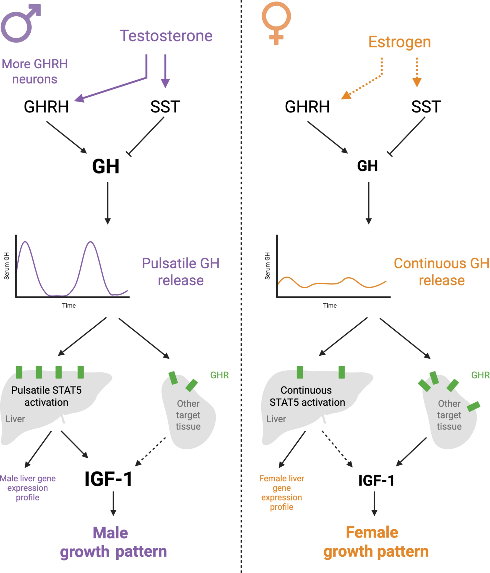
The differential activation pattern of the GH signaling pathway in the liver also elicits sex-dependent gene expression and participates in the sexually dimorphic identity of the liver (Lefebvre and Staels, 2021). In the male liver, pulsatile GH secretion yields intermittent activity of the transcription factor STAT5, while in the female liver, continuous GH secretion is associated with persistent STAT5 activity (Zhang et al., 2012). This different activation pattern leads to sex-specific STAT5 binding pattern and contributes to the liver sex-biased genetic program (ibid.). Strikingly, hepatic male-biased genes are similarly expressed in males and females at post-natal day 28 (Nikkanen et al., 2022), suggesting that male-specific gene expression patterns are acquired after this time point. Importantly, sex-dependent GH secretion patterns and associated liver dimorphism regulate other key physiological functions such as lipid metabolism, drug metabolism and immunity (Lefebvre and Staels, 2021; Nikkanen et al., 2022).

GH secretion is regulated by two hypothalamic neuropeptides: Growth Hormone Releasing Hormone (GHRH), which stimulates GH production, and somatostatin (SST), which inhibits GH production. The regulation of these two signals is intrinsically sexually dimorphic. For example, GHRH neurons are more numerous in males than in females from post-natal day 20 (Nurhidayat et al., 1999), which might contribute to male-specific GH secretion patterns. In addition, male but not female Sst-KO mice have altered GH secretory patterns and expression of sexually dimorphic genes in the liver (Adams et al., 2015), suggesting that the role of SST in regulating the somatotropic axis is more prominent in males than in females.

Interestingly, injection of testosterone into prepubertal boys with delayed sexual maturation was sufficient to elicit an increase in GH release and pulsatility (Link et al., 1986). In addition, gonadectomy before puberty decreased the growth of males but not females (Klappenbach et al., 2023). This suggests a central role for androgens in mediating sex-dependent growth phenotypes, and notably in regulating GH sensitivity and secretion in males. In line with that, orchidectomy decreases Ghrh and Sst expression in the hypothalamus, which is reversed by testosterone administration (Chowen-Breed et al., 1989; Zeitler et al., 1990), demonstrating a role of testosterone in regulating GH production. Overall, androgens appear to be pivotal in the regulation of the somatotropic axis, influencing both GH secretion and hepatic sensitivity to GH (Tenuta et al., 2021). The action of estrogen on the GH/IGF-1 pathway is also complex and depends on the developmental window considered (for a review on this topic, see (K.-C. Leung et al., 2004)). While estrogen also stimulates GH release (Marin et al., 1994), it plays an inhibitory role on GH signaling and liver IGF-1 output (K. C. Leung et al., 2003).

Interestingly, mice deficient for Igf1 in the liver displayed a sex-specific response to GH stimulation, with females growing better than males (Liu et al., 2000). In this model, the expression of GH receptor mRNA was decreased in the liver of females and increased in their white adipose tissue compared to males (ibid.). This suggests that the growth of males mainly depends on liver IGF-1 production, whereas the growth of females might be mediated by extra-hepatic GH signaling and sources of IGF-1. This sex-dependent phenotype was not affected by gonadectomy in both sexes and therefore likely not due to sex hormones (ibid.).

Overall, sex differences are evident at every level of the GH/IGF-1 somatotropic growth axis and drive sexually dimorphic linear growth patterns. Although the contributions of sex hormones to these processes are complex, they highlight the tight integration between the somatotropic and gonadotropic axes.

2.3. Juvenile protein malnutrition and growth

Sex-specific regulation of linear growth implies that responses to perturbations of the somatotropic axis may likewise be sexually dimorphic. In the context of malnutrition, it has been recently described that child sex contributes to vulnerability to stunting (Mertens et al., 2023; Slemming et al., 2017; Thompson, 2021; Thurstans et al., 2020; USAID Scaling Up Nutrition Learning and Evaluation (SUN LE), National Food and Nutrition Commission (NFNC), 2020). A meta-analysis of 38 studies examining stunting concluded that boys have 29% higher odds of being stunted than girls (Thurstans et al., 2020), despite regional exceptions where girls are more at risk than boys (Baig-Ansari et al., 2006; Mittal et al., 2007). Consistently, the mean height-for-age z-score in stunted children is lower for males than for females (Bork and Diallo, 2017; Wamani et al., 2007). It has been established that dietary protein restriction in juvenile mice also triggers stunting in male animals (Borck et al., 2017; Ozaki et al., 2015; Schwarzer, Gautam, et al., 2023; Soriano et al., 2010; Thoumas et al., 2025). In a recent study, we found that juvenile female mice exhibited minimal growth retardation compared to males when fed a low-protein diet after weaning (Joly et al., 2025), consistent with human data.

Several studies have demonstrated that adult female mice are relatively less sensitive to the physiological consequences of dietary protein dilution than males, displaying no major changes in body weight and glucose metabolism compared to males (Green et al., 2022; Larson et al., 2017). On the contrary, a systematic review concluded that protein restriction during prenatal life did not yield significant sexually dimorphic adaptation (Christians et al., 2021), suggesting that the sex-dependent growth response to protein malnutrition might depend on the developmental time window.

Stunted children exhibit disruption of the somatotropic axis, with reduced IGF-1 circulating levels together with elevated GH production, which is typical of a GH resistant state (Olusi et al., 1977; A. T. Soliman et al., 1986; Fazeli and Klibanski, 2014). This alteration of the somatotropic axis in response to juvenile malnutrition has also been reproduced in rodent models (Ozaki et al., 2015; Schwarzer, Makki, et al., 2016; Takenaka, Oki, et al., 2000; Takenaka, Hirosawa, et al., 1993). Several leads have been explored to explain this GH resistance.

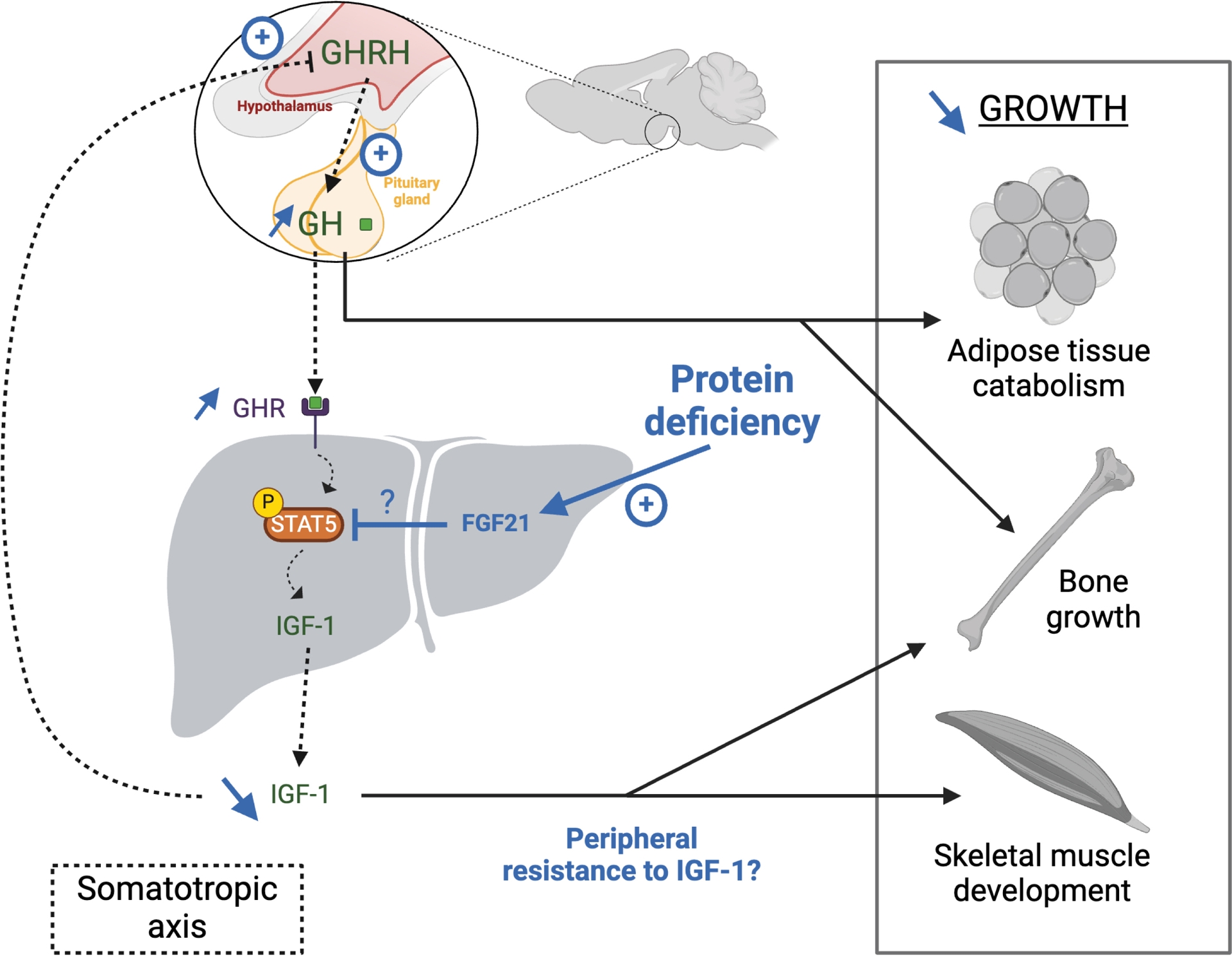
Fibroblast Growth Factor 21 (FGF21) is a liver-derived hormone which secretion is triggered by protein restriction—among other signals—in both humans and mice (Laeger et al., 2014; Na and Park, 2024). Interestingly, FGF21 induction is reproducibly lower in female mice compared to males in response to protein restriction (Green et al., 2022; Joly et al., 2025; Larson et al., 2017). Experiments in Fgf21-transgenic mice have demonstrated that Fgf21 overexpression is associated with impaired growth, GH resistance, decreased serum IGF-1 and STAT5 phosphorylation (Ding et al., 2012; Inagaki et al., 2008). Moreover, whole-body Fgf21 KO juvenile mice had better body and bone growth in response to calorie restriction than their WT counterparts (Kubicky et al., 2012). Since actived STAT5 is a major mediator of GH effect in the liver, it has been hypothesized that FGF21 attenuates GH signaling by decreasing STAT5 activation (Figure 2). However, a recent study challenged this view by demonstrating that inhibition of Fgf21 expression did not prevent GH resistance and that FGF21 alone could not impair STAT5 phosphorylation in cellulo (Saito et al., 2024). Consistent with these observations, we found that liver-specific deletion of Fgf21 did not prevent growth delay, nor decreased IGF-1 circulating levels in malnourished male mice (Joly et al., 2025), challenging the idea that liver-derived FGF21 could be a regulator of linear growth.

Figure 2.

Somatotropic axis alterations during juvenile protein malnutrition. Dysregulations induced by protein scarcity are indicated in blue. GH: Growth Hormone, GHRH: GH Releasing Hormone, GHR: GH receptor, STAT5: Signal transducer and activator of transcription 5, FGF21; Fibroblast Growth Factor 21, IGF-1: Insulin-like Growth Factor 1. This illustration was created with Biorender.com.
Other molecular mediators, such as the histone deacetylase Sirtuin 1, insulin or ghrelin, have been suggested as possible determinants of GH resistance upon protein restriction (Fazeli and Klibanski, 2014; Kojima et al., 2001).

3. Sexually dimorphic effects of nutritional stress on puberty

3.1. A brief overview of sexual maturation

The juvenile period, from weaning to young adulthood, is a critical developmental window that notably determines the onset of puberty. In mammals, pubertal development is regulated by the hypothalamic-pituitary-gonadal axis (Anderson et al., 2024). Puberty is initiated by the release of gonadotropin-releasing hormone (GnRH) from the hypothalamus, which drives the secretion of two gonadotropins: luteinizing hormone (LH) and follicle-stimulating hormone (FSH) from the pituitary gland. LH and FSH then activate maturation of the gonads and production of sex steroids (Figure 3).

Figure 3.

Juvenile protein malnutrition and female puberty. Putative disregulations induced by protein restriction are indicated in blue. AVPV: Anteroventral Periventricular nucleus, SCN: Suprachiasmatic nucleus, ARC: Arcuate nucleus, Avp: Vasopressin, Kiss1: Kisspeptin, GnRH: Gonadotropin-releasing hormone, LH: luteinizing hormone, FSH: Follicle stimulating hormone. This illustration was created with Biorender.com.
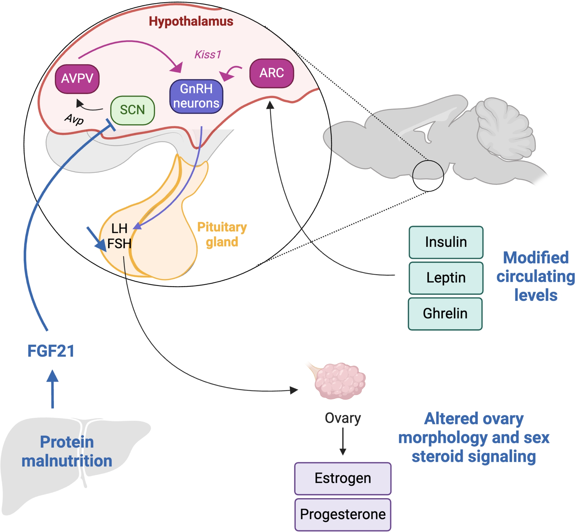
The timing of activation of this hypothalamic-pituitary-gonadal axis, particularly the timing of initiation of the GnRH pulses, determines the timing of puberty in both males and females. This timing is tightly regulated by many signals, especially those related to nutrition (Connor, 2011). In particular, GnRH release is controlled by neurons producing kisspeptin, which are located in two nuclei of the hypothalamus: the arcuate nucleus (ARC) and the anteroventral periventricular nucleus (AVPV). Kisspeptin neurons of the ARC are sensitive to insulin, ghrelin, and leptin, while those of the AVPV are only found in females and are regulated by sex steroids (Navarro, 2020) (Figure 3).

3.2. Nutritional effect on sexual maturation

In 1963, Kennedy and Mitra demonstrated that body weight, and therefore nutrition, is a major determinant of the timing of puberty (Kennedy and Mitra, 1963). To this aim, they modulated the size of rat litters to create conditions of overnutrition and undernutrition and observed that the onset of puberty was delayed in larger/malnourished litters. Further studies in rodents and humans have allowed to establish that a sufficient energy store must be available to allow puberty initiation (Anderson et al., 2024). Consistently, obesity during childhood has been associated with earlier puberty (Reinehr and Roth, 2019). In addition to puberty onset, sufficient food supply is required for the maintenance of female fertility and ovulatory cycles (Frisch and McArthur, 1974). The relationship between food availability and fertility has been established across kingdoms, including in mice, Caenorhabditis elegans (Gerisch et al., 2001), Drosophila melanogaster (Gorter et al., 2016) and in birds such as Melozone alberti (Davies et al., 2015). It is evaluated that nutritional status during childhood can account for up to 25% of the variation in the onset of puberty (A. Soliman, De Sanctis and Elalaily, 2014).

Leptin produced by the adipose tissue appears to be a key regulator of pubertal timing. Indeed, leptin-deficient obese mice are sterile (Ingalls et al., 1950), which can be prevented by injection of recombinant leptin (Chehab et al., 1996). Similarly, injection of leptin into wild-type mice accelerates the onset of puberty (Ahima et al., 1997). One possible interpretation is that leptin indicates to the brain that energy stores are sufficient to support the energy demand of puberty and of future reproduction (Mauvais-Jarvis, 2024). Indeed, specific deletion of the leptin receptor in target neuronal populations has demonstrated that leptin signals to agouti-related peptide (AgRP)/neuropeptide Y (NPY)-expressing neurons of the hypothalamus to regulate sexual maturation (Egan et al., 2017).

Consistently, the stomach-derived orexigenic and adipogenic hormone ghrelin seems to be an antagonist of sexual maturation. Notably, ghrelin might participate in the fetal programming of reproduction, since the administration of a ghrelin antagonist in pregnant mice advanced the puberty of male and female offspring (Torres et al., 2018). In addition, plasma ghrelin levels have been negatively correlated with FSH, LH and testosterone levels (El-Eshmawy et al., 2010).

Interestingly, humans with poorly controlled type 1 diabetes might have impaired puberty (Anderson et al., 2024). On the contrary, hyperinsulinemia in female mice is associated with early puberty (Saleh et al., 2022). These data suggest that insulin also regulates pubertal timing. Indeed, brain-specific knockout of the insulin receptor is associated with impaired sexual maturation, due to dysregulation of LH secretion (Brüning et al., 2000).

Overall, nutritional signals regulate endocrine cues to modulate the timing of puberty, with calorie restriction impairing or delaying reproductive development. Consistently, endocrine signals of low energy resources, such as low leptin or insulin and ghrelin, tend to block reproductive maturation (Figure 3). Interestingly, signaling of those nutrition-associated hormones is highly dysregulated during protein juvenile malnutrition (Kasai et al., 2012; Volcko et al., 2024; Yamada et al., 2019). In the next paragraph, we will explore how dietary protein deficiency affects reproductive maturation.

3.3. Juvenile protein malnutrition and reproductive maturation

In rodent models, juvenile mice fed a low-protein diet have altered reproductive development compared to animals fed an isocaloric diet with normal protein content. This suggests that protein deficiency itself might affect reproductive maturation, independently of calorie restriction. For example, weanling female rats receiving a low-protein diet displayed altered ovarian morphology, as well as reduced sex steroid receptors expression in the ovaries (de Morais Oliveira et al., 2021). Similarly, malnourished juvenile male rats had decreased testosterone levels, hypothalamic androgen receptor expression and testis weight (Karaca et al., 2003; Oliveira et al., 2018). In a recent study, we directly compared the effect of low-protein diet after weaning on the puberty of juvenile male and female littermates. While females exhibited a strong impairment of reproductive development (delayed vaginal opening and first estrus, reduced uterus weight), males had an almost normal onset of puberty, measured as the occurrence of balano-preputial separation (Joly et al., 2025). Malnourished males also had normal sperm count and seminal vesicle weight (when corrected for body weight), but reduced testis weight.

Interestingly, maternal protein restriction during pregnancy and/or early post-natal life has been strongly connected to a perturbation of puberty onset in offspring of both sexes (Connor, 2011). In rats, maternal malnutrition induces a delayed puberty onset (Brasil et al., 2005) and impaired ovarian function (Guzmán et al., 2014) of female offspring. Similarly, male rats underfed during fetal and post-natal life displayed reduced testicular weight and altered testicular structure (Genovese et al., 2010).

Malnourished children are thought to display delayed sexual development (A. Soliman, De Sanctis, Alaaraj, et al., 2021; A. Soliman, De Sanctis and Elalaily, 2014). However, to our knowledge, only one large study directly compared puberty onset between malnourished and well-fed children (Kulin et al., 1982). In this study, malnourished Kenyan girls and boys displayed a delay in the occurrence of early stages of sexual maturation, but could catch up in later stages (ibid.). Similarly, a more recent study assessing reproductive development using Tanner Stages in children from Pakistan concluded that malnourished children enter puberty at a slightly older age (0.5–0.7 year delay compared to non-stunted children), but do not display a delay in puberty completion (Campisi et al., 2021). Interestingly, a follow-up study in Barbadian children demonstrated that malnutrition during the first year of life was associated with delayed puberty in girls, but not in boys (Galler et al., 1985), suggesting putative sex differences in the impact of stunting on puberty onset in humans, consistent with our own observations in mice.

These sex-dependent effects of malnutrition on pubertal development may, in part, be mediated by FGF21. Indeed, studies in mice with a transgenic overexpression of FGF21 have demonstrated that females, but not males, display infertility (Inagaki et al., 2008). In addition, FGF21 overexpression induces delayed puberty, impaired mating and hypothalamic hypogonadism in females (Owen et al., 2013). In this model, FGF21 indirectly impairs kisspeptin production from neurons of the anteroventral periventricular (AVPV) nucleus of the hypothalamus, which normally stimulates GnRH release (ibid.). Since AVPV kisspeptin neurons are absent in males (Navarro, 2020), this would explain the sex-specific effect of FGF21 on sexual maturation (Figure 3). Consistent with the idea that FGF21 could act as a brake on female puberty, we found that the liver-specific deletion of Fgf21—which abolishes FGF21 circulating levels—partially rescued the puberty delay in malnourished females but had no effect on the puberty of malnourished males (Joly et al., 2025).

However, the role of FGF21 in regulating female fertility and puberty is still debated. In particular, a 2016 study found that the effects of FGF21 on fertility might be indirect (Singhal et al., 2016). Indeed, mice fed a ketogenic diet, and thus having elevated levels of circulating FGF21, display normal puberty and fertility, and direct infusion of FGF21 in the brain does not impair fertility (ibid.). One possibility is that the delayed puberty and infertility observed in FGF21-overexpressing mice might be related to an increase in energy expenditure induced by FGF21 and the subsequent increase in energy requirements.

Therefore, a protein deficit in the absence of a calorie deficit is sufficient to perturb the puberty onset, at least in mice. In addition, the effects of protein restriction on pubertal development might be sex-specific and FGF21-associated.

4. From male physiology to physiology

The effects of protein and calorie deficiency on child development have been investigated for decades in both humans and rodent models. In contrast, the systematic study of sex differences in this context has only emerged more recently. Among biological processes, pubertal onset and reproductive maturation—owing to their pronounced sexual dimorphism—have been the most extensively examined in both sexes. Beyond reproduction, however, sex differences have historically received far less attention. It is now increasingly recognized that the physiology of non-reproductive organs, including the gut and liver, differs substantially between males and females (Ameku et al., 2025; Miguel-Aliaga, 2022). Together with growing evidence of sex-specific outcomes in multiple areas of biomedical research (Waltz et al., 2021), these findings underscore the need for more rigorous consideration of sex as a biological variable in both pre-clinical and clinical studies.

The idea that estrous/menstrual cycles increase variability and introduce bias when analyzing data is a misconception (Levy et al., 2023; Zucker et al., 2022). Indeed, several reports have shown that variability in females, regardless of the stage of the estrous cycle, is not greater than normal variability in males (Zucker et al., 2022). This assumption has led to a preferential use of male animals in preclinical research (Woitowich et al., 2020; Zucker et al., 2022) as well as in clinical trials (Geller et al., 2018). The National Institutes of Health (NIH) required the inclusion of women in clinical trials from 1993 (Chen et al., 2022) and released in 2015 a policy of “sex as a biological variable” requiring scientists to address sex-biased responses in their research (Waltz et al., 2021). Guidelines now exist to favor better research practices for the inclusion of both sexes (Rich-Edwards and Maney, 2023). However, in 2020, only 75% of American scientists declared that they report the sex of the animals they use, and only half declared that they analyze their data by sex (Waltz et al., 2021). A follow-up study comparing papers published in different fields of biology in 2009 and 2019 found that the proportion of studies including both sexes increased from 28% to only 49% within 10 years (Woitowich et al., 2020), meaning that, in 2019, half of the studies surveyed still used only one sex or did not report sex. Consistently, another analysis conducted on papers published in surgery-related journals in 2011 and 2012 found that 80% of the studies included only males (Yoon et al., 2014). In the field of physiology, the proportion of studies including both sexes jumped from 13% to 36% between 2009 and 2019. However, half of the studies surveyed in this field were still conducted on males only (Woitowich et al., 2020). Therefore, the knowledge generated by half of the studies published in 2019 is restricted to male physiology, despite often being interpreted as broadly applicable. While female inclusion has increased, most studies still fail to conduct sex-based analyses (50% in 2009, 42% in 2019) (Woitowich et al., 2020; Zucker et al., 2022), meaning that female biology remains underreported.

Despite its obvious importance, the research community often fails to include sex as a biological variable and to report for both sexes. How to explain this phenomenon? Two of the main explanations to justify the lack of utilization of females or of sex-based analyses were that “[only males were used] to minimize possible variability resulting from sex differences” and that “[the authors] did not have the power to examine for sex effects” (Woitowich et al., 2020). In addition, a common belief is that taking into account sex as a biological variable will double the cost of research, while it might only need to choose the right experimental design or statistical analysis (Miguel-Aliaga, 2022; Zucker et al., 2022). Interestingly, a 2019 bibliometric analysis revealed that reporting sex was associated with publications in journals with lower impact factors (Sugimoto et al., 2019) and that, when the first and last author of a paper were females, there was increased probability of sex-reporting (ibid.).

Taken together, these considerations highlight that our current understanding of sex differences in biology remains sparse and incomplete. A deeper investigation of sexual dimorphism is therefore essential to strengthen the validity and translational relevance of both preclinical and clinical research.

5. Conclusion

Juvenile development under nutritional stress is shaped by profound sexual dimorphisms that extend beyond reproductive biology. While pubertal development represents a clear example of such dimorphism, linear growth is also a fundamentally sex-dependent process, whose regulation and adaptive responses differ between males and females. Both processes are differentially impacted by juvenile protein malnutrition in males and females, a pattern that might also apply to other forms of nutritional stress.

Despite the increasing recognition of sex as a biological variable, major gaps remain in our understanding of sex-specific developmental biology. Identifying regulatory pathways that are shared between sexes but drive sex-specific adaptations provides a powerful framework for disentangling sexual dimorphisms in physiology. Dissecting these key nutritional, endocrine and metabolic mechanisms is essential to fully capture the complexity of sex-dependent developmental trajectories and, ultimately, to improve the interpretation and translational relevance of preclinical and clinical studies.

Declaration of interests

The authors do not work for, adivse, own shares in, or receive funds from any organization that could benefit from this article, and have declared no affiliation other than their research organisations.


Bibliographie

[Adams et al., 2015] J. M. Adams; V. Otero-Corchon; G. L. Hammond; J. D. Veldhuis; N. Qi; M. J. Low Somatostatin is essential for the sexual dimorphism of GH secretion, corticosteroid-binding globulin production, and corticosterone levels in mice, Endocrinology, Volume 156 (2015) no. 3, pp. 1052-1065 | DOI

[Ahima et al., 1997] R. S. Ahima; J. Dushay; S. N. Flier; D. Prabakaran; J. S. Flier Leptin accelerates the onset of puberty in normal female mice, J. Clin. Investig., Volume 99 (1997) no. 3, pp. 391-395 | DOI

[Ameku et al., 2025] T. Ameku; A. Laddach; H. Beckwith; A. Milona; L. S. Rogers; C. Schwayer; E. Nye; I. R. Tough; J.-L. Thoumas; U. K. Gautam; Y.-F. Wang; S. Jha; A. Castano-Medina; C. Amourda; P. M. Vaelli; S. Gevers; E. E. Irvine; L. Meyer; I. Andrew; K. L. Choi; B. Patel; A. J. Francis; C. Studd; L. Game; G. Young; K. G. Murphy; B. Owen; D. J. Withers; M. Rodriguez-Colman; H. M. Cox; P. Liberali; M. Schwarzer; F. Leulier; V. Pachnis; N. W. Bellono; I. Miguel-Aliaga Growth of the maternal intestine during reproduction, Cell, Volume 188 (2025) no. 10, p. 2738-2756.e22 | DOI

[Anderson et al., 2024] G. M. Anderson; J. W. Hill; U. B. Kaiser; V. M. Navarro; K. K. Ong; J. R. B. Perry; V. Prevot; M. Tena-Sempere; C. F. Elias Metabolic control of puberty: 60 years in the footsteps of Kennedy and Mitra’s seminal work, Nat. Rev. Endocrinol., Volume 20 (2024) no. 2, pp. 111-123 | DOI

[Baig-Ansari et al., 2006] N. Baig-Ansari; M. H. Rahbar; Z. A. Bhutta; S. H. Badruddin Child’s gender and household food insecurity are associated with stunting among young Pakistani children residing in urban squatter settlements, Food Nutr. Bull., Volume 27 (2006) no. 2, pp. 114-127 | DOI

[Bhutta et al., 2017] Z. A. Bhutta; J. A. Berkley; R. H. J. Bandsma; M. Kerac; I. Trehan; A. Briend Severe childhood malnutrition, Nat. Rev. Dis. Primers, Volume 3 (2017) no. 1, 17067 | DOI

[Borck et al., 2017] P. C. Borck; T. M. Batista; J. F. Vettorazzi; R. L. Camargo; A. C. Boschero; E. Vieira; E. M. Carneiro Protein malnutrition after weaning disrupts peripheral clock and daily insulin secretion in mice, J. Nutr. Biochem., Volume 50 (2017), pp. 54-65 | DOI

[Bork and Diallo, 2017] K. A. Bork; A. Diallo Boys Are more stunted than girls from early infancy to 3 years of age in rural Senegal, J. Nutr., Volume 147 (2017) no. 5, pp. 940-947 | DOI

[Brasil et al., 2005] F. B. Brasil; T. S. Faria; W. S. Costa; F. J. B. Sampaio; C. F. Ramos The pups’ endometrium morphology is affected by maternal malnutrition during suckling, Maturitas, Volume 51 (2005) no. 4, pp. 405-412 | DOI

[Brüning et al., 2000] J. C. Brüning; D. Gautam; D. J. Burks; J. Gillette; M. Schubert; P. C. Orban; R. Klein; W. Krone; D. Müller-Wieland; C. R. Kahn Role of brain insulin receptor in control of body weight and reproduction, Science, Volume 289 (2000) no. 5487, pp. 2122-2125 | DOI

[Campisi et al., 2021] S. C. Campisi; K. N. Humayun; Y. Wasan; S. B. Soofi; M. Islam; A. Hussain; A. Shakeel; A. Vandermorris; O. Söder; Z. A. Bhutta The relationship between pubertal timing and under-nutrition in rural Pakistan, J. Adolesc., Volume 88 (2021) no. 1, pp. 58-66 | DOI

[Chehab et al., 1996] F. F. Chehab; M. E. Lim; R. Lu Correction of the sterility defect in homozygous obese female mice by treatment with the human recombinant leptin, Nat. Genet., Volume 12 (1996) no. 3, pp. 318-320 | DOI

[Chen et al., 2022] Y. Chen; M. Kim; S. Paye; B. A. Benayoun Sex as a biological variable in nutrition research: from human studies to animal models, Ann. Rev. Nutr., Volume 42 (2022), pp. 227-250 | DOI

[Chowen-Breed et al., 1989] J. A. Chowen-Breed; R. A. Steiner; D. K. Clifton Sexual dimorphism and testosterone-dependent regulation of somatostatin gene expression in the periventricular nucleus of the rat brain, Endocrinology, Volume 125 (1989) no. 1, pp. 357-362 | DOI

[Christians et al., 2021] J. K. Christians; H. K. Shergill; A. Y. K. Albert Sex-dependent effects of prenatal food and protein restriction on offspring physiology in rats and mice: systematic review and meta-analyses, Biol. Sex Differ., Volume 12 (2021), 21 | DOI

[Connor, 2011] N. E. Connor Impact of fetal and neonatal malnutrition on the onset of puberty and associated noncommunicable disease risks, Adolesc. Health Med. Ther., Volume 2 (2011), pp. 15-25 | DOI

[Davies et al., 2015] S. Davies; T. Cros; D. Richard; S. L. Meddle; K. Tsutsui; P. Deviche Food availability, energetic constraints and reproductive development in a wild seasonally breeding songbird, Funct. Ecol., Volume 29 (2015) no. 11, pp. 1421-1434 | DOI

[de Morais Oliveira et al., 2021] D. A. de Morais Oliveira; L. A. Lupi; H. S. Silveira; L. G. de Almeida Chuffa Protein restriction during puberty alters nutritional parameters and affects ovarian and uterine histomorphometry in adulthood in rats, Int. J. Exp. Pathol., Volume 102 (2021) no. 2, pp. 93-104 | DOI

[de Onis and Branca, 2016] M. de Onis; F. Branca Childhood stunting: a global perspective: Childhood stunting: a global perspective, Matern. Child Nutr., Volume 12 (2016), pp. 12-26 | DOI

[Ding et al., 2012] X. Ding; J. Boney-Montoya; B. M. Owen; A. L. Bookout; K. C. Coate; D. J. Mangelsdorf; S. A. Kliewer βKlotho is required for fibroblast growth factor 21 effects on growth and metabolism, Cell Metab., Volume 16 (2012) no. 3, pp. 387-393 | DOI

[Egan et al., 2017] O. K. Egan; M. A. Inglis; G. M. Anderson Leptin signaling in AgRP neurons modulates puberty onset and adult fertility in mice, J. Neurosci., Volume 37 (2017) no. 14, pp. 3875-3886 | DOI

[El-Eshmawy et al., 2010] M. M. El-Eshmawy; I. A. A. Aal; A. K. El hawary Association of ghrelin and leptin with reproductive hormones in constitutional delay of growth and puberty, Reprod. Biol. Endocrinol., Volume 8 (2010), 153 | DOI

[Endrinikapoulos et al., 2023] A. Endrinikapoulos; D. N. Afifah; M. Mexitalia; R. Andoyo; I. Hatimah; N. Nuryanto Study of the importance of protein needs for catch-up growth in Indonesian stunted children: a narrative review, SAGE Open Med., Volume 11 (2023), 20503121231165562 | DOI

[Fazeli and Klibanski, 2014] P. K. Fazeli; A. Klibanski Determinants of GH resistance in malnutrition, J. Endocrinol., Volume 220 (2014) no. 3, p. R57-R65 | DOI

[Frisch and McArthur, 1974] R. E. Frisch; J. W. McArthur Menstrual cycles: fatness as a determinant of minimum weight for height necessary for their maintenance or onset, Science, Volume 185 (1974) no. 4155, pp. 949-951 | DOI

[Galler et al., 1985] J. R. Galler; F. Ramsey; G. Solimano A follow-up study of the effects of early malnutrition on subsequent development. I. Physical growth and sexual maturation during adolescence, Pediatr. Res., Volume 19 (1985) no. 6, pp. 518-523 | DOI

[Geller et al., 2018] S. E. Geller; A. R. Koch; P. Roesch; A. Filut; E. Hallgren; M. Carnes The more things change, the more they stay the same: a study to evaluate compliance with inclusion and assessment of women and minorities in randomized controlled trials, Acad. Med., Volume 93 (2018) no. 4, pp. 630-635 | DOI

[Genovese et al., 2010] P. Genovese; M. E. Núñez; C. Pombo; A. Bielli Undernutrition during foetal and post-natal life affects testicular structure and reduces the number of sertoli cells in the adult rat, Reprod. Domest. Anim., Volume 45 (2010) no. 2, pp. 233-236 | DOI

[Gerisch et al., 2001] B. Gerisch; C. Weitzel; C. Kober-Eisermann; V. Rottiers; A. Antebi A hormonal signaling pathway influencing C. elegans metabolism, reproductive development, and life span, Dev. Cell, Volume 1 (2001) no. 6, pp. 841-851 | DOI

[Gorter et al., 2016] J. A. Gorter; S. Jagadeesh; C. Gahr; J. J. Boonekamp; J. D. Levine; J.-C. Billeter The nutritional and hedonic value of food modulate sexual receptivity in Drosophila melanogaster females, Sci. Rep., Volume 6 (2016) no. 1, 19441 | DOI

[Green et al., 2022] C. L. Green; H. H. Pak; N. E. Richardson; V. Flores; D. Yu; J. L. Tomasiewicz; S. N. Dumas; K. Kredell; J. W. Fan; C. Kirsh; K. Chaiyakul; M. E. Murphy; R. Babygirija; G. A. Barrett-Wilt; J. Rabinowitz; I. M. Ong; C. Jang; J. Simcox; D. W. Lamming Sex and genetic background define the metabolic, physiologic, and molecular response to protein restriction, Cell Metab., Volume 34 (2022) no. 2, p. 209-226.e5 | DOI

[Guzmán et al., 2014] C. Guzmán; R. García-Becerra; M. A. Aguilar-Medina; I. Méndez; H. Merchant-Larios; E. Zambrano Maternal protein restriction during pregnancy and/or lactation negatively affects follicular ovarian development and steroidogenesis in the prepubertal rat offspring, Arch. Med. Res., Volume 45 (2014) no. 4, pp. 294-300 | DOI

[Hedrick and Temeles, 1989] A. V. Hedrick; E. J. Temeles The evolution of sexual dimorphism in animals: Hypotheses and tests, Trends Ecol. Evol., Volume 4 (1989) no. 5, pp. 136-138 | DOI

[Inagaki et al., 2008] T. Inagaki; V. Y. Lin; R. Goetz; M. Mohammadi; D. J. Mangelsdorf; S. A. Kliewer Inhibition of growth hormone signaling by the fasting-induced hormone FGF21, Cell Metab., Volume 8 (2008) no. 1, pp. 77-83 | DOI

[Ingalls et al., 1950] A. M. Ingalls; M. M. Dickie; G. D. Snell Obese, a new mutation in the house mouse, J. Hered., Volume 41 (1950) no. 12, pp. 317-318 | DOI

[Jansson et al., 1985] J. O. Jansson; S. Edén; O. Isaksson Sexual dimorphism in the control of growth hormone secretion, Endocr. Rev., Volume 6 (1985) no. 2, pp. 128-150 | DOI

[Jiang et al., 2023] N Jiang; C. J. Cheng; J. Gelfond; R. Strong; V. Diaz; J. F. Nelson Prepubertal castration eliminates sex differences in lifespan and growth trajectories in genetically heterogeneous mice, Aging Cell, Volume 22 (2023) no. 8, e13891 | DOI

[Joly et al., 2025] A. Joly; L. Rebiffé; A. Lambert; E. Caillon; S. Hughes; B. Gillet; I. Rahioui; P. Da Silva; J. Bruse; A. Fougerat; F. De Vadder; F. Leulier Liver FGF21 mediates sex-specific adaptation to juvenile protein malnutrition (2025) | DOI

[Karaca et al., 2003] F. Karaca; H. Dönmez; M. Karsli Effects of protein deficiency on testosterone levels, semen quality and testicular histology in the developing male rat, Scand. J. Lab. Anim. Sci., Volume 30 (2003), pp. 7-13

[Kasai et al., 2012] A. Kasai; P. Gama; E. P. Alvares Protein restriction inhibits gastric cell proliferation during rat postnatal growth in parallel to ghrelin changes, Nutrition, Volume 28 (2012) no. 6, pp. 707-712 | DOI

[Kennedy and Mitra, 1963] G. C. Kennedy; J. Mitra Body weight and food intake as initiating factors for puberty in the rat, J. Physiol., Volume 166 (1963) no. 2, pp. 408-418 | DOI

[Klappenbach et al., 2023] C. M. Klappenbach; Q. Wang; A. L. Jensen; N. C. Glodosky; K. Delevich Sex and timing of gonadectomy relative to puberty interact to influence weight, body composition, and feeding behaviors in mice, Horm. Behav., Volume 151 (2023), 105350 | DOI

[Kojima et al., 2001] M. Kojima; H. Hosoda; H. Matsuo; K. Kangawa Ghrelin: discovery of the natural endogenous ligand for the growth hormone secretagogue receptor, Trends Endocrinol. Metab., Volume 12 (2001) no. 3, pp. 118-122 | DOI

[Kubicky et al., 2012] R. A. Kubicky; S. Wu; A. Kharitonenkov; F. De Luca Role of fibroblast growth factor 21 (FGF21) in undernutrition-related attenuation of growth in mice, Endocrinology, Volume 153 (2012) no. 5, pp. 2287-2295 | DOI

[Kulin et al., 1982] H. E. Kulin; N. Bwibo; D. Mutie; S. J. Santner The effect of chronic childhood malnutrition on pubertal growth and development, Am. J. Clin. Nutr., Volume 36 (1982) no. 3, pp. 527-536 | DOI

[Laeger et al., 2014] T. Laeger; T. M. Henagan; D. C. Albarado; L. M. Redman; G. A. Bray; R. C. Noland; H. Münzberg; S. M. Hutson; T. W. Gettys; M. W. Schwartz; C. D. Morrison FGF21 is an endocrine signal of protein restriction, J. Clin. Investig., Volume 124 (2014) no. 9, pp. 3913-3922 | DOI

[Larson et al., 2017] K. R. Larson; K. A. Russo; Y. Fang; N. Mohajerani; M. L. Goodson; K. K. Ryan Sex differences in the hormonal and metabolic response to dietary protein dilution, Endocrinology, Volume 158 (2017) no. 10, pp. 3477-3487 | DOI

[Lefebvre and Staels, 2021] P. Lefebvre; B. Staels Hepatic sexual dimorphism—implications for non-alcoholic fatty liver disease, Nat. Rev. Endocrinol., Volume 17 (2021) no. 11, pp. 662-670 | DOI

[Leung et al., 2003] K. C. Leung; N. Doyle; M. Ballesteros; K. Sjogren; C. K. W. Watts; T. H. Low; G. M. Leong; R. J. M. Ross; K. K. Y. Ho Estrogen inhibits GH signaling by suppressing GH-induced JAK2 phosphorylation, an effect mediated by SOCS-2, Proc. Natl. Acad. Sci. USA, Volume 100 (2003) no. 3, pp. 1016-1021 | DOI

[Leung et al., 2004] K.-C. Leung; G. Johannsson; G. M. Leong; K. K. Y. Ho Estrogen regulation of growth hormone action, Endocr. Rev., Volume 25 (2004) no. 5, pp. 693-721 | DOI

[Levy et al., 2023] D. R. Levy; N. Hunter; S. Lin; E. M. Robinson; W. Gillis; E. B. Conlin; R. Anyoha; R. M. Shansky; S. R. Datta Mouse spontaneous behavior reflects individual variation rather than estrous state, Curr. Biol., Volume 33 (2023) no. 7, p. 1358-1364.e4 | DOI

[Link et al., 1986] K. Link; R. M. Blizzard; W. S. Evans; D. L. Kaiser; M. W. Parker; A. D. Rogol The effect of androgens on the pulsatile release and the twenty-four-hour mean concentration of growth hormone in peripubertal males, J. Clin. Endocrinol. Metab., Volume 62 (1986) no. 1, pp. 159-164 | DOI

[Liu et al., 2000] J. L. Liu; S. Yakar; D. LeRoith Mice deficient in liver production of insulin-like growth factor I display sexual dimorphism in growth hormone-stimulated postnatal growth, Endocrinology, Volume 141 (2000) no. 12, pp. 4436-4441 | DOI

[Lu et al., 2014] D. Lu; C. Zhou; W. B. Liao Sexual size dimorphism lacking in small mammals, North-West. J. Zool., Volume 10 (2014), pp. 53-59 (article no. 131707)

[MacLeod et al., 1991] J. N. MacLeod; N. A. Pampori; B. H. Shapiro Sex differences in the ultradian pattern of plasma growth hormone concentrations in mice, J. Endocrinol., Volume 131 (1991) no. 3, pp. 395-399 | DOI

[Marin et al., 1994] G. Marin; H. M. Domené; K. M. Barnes; B. J. Blackwell; F. G. Cassorla; G. B. Cutler The effects of estrogen priming and puberty on the growth hormone response to standardized treadmill exercise and arginine-insulin in normal girls and boys, J. Clin. Endocrinol. Metab., Volume 79 (1994) no. 2, pp. 537-541 | DOI

[Mauvais-Jarvis, 2024] F. Mauvais-Jarvis Sex differences in energy metabolism: natural selection, mechanisms and consequences, Nat. Rev. Nephrol., Volume 20 (2024), pp. 56-69 | DOI

[Mertens et al., 2023] A. Mertens; J. Benjamin-Chung; J. M. Colford; J. Coyle; M. J. van der Laan; A. E. Hubbard; S. Rosete; I. Malenica; N. Hejazi; O. Sofrygin; W. Cai; H. Li; A. Nguyen; N. N. Pokpongkiat; S. Djajadi; A. Seth; E. Jung; E. O. Chung; W. Jilek; V. Subramoney; R. Hafen; J. Häggström; T. Norman; K. H. Brown; P. Christian; B. F. Arnold Causes and consequences of child growth faltering in low-resource settings, Nature, Volume 621 (2023) no. 7979, pp. 568-576 | DOI

[Miguel-Aliaga, 2022] I. Miguel-Aliaga Let’s talk about (biological) sex, Nat. Rev. Mol. Cell Biol., Volume 23 (2022) no. 4, pp. 227-228 | DOI

[Mittal et al., 2007] A. Mittal; J. Singh; S. K. Ahluwalia Effect of maternal factors on nutritional status of 1-5-year-old children in urban slum population, Indian J. Community Med., Volume 32 (2007) no. 4, pp. 264-267 | DOI

[Moore, 2024] S. E. Moore Sex differences in growth and neurocognitive development in infancy and early childhood, Proc. Nutr. Soc., Volume 83 (2024) no. 4, pp. 221-228 | DOI

[Na and Park, 2024] K. Na; Y. J. Park Protein restriction in metabolic health: lessons from rodent models, Nutrients, Volume 16 (2024) no. 2, 229 | DOI

[Navarro, 2020] V. M. Navarro Metabolic regulation of kisspeptin—the link between energy balance and reproduction, Nat. Rev. Endocrinol., Volume 16 (2020) no. 8, pp. 407-420 | DOI

[Nieves et al., 2005] J. W. Nieves; C. Formica; J. Ruffing; M. Zion; P. Garrett; R. Lindsay; F. Cosman Males have larger skeletal size and bone mass than females, despite comparable body size, J. Bone Miner. Res.: Official J. Am. Soc. Bone Miner. Res., Volume 20 (2005) no. 3, pp. 529-535 | DOI

[Nikkanen et al., 2022] J. Nikkanen; Y. A. Leong; W. C. Krause; D. Dermadi; J. A. Maschek; T. V. Ry; J. E. Cox; E. J. Weiss; O. Gokcumen; A. Chawla; H. A. Ingraham An evolutionary trade-off between host immunity and metabolism drives fatty liver in male mice, Science, Volume 378 (2022) no. 6617, pp. 290-295 | DOI

[Nurhidayat et al., 1999] Y. T. Nurhidayat; K. Sigit; F. Sasaki Sex differentiation of growth hormone-releasing hormone and somatostatin neurons in the mouse hypothalamus: an immunohistochemical and morphological study, Brain Res., Volume 821 (1999) no. 2, pp. 309-321 | DOI

[Oliveira et al., 2018] J. C. de Oliveira; E. G. de Moura; R. A. Miranda; A. M. P. de Moraes; L. F. Barella; E. P. S. da Conceição; R. M. Gomes; T. A. Ribeiro; A. Malta; I. P. Martins; C. C. da Silva Franco; P. C. Lisboa; P. C. de Freitas Mathias Low-protein diet in puberty impairs testosterone output and energy metabolism in male rats, J. Endocrinol., Volume 237 (2018) no. 3, pp. 243-254 | DOI

[Olusi et al., 1977] S. O. Olusi; D. H. Orrell; P. M. Morris; H. McFarlane A study of endocrine function in protein-energy malnutrition, Clin. Chim. Acta, Volume 74 (1977), pp. 261-269 | DOI

[Owen et al., 2013] B. M. Owen; A. L. Bookout; X. Ding; V. Y. Lin; S. D. Atkin; L. Gautron; S. A. Kliewer; D. J. Mangelsdorf FGF21 contributes to neuroendocrine control of female reproduction, Nat. Med., Volume 19 (2013) no. 9, pp. 1153-1156 | DOI

[Ozaki et al., 2015] Y. Ozaki; K. Saito; K. Nakazawa; M. Konishi; N. Itoh; F. Hakuno; S.-I. Takahashi; H. Kato; A. Takenaka Rapid increase in fibroblast growth factor 21 in protein malnutrition and its impact on growth and lipid metabolism, Br. J. Nutr., Volume 114 (2015) no. 9, pp. 1410-1418 | DOI

[Reinehr and Roth, 2019] T. Reinehr; C. L. Roth Is there a causal relationship between obesity and puberty?, Lancet Child Adolesc. Health, Volume 3 (2019) no. 1, pp. 44-54 | DOI

[Rich-Edwards and Maney, 2023] J. W. Rich-Edwards; D. L. Maney Best practices to promote rigor and reproducibility in the era of sex-inclusive research, eLife, Volume 12 (2023), e90623 | DOI

[Ruff et al., 2017] J. S. Ruff; D. H. Cornwall; L. C. Morrison; J. W. Cauceglia; A. C. Nelson; S. M. Gaukler; S. Meagher; L. S. Carroll; W. K. Potts Sexual selection constrains the body mass of male but not female mice, Ecol. Evol., Volume 7 (2017) no. 4, pp. 1271-1275 | DOI

[Saito et al., 2024] M. Saito; H. Nishi; S.-I. Takahashi; F. Hakuno; I. Miyata Growth hormone resistance induced by amino acid deprivation in fao cells is independent of FGF21, Biochem. Biophys. Res. Commun., Volume 709 (2024), 149811 | DOI

[Saleh et al., 2022] F. L. Saleh; A. A. Joshi; A. Tal; P. Xu; J. R. Hens; S. L. Wong; C. A. Flannery Hyperinsulinemia induces early and dyssynchronous puberty in lean female mice, J. Endocrinol., Volume 254 (2022) no. 3, pp. 121-135 | DOI

[Schwarzer et al., 2016] M. Schwarzer; K. Makki; G. Storelli; I. Machuca-Gayet; D. Srutkova; P. Hermanova; M. E. Martino; S. Balmand; T. Hudcovic; A. Heddi; J. Rieusset; H. Kozakova; H. Vidal; F. Leulier Lactobacillus plantarum strain maintains growth of infant mice during chronic undernutrition, Science, Volume 351 (2016) no. 6275, pp. 854-857 | DOI

[Schwarzer et al., 2023] M. Schwarzer; U. K. Gautam; K. Makki; A. Lambert; T. Brabec; A. Joly; D. Šrůtková; P. Poinsot; T. Novotná; S. Geoffroy; P. Courtin; P. P. Hermanová; R. C. Matos; J. J. M. Landry; C. Gérard; A.-L. Bulteau; T. Hudcovic; H. Kozáková; D. Filipp; M.-P. Chapot-Chartier; M. Šinkora; N. Peretti; I. G. Boneca; M. Chamaillard; H. Vidal; F. De Vadder; F. Leulier Microbe-mediated intestinal NOD2 stimulation improves linear growth of undernourished infant mice, Science, Volume 379 (2023) no. 6634, pp. 826-833 | DOI

[Schäppi et al., 2022] J. Schäppi; S. Stringhini; I. Guessous; K. Staub; K. L. Matthes Body height in adult women and men in a cross-sectional population-based survey in Geneva: temporal trends, association with general health status and height loss after age 50, BMJ Open, Volume 12 (2022) no. 7, e059568 | DOI

[Shingleton and Vea, 2023] A. W. Shingleton; I. M. Vea Sex-specific regulation of development, growth and metabolism, Semin. Cell Dev. Biol., Volume 138 (2023), pp. 117-127 | DOI

[Singhal et al., 2016] G. Singhal; N. Douris; A. J. Fish; X. Zhang; A. C. Adams; J. S. Flier; P. Pissios; E. Maratos-Flier Fibroblast growth factor 21 has no direct role in regulating fertility in female mice, Mol. Metab., Volume 5 (2016) no. 8, pp. 690-698 | DOI

[Slemming et al., 2017] W. Slemming; J. Kagura; H. Saloojee; L. M. Richter Early life risk exposure and stunting in urban South African 2-year old children, J. Dev. Orig. Health Dis., Volume 8 (2017) no. 3, pp. 301-310 | DOI

[Soliman et al., 1986] A. T. Soliman; A. E. H. I. Hassan; M. K. Aref; R. L. Hintz; R. G. Rosenfeld; A. D. Rogol Serum insulin-like growth factors I and II concentrations and growth hormone and insulin responses to arginine infusion in children with protein-energy malnutrition before and after nutritional rehabilitation, Pediatr. Res., Volume 20 (1986), pp. 1122-1130 | DOI

[Soliman et al., 2014] A. Soliman; V. De Sanctis; R. Elalaily Nutrition and pubertal development, Indian J. Endocrinol. Metab., Volume 18 (2014) no. Suppl 1, p. S39-S47 | DOI

[Soliman et al., 2021] A. Soliman; V. De Sanctis; N. Alaaraj; S. Ahmed; F. Alyafei; N. Hamed; N. Soliman Early and long-term consequences of nutritional stunting: from childhood to adulthood, Acta Bio Med.: Atenei Parm., Volume 92 (2021) no. 1, e2021168 | DOI

[Solon-Biet et al., 2015] S. M. Solon-Biet; S. J. Mitchell; S. C. P. Coogan; V. C. Cogger; R. Gokarn; A. C. McMahon; D. Raubenheimer; R. de Cabo; S. J. Simpson; D. G. Le Couteur Dietary protein to carbohydrate ratio and caloric restriction: comparing metabolic outcomes in mice, Cell Rep., Volume 11 (2015) no. 10, pp. 1529-1534 | DOI

[Soofi et al., 2023] S. B. Soofi; A. Khan; S. Kureishy; I. Hussain; M. A. Habib; M. Umer; S. Ariff; M. Sajid; A. Rizvi; I. Ahmed; J. Iqbal; K. M. Ahmed; A. B. K. Achakzai; Z. A. Bhutta Determinants of stunting among children under five in Pakistan, Nutrients, Volume 15 (2023) no. 15, 3480 | DOI

[Soriano et al., 2010] S. Soriano; A. Gonzalez; L. Marroquí; E. Tudurí; E. Vieira; A. G. Amaral; T. M. Batista; A. Rafacho; A. C. Boschero; A. Nadal; E. M. Carneiro; I. Quesada Reduced insulin secretion in protein malnourished mice is associated with multiple changes in the β-cell stimulus-secretion coupling, Endocrinology, Volume 151 (2010) no. 8, pp. 3543-3554 | DOI

[Sugimoto et al., 2019] C. R. Sugimoto; Y.-Y. Ahn; E. Smith; B. Macaluso; V. Larivière Factors affecting sex-related reporting in medical research: a cross-disciplinary bibliometric analysis, Lancet (London, England), Volume 393 (2019) no. 10171, pp. 550-559 | DOI

[Takenaka et al., 1993] A. Takenaka; M. Hirosawa; M. Mori; S. Yamada; Y. Miura; H. Kato; S.-I. Takahashi; T. Noguchi Effect of protein nutrition on the mRNA content of insulin-like growth factor-binding protein-1 in liver and kidney of rats, Br. J. Nutr., Volume 69 (1993) no. 1, pp. 73-82 | DOI

[Takenaka et al., 2000] A. Takenaka; N. Oki; S. I. Takahashi; T. Noguchi Dietary restriction of single essential amino acids reduces plasma insulin-like growth factor-I (IGF-I) but does not affect plasma IGF-binding protein-1 in rats, J. Nutr., Volume 130 (2000) no. 12, pp. 2910-2914 | DOI

[Tannenbaum and Martin, 1976] G. S. Tannenbaum; J. B. Martin Evidence for an endogenous ultradian rhythm governing growth hormone secretion in the rat, Endocrinology, Volume 98 (1976) no. 3, pp. 562-570 | DOI

[Tenuta et al., 2021] M. Tenuta; F. Carlomagno; B. Cangiano; G. Kanakis; C. Pozza; E. Sbardella; A. M. Isidori; C. Krausz; D. Gianfrilli Somatotropic-testicular axis: a crosstalk between GH/IGF-I and gonadal hormones during development, transition, and adult age, Andrology, Volume 9 (2021) no. 1, pp. 168-184 | DOI

[Thompson, 2021] A. L. Thompson Greater male vulnerability to stunting? Evaluating sex differences in growth, pathways and biocultural mechanisms, Ann. Hum. Biol., Volume 48 (2021) no. 6, pp. 466-473 | DOI

[Thoumas et al., 2025] J.-L. Thoumas; A. Cavaroc; D. Sery; F. Leulier; F. De Vadder Suboptimal refeeding compensates stunting in a mouse model of juvenile malnutrition, J. Nutr., Volume 155 (2025) no. 3, pp. 849-861 (PMID: 39756681) | DOI

[Thurstans et al., 2020] S. Thurstans; C. Opondo; A. Seal; J. Wells; T. Khara; C. Dolan; A. Briend; M. Myatt; M. Garenne; R. Sear; M. Kerac Boys are more likely to be undernourished than girls: a systematic review and meta-analysis of sex differences in undernutrition, BMJ Glob. Health, Volume 5 (2020) no. 12, e004030 | DOI

[Tombak et al., 2024] K. J. Tombak; S. B. S. W. Hex; D. I. Rubenstein New estimates indicate that males are not larger than females in most mammal species, Nat. Commun., Volume 15 (2024) no. 1, 1872 | DOI

[Torres et al., 2018] P. J. Torres; E. M. Luque; M. F. Ponzio; V. Cantarelli; M. Diez; S. Figueroa; L. M. Vincenti; V. P. Carlini; A. C. Martini The role of intragestational ghrelin on postnatal development and reproductive programming in mice, Reproduction, Volume 156 (2018) no. 4, pp. 331-341 | DOI

[United Nations Children’s Fund (UNICEF), World Health Organization, International Bank for Reconstruction and Development/The World Bank, 2025] United Nations Children’s Fund (UNICEF), World Health Organization, International Bank for Reconstruction and Development/The Wo Levels and trends in child malnutrition: UNICEF / WHO / World Bank Group Joint Child Malnutrition Estimates. Key findings of the 2025 edition, World Health Organization, Geneva, 2025 (Licence: CC BY-NC-SA 3.0 IGO)

[USAID Scaling Up Nutrition Learning and Evaluation (SUN LE), National Food and Nutrition Commission (NFNC), 2020] USAID Scaling Up Nutrition Learning and Evaluation (SUN LE), National Food and Nutrition Commission (NFNC) 2019 Baseline Survey of the First 1000 Most Critical Days Programme (MCDP) II, Lusaka, Zambia (2020) https://pdf.usaid.gov/pdf_docs/PA00X5VQ.pdf (Accessed 2026-08-20)

[Volcko et al., 2024] K. L. Volcko; H. Taghipourbibalan; J. E. McCutcheon Intermittent protein restriction elevates food intake and plasma ghrelin in male mice, Appetite, Volume 203 (2024), 107671 (PMID: 39265824) | DOI

[Waltz et al., 2021] M. Waltz; K. W. Saylor; J. A. Fisher; R. L. Walker Biomedical researchers’ perceptions of the NIH’s sex as a biological variable policy for animal research: results from a U.S. national survey, J. Women’s Health, Volume 30 (2021) no. 10, pp. 1395-1405 | DOI

[Wamani et al., 2007] H. Wamani; A. N. Åstrøm; S. Peterson; J. K. Tumwine; T. Tylleskär Boys are more stunted than girls in Sub-Saharan Africa: a meta-analysis of 16 demographic and health surveys, BMC Pediatr., Volume 7 (2007) no. 1, 17 | DOI

[WHO and de Onis, 2006] WHO; M. de Onis WHO child growth standards based on length/height, weight and age, Acta Paediatr., Volume 95 (2006) no. S450, pp. 76-85 | DOI

[Woitowich et al., 2020] N. C. Woitowich; A. Beery; T. Woodruff A 10-year follow-up study of sex inclusion in the biological sciences, eLife, Volume 9 (2020), e56344 | DOI

[Yakar and Isaksson, 2016] S. Yakar; O. Isaksson Regulation of skeletal growth and mineral acquisition by the GH/IGF-1 axis: lessons from mouse models, Growth Horm. IGF Res., Volume 28 (2016), pp. 26-42 | DOI

[Yamada et al., 2019] R. Yamada; S. Odamaki; M. Araki; T. Watanabe; K. Matsuo; K. Uchida; T. Kato; Y. Ozaki-Masuzawa; A. Takenaka Dietary protein restriction increases hepatic leptin receptor mRNA and plasma soluble leptin receptor in male rodents, PLOS One, Volume 14 (2019) no. 7, e0219603 | DOI

[Yoon et al., 2014] D. Y. Yoon; N. A. Mansukhani; V. C. Stubbs; I. B. Helenowski; T. K. Woodruff; M. R. Kibbe Sex bias exists in basic science and translational surgical research, Surgery, Volume 156 (2014) no. 3, pp. 508-516 | DOI

[Zeitler et al., 1990] P. Zeitler; L. Vician; J. A. Chowen-Breed; J. Argente; G. S. Tannenbaum; D. K. Clifton; R. A. Steiner Regulation of somatostatin and growth hormone-releasing hormone gene expression in the rat brain, Metabolism, Volume 39 (1990) no. 9, Supplement 2, pp. 46-49 | DOI

[Zhang et al., 2012] Y. Zhang; E. V. Laz; D. J. Waxman Dynamic, sex-differential STAT5 and BCL6 binding to sex-biased, growth hormone-regulated genes in adult mouse liver, Mol. Cell. Biol., Volume 32 (2012) no. 4, pp. 880-896 | DOI

[Zucker et al., 2022] I. Zucker; B. J. Prendergast; A. K. Beery Pervasive neglect of sex differences in biomedical research, Cold Spring Harb. Perspect. Biol., Volume 14 (2022) no. 4, a039156 | DOI


Commentaires - Politique