Evaluation, in vitro, of the radioprotection of DNA from γ-rays by naphazoline
[Étude in vitro de l'effet radioprotecteur vis-à-vis de l'ADN par la naphazoline]
1 Laboratoire ‘Hétérochimie fondamentale et appliquée’, UMR 5069–CNRS/UPS, université Paul-Sabatier, 118, route de Narbonne, 31062 Toulouse cedex 9, France 2 Laboratoire ‘Interactions moléculaires Réactivité chimique et photochimique’, UMR 5623–CNRS/UPS, université Paul-Sabatier, 118, route de Narbonne, 31062 Toulouse cedex 9, France
Comptes Rendus. Biologies, Volume 329 (2006) no. 3, pp. 196-199.
Design of compounds that can protect efficiently against γ-rays irradiation is a great challenge. An ionizing event can cause variety of DNA damage scenarios leading to mutagenesis, cell death. 2-(1-Naphthylmethyl)-2-imidazoline (naphazoline, NP) is a drug belonging to the vasoregulator class, which was shown to be a very interesting compound in radioprotection. In order to highlight the NP radioprotective activity, a comparison of its ability to protect DNA against either γ-irradiation or radicals generated by Fenton's reaction was made. Results show that NP inhibits efficiently the generation of DNA single-strand breaks and that NP is a potent radioprotector and also an hydroxyl radical scavenger.
Les rayonnements induisent de nombreux dommages, notamment au niveau de l'ADN. La 2-(1-naphthylméthyl)-2-imidazoline (naphazoline, NP) est un composé α-adrenergique possédant une activité radioprotectrice intéressante chez la souris. Afin de mieux comprendre l'activité radioprotectrice de ce composé in vitro, nous avons réalisé une étude comparative de l'effet protecteur de l'ADN à la fois contre les rayonnements gamma et les radicaux issus de la réaction de Fenton. Les résultats obtenus montrent que la NP empêche efficacement la formation des coupures de l'ADN. La NP est un radioprotecteur potentiel ainsi qu'un antioxydant dès la concentration de 500 μM.
1 Laboratoire ‘Hétérochimie fondamentale et appliquée’, UMR 5069–CNRS/UPS, université Paul-Sabatier, 118, route de Narbonne, 31062 Toulouse cedex 9, France 2 Laboratoire ‘Interactions moléculaires Réactivité chimique et photochimique’, UMR 5623–CNRS/UPS, université Paul-Sabatier, 118, route de Narbonne, 31062 Toulouse cedex 9, France
@article{CRBIOL_2006__329_3_196_0,
     author = {Caroline Prouillac and Beno{\^\i}t C\'elari\`es and Patricia Vicendo and Ghassoub Rima},
     title = {Evaluation, in vitro, of the radioprotection of {DNA} from \ensuremath{\gamma}-rays by naphazoline},
     journal = {Comptes Rendus. Biologies},
     pages = {196--199},
     publisher = {Elsevier},
     volume = {329},
     number = {3},
     year = {2006},
     doi = {10.1016/j.crvi.2006.01.002},
     language = {en},
}
TY  - JOUR
AU  - Caroline Prouillac
AU  - Benoît Célariès
AU  - Patricia Vicendo
AU  - Ghassoub Rima
TI  - Evaluation, in vitro, of the radioprotection of DNA from γ-rays by naphazoline
JO  - Comptes Rendus. Biologies
PY  - 2006
SP  - 196
EP  - 199
VL  - 329
IS  - 3
PB  - Elsevier
DO  - 10.1016/j.crvi.2006.01.002
LA  - en
ID  - CRBIOL_2006__329_3_196_0
ER  -
%0 Journal Article
%A Caroline Prouillac
%A Benoît Célariès
%A Patricia Vicendo
%A Ghassoub Rima
%T Evaluation, in vitro, of the radioprotection of DNA from γ-rays by naphazoline
%J Comptes Rendus. Biologies
%D 2006
%P 196-199
%V 329
%N 3
%I Elsevier
%R 10.1016/j.crvi.2006.01.002
%G en
%F CRBIOL_2006__329_3_196_0
Caroline Prouillac; Benoît Célariès; Patricia Vicendo; Ghassoub Rima. Evaluation, in vitro, of the radioprotection of DNA from γ-rays by naphazoline. Comptes Rendus. Biologies, Volume 329 (2006) no. 3, pp. 196-199. doi : 10.1016/j.crvi.2006.01.002. https://comptes-rendus.academie-sciences.fr/biologies/articles/10.1016/j.crvi.2006.01.002/
Gamma irradiation may interact with biological medium to cause damage, either directly by disrupting critical molecules (such as enzymes, DNA or RNA), or indirectly by producing free radicals such as hydroxyl radicals [1]. The design of compounds that can protect efficiently against γ-irradiation is a great challenge. 2-(1-Naphthylmethyl)-2-imidazoline (naphazoline, NP), a drug belonging to the vasoregulator class, is a very interesting compound used in therapeutics as a peripheric α-adrenergic agonist [2]; see Fig. 1 for its structure. Several years ago, NP was reported to have a good radioprotective effect even at low dose [3]. Further, NP appears to be, in vivo, an efficient protector agent against ionizing radiation with a Dose Reduction Factor (DRF) equal to 1.5, when it is injected at 30 mg/kg, 15 min before irradiation [4].
Laval et al. have demonstrated that the association of NP with amifostine (WR-2721, S-2-(3-aminopropylaminoethyl)-phosphorothioic acid), the most effective radioprotector currently known, enhanced the radioprotective activity on γ-irradiated mice without increasing the toxicity of each compound [4]. Phosphorothioates are known to protect tissues or cells by free-radical trapping, hydrogen-atom donation and induction of hypoxia [5]. The mechanism of radioprotection of NP is still unknown. The aim of this work was to get a further insight into this unexpected activity of NP against γ-irradiation via in vitro experiments. In order to highlight the NP radioprotective activity, a comparison of its ability to inhibit DNA strand breaks produced, either by γ-irradiation or Fenton's reaction [6], was performed.
First, in order to observe the protective effect of NP against γ-ray-induced DNA damage, solutions of 0.5 μg of ΦX 174 plasmidic DNA (4361 bp, Amersham Pharmacia Biotech Inc) in 30 μl phosphate buffer 5 mM (pH 7.4, NaCl 10 mM) were submitted to various γ-radiation doses ( γ-rays at 25 °C; 50 Gy/h) aerobically in the absence or in the presence of different concentrations of 2-(1-naphthylmethyl)-2-imidazoline hydrochloride (Sigma Aldrich) (0.5, 1, 5, 10 mM). The doses applied in these experiments were similar to those involved in cellular tests (4, 5, 7 Gy) [7]. The supercoiled (Form I) and open circular forms (Form II) of DNA were separated by electrophoresis on an 0.8% agarose gel containing 25 μl ethidium bromide (10 mg/ml) after addition of 10 μl of bromophenol blue (75% glycerol, 24.95% Tris buffer, 0.05% bromophenol blue) in each sample. The number of single-strand breaks per mole of DNA generated by γ-irradiation was calculated from the relative percentage of forms I and II. The Student's t-test was used to determine effect of NP on the protection of DNA.
As shown Fig. 2A, γ-rays induced the formation of single-strand breaks (SSB), which increased with the dose. The yield of single-strand breaks (SSB) was evaluated around , and for 4, 5 and 7 Gy irradiation respectively (significantly different, drug + irradiation vs untreated control).
Fig. 2
(A) Agarose electrophoresis gel pattern of ΦX 174 DNA exposed to γ-rays (7 Gy) in the presence or in the absence of NP. Lane 1: Untreated DNA, lane 2 γ-irradiated DNA (7 Gy), lanes 3, 4, 5, 6: γ-irradiated DNA (7 Gy) in the presence of NP (0.5; 1; 5; 10 mM). SC: supercoiled form; RF: relaxed form. (B) Percentage of DNA SSB generated by different doses (□ 0, ▪ 4, 5, 7 Gy) of γ-rays in the presence or in the absence of NP (0.5; 1; 5; 10 mM). All the samples are taken in triplicates and values are expressed as mean ± S.D. Drug + irradiation vs. untreated control; ; . Masquer
(A) Agarose electrophoresis gel pattern of ΦX 174 DNA exposed to γ-rays (7 Gy) in the presence or in the absence of NP. Lane 1: Untreated DNA, lane 2 γ-irradiated DNA (7 Gy), lanes 3, 4, 5, 6: γ-irradiated DNA ... Lire la suite
From Fig. 2B, we could observe that the control ΦX 174 contained mostly supercoiled DNA and only a small amount of the relaxed form evaluated to . The addition of NP in the reaction medium induced a decrease of the amount of single-strand breaks after γ-irradiation (Fig. 2). This effect increased with the concentration of NP. At a concentration of 5 mM, NP inhibited almost of the SSB formation. NP protects efficiently DNA against γ-rays.
The ability of NP to inhibit the deleterious effect of OH radical on DNA was investigated. For this purpose, Fenton reactions were performed. Indeed, the Fenton reaction allows us to simulate the indirect effect of γ-radiation called radiolysis of water, which leads to the production of . Hydrogen peroxide and Fe(II) can produce hydroxyl radicals according to the following reaction:
Aerated aqueous solutions of DNA (16.7 μg/μl) in phosphate buffer 5 mM (pH 7.4, NaCl 10 mM) were treated with freshly prepared NP solutions (0; 0.5; 1; 5; 10 mM final concentrations) and then with freshly prepared Mohr salt solution (0–100 μM final concentrations) in the presence of equimolar amount of ethylenediaminetetraacetic acid (EDTA). Hydrogen peroxide was finally added (final concentration: 0–100 μM). The reaction mixture was kept at 37 °C for 12 min. Fenton's reaction was stopped by addition of desferroxamine mesylate solution (final concentration: 0.18 mM). The supercoiled and open circular forms of DNA were separated by 0.8% agarose gel electrophoresis [6,8].
As shown in Fig. 3, radical generated induced the formation of SSB whereas 10 mM NP does not. In these conditions of supercoiled DNA is converted into the relaxed form by the radicals attack, whereas of relaxed form is present in untreated DNA (Fig. 3). This difference is significant. Addition of NP in the reaction medium induced a drastic decrease of the yield of SSB starting at 0.5 mM. At this concentration, around 46% of the formation of SSB was inhibited. This inhibitory effect increased with the concentration of NP. The maximum effect was reached at 5 mM, when DNA was almost completely protected. At this concentration, the amount of single-strand breaks corresponds to of the whole plasmidic DNA as in non-treated DNA (difference not significant). This concentration of NP is fairly realistic and is similar to that used for other radioprotectors such as verbascoside [8] to protect DNA against hydroxyl radical attack.
Fig. 3
(A) Agarose electrophoresis gel pattern of ΦX 174 DNA exposed to generated by Fenton reaction in the presence and absence of NP (0.5; 1; 5; 10 mM). Lane 1: untreated DNA, lane 2: DNA exposed to , lane 3: DNA treated with NP 10 mM. DNA exposed to and treated by NP: lane 4: 0.5 mM, lane 5: 1 mM, lane 6: 5 mM, lane 7: 10 mM. SC: supercoiled; RF: relaxed form. (B) Percentage of DNA SSB generated by Fenton's reaction in the presence or in the absence of NP at various concentrations (0.5; 1; 5; 10 mM). All the samples are taken in triplicates and values are expressed as mean ± S.D. **Drug + Fenton reaction vs. untreated control; p<0.01. Masquer
(A) Agarose electrophoresis gel pattern of ΦX 174 DNA exposed to generated by Fenton reaction in the presence and absence of NP (0.5; 1; 5; 10 mM). Lane 1: untreated DNA, lane 2: ... Lire la suite
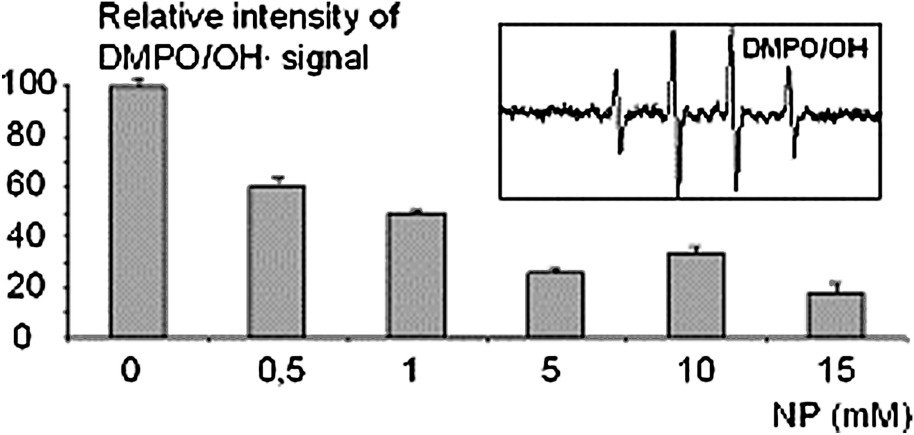
This result accords with the radioprotective effect of NP observed on γ-rays irradiated DNA and mice. Moreover, all of these experiments strongly suggest that NP acts as an radical scavenger and so this property may partly contribute to the inhibition of the indirect effect of γ-rays. To confirm this last assumption and to evaluate the capability of NP to scavenge radicals, electron-spin resonance spectrometry studies were performed by using 5,5-dimethyl-2-pyrolidine-1-oxide (DMPO, 150 mM, Sigma Aldrich) as a spin trap [9]. Hydroxyl radicals were generated by Fenton's reaction as described above. All reactions were carried out in phosphate buffer 5 mM (pH 7.4, NaCl 10 mM) in a final volume of 100 μl. In each experiment, reactants were added in the same order: EDTA (100 μM), hydrogen peroxide (100 μM), NP (0; 0.5; 1; 5; 10 mM), ferrous sulphate solution (100 μM) and DMPO (150 mM). ESR measurements were performed on a Bruker EFP 300e spectrometer at room temperature. The intensity of the signal obtained at different concentrations of NP, was compared with the signal of DMPO/OH.
In the absence of NP, an ESR signal corresponding to (, ) was detected (Fig. 4). Addition of NP at various concentrations (0.5, 1, 5, 10 mM) induced a drastic decrease of the ESR signal attributed to OH radicals. This effect increased with the concentration of NP to raise a plateau at 5 mM (Fig. 4) as in DNA radioprotection experiments. This result confirms that NP is a potential antioxidant.
Fig. 4
EPR investigation of the OH radical scavenging by NP at various concentrations. Percentage of the signal intensity of adduct produced in the presence of NP by comparison with a control without NP. Insert: signal of the adduct.
Radical trapping is one of the most-known mechanisms of chemical radioprotection. However, one physiological mechanism of radioprotectors known is the induction of hypoxia in different tissues. For example, the 5-hydroxytryptamine is described as a radioprotector, one of the mechanisms of action of which is tissue hypoxia as a consequence of vasoconstriction [10]. The reduction of oxygen tension of the blood-forming organs decreases the sensitivity of these organs towards radiation. NP possesses peripheric α-adrenergic property that induces hypertension. This pharmacological activity is effective when NP is administrated at high doses as in in vivo tests of radioprotection [3,4]. Our study shows clearly that NP has also antioxidant properties at high concentration. The activity of NP as radioprotector may result from a combination of both radical scavenging efficiency and vaso-constrictive property.
Conclusion
In conclusion, this work points out for the first time that NP may act as an antioxidant, thus preventing the deleterious effects of γ-radiation. Moreover, this effect can be increased by the cationic charge of NP at physiological pH, which can facilitate interactions with cell membranes and anionic proteins in the serum.
Acknowledgements
The authors wish to thank the ‘Délégation générale pour l'armement’ (DGA), the ‘Ministère de la Défense nationale’, France, and the ‘Office national d’études et de recherches aérospatiales', Centre de Toulouse (ONERA), France.
Bibliographie
[1] C. Martyn; R. Symons Direct and indirect damage to DNA by ionizing radiation, Radiat. Phys. Chem., Volume 43 (1994) no. 4, pp. 403-405
[2] B. Szabo Imidazoline antihypertensive drugs: A critical review on their mechanism of action, Pharmacol. Ther., Volume 93 (2002) no. 1, pp. 1-35
[3] R. Rinaldi; Y. Bernard; M. Guilhermet Radioprotective action of nitrogenated heterocyclic compound derivatives of imidazole and benzimidazole, C. R. Acad. Sci. Paris, Ser. D, Volume 261 (1965) no. 2, pp. 570-572
[4] J.-D. Laval; V. Roman; J. Laduranty; L. Miginac; C. Lion; H. Sentenac-Roumanou; M. Fatôme Radioprotective effect of low doses of 2-(1-naphthylmethyl)-2-imidazoline alone or associated with phosphorothioates, Eur. J. Med. Chem., Volume 28 (1993) no. 9, pp. 709-713
[5] E.S. Copeland Mechanisms of radioprotection – a review, Photochem. Photobiol., Volume 28 (1978) no. 4–5, pp. 839-944
[6] S. Frelon; T. Douki; A. Favier; J. Cadet Comparative study of base damage induced by gamma radiation and Fenton reaction in isolated DNA, J. Chem. Soc., Perkin Trans., Volume 1 (2002) no. 24, pp. 2866-2870
[7] S.S. Kumar; R.C. Chaubey; T.P.A. Devasagayam; K.I. Priyadarsini; P.S. Chauhan Inhibition of radiation-induced DNA damage in plasmid pBR322 by chlorophyllin and possible mechanism(s) of action, Mutat. Res., Volume 425 (1999) no. 1, pp. 71-79
[8] C. Zhao; G. Dodin; C. Yuan; H. Chen; R. Zheng; Z. Jia; B.-T. Fan ‘In vitro’ protection of DNA from Fenton reaction by plant polyphenol verbascoside, Biochim. Biophys. Acta, Volume 1723 (2005) no. 1–3, pp. 114-123
[9] S.A. Cheng; W.-K. Fung; K.-Y. Chan; P.P. Shen Optimizing electron spin resonance detection of hydroxyl radical in water, Chemosphere, Volume 52 (2003), pp. 1797-1805
[10] C. Van der Meer; D.W. van Bekkum A study on the mechanism of radiation protection by 5-hydroxytryptamine and tryptamine, Int. J. Radiat. Biol., Volume 4 (1961) no. 1, pp. 105-110
Cité par
Muhammed Amanat; Swati Gautam; Rishabh Chalotra; Kanhaiya Lal; Tanya Gupta; Rohini Agrawal; Somdutt Mojwar; Randhir Singh Zingiber roseum Roscoe. (Zingiberaceae): Current and future perspective, Pharmacological Research - Modern Chinese Medicine, Volume 7 (2023), p. 100258 | DOI:10.1016/j.prmcm.2023.100258
Valerii S. Ivanov; Aleksei B. Seleznev; Evgenii V. Raguzin; Evgenii V. Ivchenko; Tatyana B. Pechurina; Igor M. Ivanov; Daniil D. Glushenko; Ruslan V. Glushakov Assessment of naphazoline effect on erythrocyte superoxide dismutase activity in rats under experimental modeling of acute radiation sickness, Science and Innovations in Medicine, Volume 8 (2023) no. 1, p. 60 | DOI:10.35693/2500-1388-2023-8-1-60-65
M. V. Vasin; I. B. Ushakov An Analysis of the Role of Bioenergetic Processes under Radioprotective Effects Mediated by Alpha1-Adrenergic Agonists, Biophysics, Volume 66 (2021) no. 3, p. 502 | DOI:10.1134/s0006350921030210
Tanmoy Mondal; Sandip Pal; Subrata Kumar Dey Quercetin Mediated Inhibition of Hydrogen Peroxide-induced Genomic DNA Damage and Toxicity, Journal of Biologically Active Products from Nature, Volume 7 (2017) no. 3, p. 200 | DOI:10.1080/22311866.2017.1329665
M. V. Vasin; I. B. Ushakov Comparative efficacy and the window of radioprotection for adrenergic and serotoninergic agents and aminothiols in experiments with small and large animals, Journal of Radiation Research, Volume 56 (2015) no. 1, p. 1 | DOI:10.1093/jrr/rru087
Amin Imani‐Nabiyyi; Mohammad H. Sorouraddin Determination of naphazoline hydrochloride in biological and pharmaceutical samples by a quantum dot‐assisted chemiluminescence system using response‐surface methodology, Luminescence, Volume 29 (2014) no. 8, p. 994 | DOI:10.1002/bio.2649
Tengku Ahmad; Zakiah Jubri; Nor Rajab; Khairuddin Rahim; Yasmin Yusof; Suzana Makpol Gelam Honey Protects against Gamma-Irradiation Damage to Antioxidant Enzymes in Human Diploid Fibroblasts, Molecules, Volume 18 (2013) no. 2, p. 2200 | DOI:10.3390/molecules18022200
Nagarajan Devipriya; Adluri Ram Sudheer; Marimuthu Srinivasan; Venugopal P. Menon Quercetin ameliorates gamma radiation-induced DNA damage and biochemical changes in human peripheral blood lymphocytes, Mutation Research/Genetic Toxicology and Environmental Mutagenesis, Volume 654 (2008) no. 1, p. 1 | DOI:10.1016/j.mrgentox.2008.03.003


Vous devez vous connecter pour continuer.
S'authentifier