

1 Introduction
Classical swine fever (CSF) caused by the classical swine fever virus (CSFV) is an important, highly contagious pig disease with great economic loss. CSFV belongs to the pestivirus genus within the Flaviviridae family [1,2]. Live-attenuated vaccines are routinely used to prevent from CSFV. The most widely used conventional vaccination is the Chinese strain, which was safe and effective, but interferes with serodiagnosis [3–5]. However, it is necessary to develop safe and effective vaccines against CSFV.
Envelope glycoprotein E2 is the major neutralizing antigen for CSFV infection [6–10]. Vaccines based on E2 protect swine from CSFV and induce neutralizing antibodies [11–13]. The structural protein E2 of classical swine fever virus (CSFV) has been expressed in different systems [13–18]. Moreover, CSFV-specific antibody detection tests and animal immunization experiments have been developed using the recombinant E2 protein [19,20]. Recently, a subunit vaccine, which contains E2 produced in insect cells by a baculovirus expression vector as a potential marker vaccine, allows discrimination between infected and vaccinated pigs [13–17,19–24].
Livestock vaccines from anti-disease plant gene engineering as bioreactors have obtained some progress owing to their advantages [1–7,25–37]. In this study, we describe the production of recombined CSFV E2 protein by tobacco chloroplast transformation and its utilization as an immunogen in animal experiments; this is one of a few reports of a method using plant chloroplasts as bioreactors for livestock vaccines in the field of anti-disease plant-gene engineering.
2 Material and methods
2.1 Plasmids, reagents and alga strains
Plasmid pGEM-E2 containing the CSFV E2 gene (1.017 kb) was provided by the Lan Zhou Veterinary Research Institute of the Chinese Academy of Agricultural Sciences (CAAS). Plasmids 16APT and pTRV, containing the aadA gene cassette and the homologous fragment of tobacco chloroplasts, respectively, were constructed in our lab. The CSFV E2 standard antigen and mouse anti-CSFV polyclonal antibodies were provided by the Chinese Institute of Veterinary Drug Control (CIVDC), and HRG-IgG was purchased from Santa Cruz Biotechnology.
2.2 Construction of recombination vector and transformation
Recombination plasmids were constructed by standard procedures [13,14,18]. The following oligonucleotides were used as primers for the amplification of the E2 gene from plasmid pGEM-E2:
E2-P1: 5'-GTTAAGCTTCCATGGGTCTAGCCTGCAAG-3'
E2-P2: 5'-GCGTCGACTTAGAAGTAATCTGAGTGGCTG-3'
The 1017-bp-long PCR product was isolated and ligated into the pBluescript-SK plastid to be sequenced. The cloned DNA fragment was then digested by HindIII and SalI, and inserted into HindIII- and SalI-digested intermediated vector 16APT, which contains the tobacco chloroplast constitutive 16S promoter, the psbA terminator, and the aadA cassette, to yield the plastid 16APT-E2. The fragment from the BamHI digested plastid 16APT-E2 was inserted into the BglII site of plastid pTRV containing the tobacco chloroplast homologous fragments trnH-psbA and trnK to give rise to the expression vector, pTRVE2. Three–four-week-old sterile tobacco leaves were transformed by biolistic bombardment with plastid pTRVE2 [32].
2.3 PCR identification of transformants
Tobacco chloroplast DNA was extracted according to the methods of Gong and Yan [7] and He et al. [13,14]. In order to identify the integration and homogeneity of the E2 gene into the genome of tobacco chloroplasts, transformed plants were used as templates to be amplified by E2-P1, P2, and CHR-P1, P2 with negative control of wild-type plants and positive control of plasmid p64E2 simultaneously. PCR was performed as follows: 94 °C for 1 min, 60 °C for 1 min, 72 °C for 2 min, 30 cycles.
2.4 Southern-blot analysis
In order to identify further integration of the E2 gene into the genome of tobacco chloroplasts, Southern-blot assays of enzyme-excised products were performed. 4.07-kb enzyme-excised 1.0-kb PCR fragments of the E2 gene, after being digested with SalI and BamHI, were electrophoresed on a 1.0% agarose gel, and transferred to a nylon filter by a Bio-Rad Model 785 vacuum blotter system. The E2 gene was hybridized by a a-dATP labelling E2 probe [13,14].
2.5 SDS-PAGE and Western blot analysis
To evaluate the antigenicity of the expressed protein, a Western-blot assay was performed. Crude proteins were extracted from tobacco chloroplasts and Western-blot assays were carried out according to the protocol of the Promega Company. The crude proteins were electrophoresed on 12% DS-PAGE gel and then transferred on a PVDF membrane. The blot was probed with antiserum against FMDV (1:500 dilution) followed by goat anti-cow secondary antibodies (1:1000 dilution) after being extracted from tuber of potato. The signal was detected by BCIP/NBT. A wild-type and three transformed plants were analyzed.
2.6 ELISA quantitative assay of expressed protein
Total soluble proteins (TSP) of tobacco leaves were extracted and an ELISA assay of E2 protein in transformed plant was conducted as described in [18]. Quantitative analysis of total soluble proteins was made according to the method of He et al. [13,14]. The microtiter plate was coated with total soluble proteins from the wild-type and transformed plants and the known CSFV E2 antigen at 4 °C overnight. The wells were washed by PBST and incubated with antiserum reactive against CSFV (1:500 dilution) and then with alkaline phosphatase-conjugated goat anti-cow IgG (1:1000). Wells were developed with pNPP substrate, and the colour reaction was terminated with 2 N H2SO4 and read at 405-nm wavelength.
2.7 Immunisation of mice
The antigens used in this study were the expressed E2 proteins in tobacco chloroplast. Mice were vaccinated with recombined E2 antigen by two different routes simultaneously (subcutaneous and intragastric immunization). Female Kunming mice (6–8 weeks old) were purchased from the Peking University Health Science Centre, China. About 10–20 ug/pose of E2 protein were given by each route. Blood samples were taken on the 10th day after the last booster. Sera taken four weeks after vaccination were tested for nAb against CSFV. Control animals were immunized with control extracts. Ten days after the last booster, the animals were bled and sera were analyzed for the presence of anti-CSFV antibody. Serum samples were collected at several time intervals from tail blood. From some groups, intestinal scrapings were collected on the day of sacrifice as described in [27].
3 Results and discussion
3.1 Selection of tobacco chloroplast transformation
The transformed plants were selected and regenerated on resistant medium containing 500 mg/L of spectinomycin according to the method described by He et al. [13,14]. Transformed plants were identified by the formation of green shoots on the selective shoot-regeneration medium. Only one resistant transformant was obtained out of eight bombarded leaf segments after selection on resistant medium. The transgenic clone was subjected to next round of spectinomycin selection.
3.2 PCR analysis of tobacco transformants
PCR results showed that a 1.017-kb fragment of E2 was obtained with primers E2-P1, P2. As shown in Fig. 1, a 1.0-kb fragment was obtained in wild type, but an obvious 3.6-kb fragment and a faint 1.0-kb fragment were obtained in incompletely homogeneous transformants with primers CHR-P1 and P2. The results indicated that the expected amplified fragments were obtained in the transformed plants and positive control; however, this was the case in untransformed plants (Fig. 2). It could be concluded primarily that the foreign gene had integrated into the genome of tobacco chloroplasts.
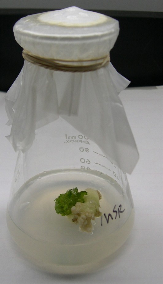
The selection of tobacco transformants on resistant medium. Only one transformed clone was obtained by selection on spectinomycin-resistant medium and regenerated.
Integration and PCR assay of foreign genes of tobacco transformants. (A) PCR assay of E2 gene in transformed tobacco chloroplast. Lane M: 1-kb DNA markers; lane 1: positive control; lane 2: wild-type sample; lanes 3, 4: independent transgenic lines. Expected 1.0-kb fragments were amplified by primer E2-P1, E2-P2. (B) Integration and homogeneity of foreign genes in tobacco transformants. Lane M: 1-kb DNA markers; lane 1: positive control; lanes 2, 3: independent transgenic lines. 3.6-kb and 1.0-kb fragments were amplified in inhomogeneous transformants (lines 2, 3) by primer CHR-P1, P2.
3.3 Southern-blot assay of tobacco transformants
Two transformed lines were investigated by comparing them to control. The results as described by Fig. 3 showed that the 1.0-kb band of E2 fragments and the 4.07-kb band of enzymed digestion, respectively, could be detected (Fig. 3) when hybridized with the E2 probe, but the expected bands did not appear in untransformed plants. It indicated that the transformed genome contained the inserted foreign gene.
Southern blot assay of transformed tobacco. The positive control (lane 1), the wild-type (lane 2) and the transformant lines (lanes 3–4) were hybridized with the 32P-labelled E2 probe. The 1.02-kb band of E2 gene in the positive control and 4.07-kb band of enzymed excision could be detected when hybridized with the 32P-labelled E2 probe (lanes 1, 3, and 4).
3.4 Western blot analysis of tobacco transformants
Analyzed by 12% SDS-PAGE, the result showed a prominently stained band corresponding to an approximate molecular mass of 51–55 kDa, which was identified, and the proteins were expressed as expected. To identify the products, Western-blot assays were performed with monoclonal antibodies of E2. Proteins were separated by 12% SDS and then transferred to the nitrocellulose membrane by electrotransferring. Western blotting showed a good antigenicity of the target protein (Fig. 4).
SDS-PAGE and Western-blot assay of E2 protein expression in tobacco transformants. (A) Line M: protein marker; line 1: wild-type; lines 2–3: transformant lines. (B) The 55-kDa E2 protein was detected by anti-CSFV polyclonal antibodies and goat anti-mouse IgG-AP. Lane 1: positive control; lane 2: wild type sample; lanes 3–4, transformant lines.
3.5 ELISA quantitative assay of the expressed protein
The expression level of the E2 gene was identified in transformed tobacco by ELISA assays by comparing to wild-type samples with CSFV standard antigen diluted grades for quantification (Fig. 5A). The specific antigen–antibody reaction happened as described above. The results suggested that the level of the expressed fused protein is up to 1–2% TSP. The OD of protein at 492-nm wavelength was determined for a standard E2 antigen concentration by the Bradford method [18].
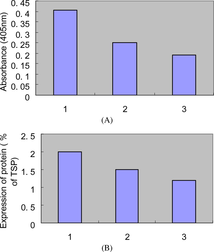
ELISA quantification assay of expressed protein in tobacco transformants. Absorbance of the total soluble protein (TSP) from the wild-type sample as control (A). The content of targeted proteins was standardized against a linear curve of a known amount of standard CSFV protein–antibody complex. The amount of E2 protein was expressed as a percentage of TSP from the transformants (B).
3.6 Induction of immune response
To determine the immunogenicity of the E2 antigen, antibody responses were measured after subcutaneous and intragastric immunization, respectively. The results showed that subcutaneous immunization of extracts of E2 induced significant serum antibody reaction against CSFV, but no immune responses was detected by oral immunization. Most probably, it was due to low antigen dose for oral immunization and degradation; so, a correlation between dose and immune response could not be determined. Antibody responses in faeces could not be detected.
4 Conclusion
Oral vaccination is regarded as an attractive alternative for injected vaccines because of its cheapness and safety, despite the low immune responses upon oral intake. Furthermore, it can be immune as food directly. However, oral vaccination is often not very effective [1–7].
In our study, the immune responses against antigen E2 after infection and oral administration of extracts are compared. Subcutaneous immunization of extracts of E2 induced significant serum antibody reaction against CSFV, but oral immunization of naïve mice with these extracts did not result in detectable serum responses. This indicated that E2 proteins expressed in tobacco chloroplasts are immunogenic and that oral administration mostly evoked low IgA responses at the local level, but not in the serum. The immune response is short lasting. Large doses of antigen and strong mucosal adjuvants such as cholera toxin or heat-labile toxin B subunits, which might be interesting candidates, are needed for the enhancement of the immune response [8–14].
In summary, oral immunization by using edible vaccines remains an attractive concept, but several problems [15–17] must be solved before an effective edible vaccine is available. First, the expression levels of recombinant proteins in plants must be increased [19,20]. Second, appropriate plants should be selected in terms of their bioreactors. Third, appropriate mucosal adjuvants should be incorporated [21–37].
Acknowledgements
We thank the Lanzhou Veterinary Research Institute of the CAAS for providing vector pGEM-E2 containing E2 gene. We also acknowledged the China Institute of Veterinary Drug Control (CIVDC) for excellent technical assistance during animal immunity experiments. This work has been supported by the Doctoral Initiation Foundation of Guizhou University.