Plan
Comptes Rendus

Modeling the 3D functional architecture of the nucleus in animal and plant kingdoms
[Modéliser l'architecture tri-dimensionnelle du noyau dans des cellules animales et végétales]
Comptes Rendus. Biologies, Volume 332 (2009) no. 11, pp. 937-946.

Résumés

Compartmentalization is one of the fundamental principles which underly nuclear function. Numerous studies describe complex and sometimes conflicting relationships between nuclear gene positioning and transcription regulation. Therefore the question is whether topological landmarks and/or organization principles exist to describe the nuclear architecture and, if existing, whether these principles are identical in the animal and plant kingdoms. In the frame of an agroBI-INRA program on nuclear architecture, we set up a multidisciplinary approach combining biological studies, spatial statistics and 3D modeling to investigate spatial organization of a nuclear compartment in both plant and animal cells in their physiological contexts. In this article, we review the questions addressed in this program and the methodology of our work.

L'organisation en compartiments est un des principes fondamentaux qui gouverne le fonctionnement du noyau. De nombreuses études montrent des relations complexes et parfois contradictoires entre la position d'un gène dans le noyau et son état transcriptionnel. Deux questions se posent : existe-t-il des principes généraux gouvernant l'organisation nucléaire, et, si oui, ces principes sont-ils analogues dans les règnes animal et végétal ? Dans le cadre d'un programme agroBI-INRA sur l'architecture nucléaire, nous avons développé une approche multidisciplinaire combinant expériences biologiques, statistiques spatiales et modélisation en 3 dimensions pour analyser l'organisation spatiale d'un compartiment nucléaire dans des cellules animales et végétales, dans leur environnement physiologique. Dans cet article, nous faisons une revue autour des questions posées dans ce programme et présentons la méthodologie de ce travail.

Métadonnées
Publié le :
DOI : 10.1016/j.crvi.2009.09.001
Keywords: Nuclear architecture, Heterochromatin, Chromocenter, Confocal imaging, Digital image analysis, 3D modeling, Spatial statistics
Mot clés : Architecture nucléaire, Hétérochromatine, Chromocentre, Imagerie confocale, Analyse d'images numériques, Reconstruction 3D, Statistique spatiale
Valérie Gaudin 1 ; Philippe Andrey 2, 3, 4 ; Eve Devinoy 5 ; Clémence Kress 5 ; Kiên Kieu 6 ; Nathalie Beaujean 7 ; Yves Maurin 2, 3 ; Pascale Debey 7

1 Laboratoire de biologie cellulaire, UR501, IJPB, route de Saint-Cyr, INRA, 78026 Versailles, France
2 INRA, UMR1197, Neurobiologie de l'olfaction et de la prise alimentaire, Domaine de Vilvert, 78350 Jouy-en-Josas, France
3 Université Paris-Sud 11, UMR1197, 91405 Orsay, France
4 UPMC, Univ Paris 06, France
5 INRA, UR1196 Génomique et physiologie de la lactation, Domaine de Vilvert, F-78350 Jouy-en-Josas, France
6 INRA, UR341, Mathématiques et informatique appliquée, Domaine de Vilvert, 78350 Jouy-en-Josas, France
7 INRA, UMR1198 Biologie du développement et de la reproduction, Domaine de Vilvert, 78350 Jouy-en-Josas, France
@article{CRBIOL_2009__332_11_937_0,
     author = {Val\'erie Gaudin and Philippe Andrey and Eve Devinoy and Cl\'emence Kress and Ki\^en Kieu and Nathalie Beaujean and Yves Maurin and Pascale Debey},
     title = {Modeling the {3D} functional architecture of the nucleus in animal and plant kingdoms},
     journal = {Comptes Rendus. Biologies},
     pages = {937--946},
     publisher = {Elsevier},
     volume = {332},
     number = {11},
     year = {2009},
     doi = {10.1016/j.crvi.2009.09.001},
     language = {en},
}
TY  - JOUR
AU  - Valérie Gaudin
AU  - Philippe Andrey
AU  - Eve Devinoy
AU  - Clémence Kress
AU  - Kiên Kieu
AU  - Nathalie Beaujean
AU  - Yves Maurin
AU  - Pascale Debey
TI  - Modeling the 3D functional architecture of the nucleus in animal and plant kingdoms
JO  - Comptes Rendus. Biologies
PY  - 2009
SP  - 937
EP  - 946
VL  - 332
IS  - 11
PB  - Elsevier
DO  - 10.1016/j.crvi.2009.09.001
LA  - en
ID  - CRBIOL_2009__332_11_937_0
ER  - 
%0 Journal Article
%A Valérie Gaudin
%A Philippe Andrey
%A Eve Devinoy
%A Clémence Kress
%A Kiên Kieu
%A Nathalie Beaujean
%A Yves Maurin
%A Pascale Debey
%T Modeling the 3D functional architecture of the nucleus in animal and plant kingdoms
%J Comptes Rendus. Biologies
%D 2009
%P 937-946
%V 332
%N 11
%I Elsevier
%R 10.1016/j.crvi.2009.09.001
%G en
%F CRBIOL_2009__332_11_937_0
Valérie Gaudin; Philippe Andrey; Eve Devinoy; Clémence Kress; Kiên Kieu; Nathalie Beaujean; Yves Maurin; Pascale Debey. Modeling the 3D functional architecture of the nucleus in animal and plant kingdoms. Comptes Rendus. Biologies, Volume 332 (2009) no. 11, pp. 937-946. doi : 10.1016/j.crvi.2009.09.001. https://comptes-rendus.academie-sciences.fr/biologies/articles/10.1016/j.crvi.2009.09.001/

Version originale du texte intégral

1 Introduction

Increasing evidence suggests that besides DNA regulatory sequences and transcription factors, the spatial and temporal organization of nuclear processes (transcription, reparation, replication, …) as well as chromosome and gene distribution within the nuclear space are central determinants of genome functions. An important goal of the post-genomic systems biology will be therefore to better understand genome function within the architectural framework of the nucleus and integrate different levels of genome regulation [1,2]. However, deciphering the three-dimensional (3D) nuclear organization in a relevant physiological context is a difficult task due to technical constraints and requirements of imaging and computational modeling.

To develop a systems biology view of the nucleus and to give standardized and statistical representations of the nuclear architecture in order to highlight possible organizational rules, we investigated the spatial organization of a nuclear compartment in both plant and animal cells. We set up a multidisciplinary approach combining biological studies, spatial statistics and 3D modeling in the frame of an agroBI-INRA program entitled “Nuclear Architecture: spatial modeling and application to a better understanding of differentiation/dedifferentiation mechanisms”. This article aims at presenting the conceptual background, the main goals and the methodology of our work.

2 Functional nuclear organization: background

2.1 Elements of the nuclear organization

Compartmentalization is one of the fundamental principles regulating organization-function crosstalk in the nucleus. As defined by Misteli, a “compartment” is “a macroscopic region within the nucleus that is morphologically and/or functionally distinct from its surrounding[3]. The compartments may be either sites highly enriched in a definite set of nuclear proteins (nucleoli, speckles, Cajal bodies, promyelocytic leukemia protein (PLM) nuclear bodies, interchromatin granules, …) or chromatin domains, which we will focus on (Fig. 1). One of the most studied compartments is “constitutive heterochromatin”, a major component of eukaryotic genomes usually formed by large blocks located near centromeres and telomeres and that has a compact organization throughout cell cycle [4–6]. It is opposed to “euchromatin”, which exhibits a more “open” conformation and corresponds to actively transcribed regions of the genome. Euchromatin can be turned temporarily into heterochromatin upon silencing, being thus termed “facultative heterochromatin”. Heterochromatin can be distinguished from euchromatin by cytological properties, compaction levels and epigenetic marks (Fig. 1). However, several studies have shown that transcription can occur in heterochromatin regions in yeast, plants and animals [7–9].

Fig. 1

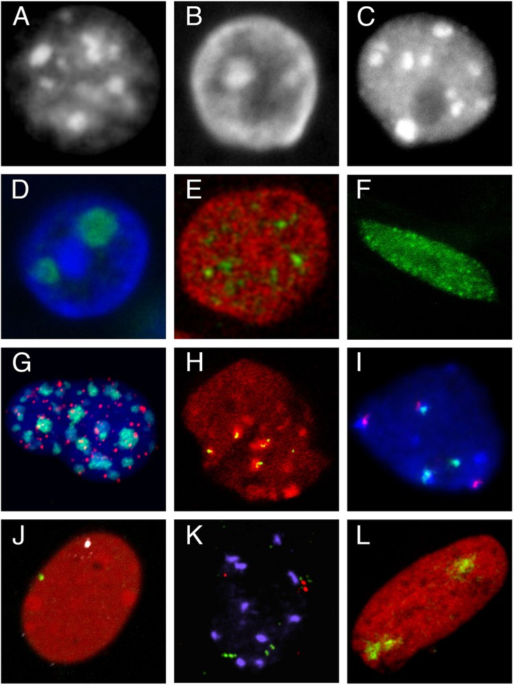
Compartmentalization of the nucleus and elements of its architecture. A–C. Heterochromatin compartments. A. Mouse fibroblast nucleus (Hoechst staining). B. Epithelial cell nucleus from a rabbit mammary gland in lactation (tissue section, DAPI staining). C. Nucleus isolated from an A. thaliana plantlet (DAPI staining). D–F. Compartments defined as regions enriched in nuclear proteins. D. Nucleoli in an epithelial cell nucleus from a rabbit mammary gland in lactation; immunolocalization of B23 protein (green), DNA counterstained with DAPI (blue) (tissue section). E. Splicing speckles in the nucleus of a mouse mammary epithelial cell (cultured HC11 cell line); immunolocalization of SC35 protein (green), DNA counterstained with propidium iodide (red). F. Nuclear localization of LHP1, the plant homolog of Heterochromatin Protein 1 (HP1) in a root cell nucleus from a transgenic line expressing the LHP1-GFP fusion protein. G–I. Chromocenters, centromeres and telomeres. G. FISH with a pancentromeric probe (green) and a LNA telomeric probe (red) in an HC11 cell nucleus; DNA counterstained with DAPI (blue). H. Immunolocalization of pericentromeres (HP1 protein, red) and centromeres (CENP proteins, green) in the nucleus of a rabbit embryo at 8-cell stage. I. 5S (green) and 45S rDNA (red) repeats in an A. thaliana leaf nucleus revealed by FISH; DNA counterstained with DAPI (blue) (kindly provided by F. Tessadori). J–L. Visualization of genomic regions of different sizes by FISH. J. Localization of genes encoding casein (green) and whey acidic protein (white) in the nucleus of HC11 cells after hybridization with gene specific BAC probes and DNA staining with propidium iodide (red). K. Localization of 3 genomic regions (about 100 kbp) with 3 BAC probes (2 in green and one in red) in an A. thaliana leaf nucleus (kindly provided by F. Tessadori). L. Territory of chromosome 15 (green) in the nucleus of a cultured rabbit embryonic fibroblast; DNA counterstained with propidium iodide (red). All images were acquired on confocal microscopes.

Furthermore, large-scale mapping of the human genome has shown a link between chromatin morphology and gene density, with gene-rich regions being present in open chromatin regions and gene-poor regions in more condensed domains, regardless of their activity status [10]. In some animal species such as mouse and, to a lesser extent human, the pericentric heterochromatin of several chromosomes assembles to form large heterochromatic domains called “chromocenters” [11,12]. In Arabidopsis thaliana, discrete and intensely stained heterochromatic compartments are referred to as chromocenters (Fig. 1). They correspond to centromeric and pericentromeric regions and were described as anchoring regions for 0.1–1.5 Mbp euchromatin loops allowing to propose a chromocenter-loop nuclear organizational model [13,14]. Chromocenters can be visualized using FISH [13], immunolocalization [15] or GFP markers [16].

Since the first laser microbeam irradiation experiments [17], it is now well demonstrated that each chromosome of an animal or plant cell's nucleus is confined to a discrete region of the interphase nucleus, referred to as a chromosome territory (CT) and corresponding to a chromatin compartment at a larger scale (Fig. 1) [18–20]. In mammalian cells, the distribution of CT appears non-random, with most gene-rich CTs preferentially localized in the nuclear interior, whereas gene-poor CTs localized toward the nuclear periphery [18,21,22]. Such arrangements are evolutionarily conserved [23,24]. Furthermore, cell or nuclear shapes and chromosome size have been observed to influence the 3D arrangement of CTs: for example, the ellipsoid human fibroblast nuclei present a chromosome size dependent pattern even if the radial distribution of gene-rich and gene-poor CTs was found to be recapitulated at the sub-chromosomal level [25–27]. In A. thaliana, CTs were described as predominantly randomly located within the nucleus. Only chromosomes 2 and 4, which bear nucleolus organizer regions (NOR), were associated more frequently than randomly [19,20]. The relationships between CTs distribution and nuclear organization require therefore further investigation. Parameters such as DNA content, gene density, and transcriptional activity of chromosomal sub-domains might all come into account and raise the question of the mechanism of chromosome positioning.

2.2 Relationship between gene positioning and transcriptional activity

Numerous studies described complex and sometimes conflicting relationships between gene positioning with respect to several compartments, and transcription regulation.

2.2.1 Relative positioning of genes with respect to nuclear compartments

Some studies address the question of comparative positioning inside the nucleus by examining different genes in a given cell, or the same gene in different cell types.

As stated above, gene-rich chromosomes are usually more centrally located than gene-poor chromosomes in human [22], chicken [28], hydra [29], and this trend is observed also for subchromosomal domains [25,30]. The nuclear periphery, which is lined in most higher eukaryotes by a peripheral ring of condensed chromatin, represents generally a repressive zone: some genes are preferentially associated with the nuclear periphery in cells where they are inactive, and more centrally located in cells where they are highly transcribed [31–34]. More generally, late replicating foci, assumed to correspond to inactive chromatin are located at the nuclear periphery. However, location of transcribed genes at the nuclear envelope has also been observed in yeast [35], possibly because of their association with proteins of the nuclear-pore complexes [36]. It is not known whether similar gene activation processes exist at the nuclear periphery in higher eukaryotes. The potential role of the nuclear periphery in genome regulation has become of particular importance in view of human diseases caused by mutations in genes encoding the architectural proteins of the lamina [37].

The relationship between location and transcriptional status of genomic sequences with respect to their CTs is the subject of conflicting reports: non-coding sequences are randomly distributed or preferentially located at the interior of the CT, whereas several genes have been found preferentially located at the periphery of the CT, irrespective of their transcriptional status [38,39]. This peripheral location might facilitate access to the transcriptional machinery located in the inter-chromosomal space [40,41]. Chromatin loops containing large gene clusters (major histocompatibility complex locus, epidermal differentiation complex) can even loop out of their CTs into these interchromosomal compartments under conditions where they are highly expressed [42,43]. On the other hand, some active genes reside in the CT interior [44], suggesting that CTs can be invaginated by infoldings of the interchromosomal compartment [41,45] (reviewed in [46]).

Proximity to centromeric heterochromatin is generally associated with gene silencing, the most striking examples being the association of T-cell specific and developmentally regulated B-cell genes with the Ikaros complex at the centromeric heterochromatin [47] and the beta-globin gene in erythroid cells [48].

2.2.2 Dynamic gene repositioning upon activation/inhibition

Studying the dynamics of genes during the differentiation process has brought other strong arguments for a relationship between nuclear positioning and transcription status [3,49]. For instance, the Hoxb1 locus loops out of its CT during transcriptional activation [50]; movement towards the nuclear periphery was observed upon inactivation [51], or towards the nuclear interior upon neural induction [33]; or gene repositioning was described away of the chromocenters upon activation [48] or towards the chromocenters upon silencing [52]. Finally, active genes widely separated in cis or located on different chromosomes can even colocalize possibly as a result of migration upon activation towards preassembled transcription sites [53,54]. Similarly, the ribosomal RNA genes coalesce in the nucleolus where RNA polymerase I accumulates [55]. More recently, interchromatin granules were suggested to actively recruit genomic loci, thus forming “enhancer hubs” for inter-chromosomal interactions [56].

2.3 Nuclear organization depends on the physiological status of the cell

Besides the above-described changes in gene location, changes in nuclear architecture itself are amongst the most dramatic hallmarks of cell differentiation, development or malignant processes [26,31,49,57,58]. For instance, condensation or morphology of CTs may vary with differentiation in chicken, mouse, or human cells [59,60]. The spatial organization of centromeric heterochromatin in haematopoeitic cells changes with cell types and is ontogenically determined [12]. Clustering of pericentric heterochromatin has been reported during terminal differentiation, e.g. during myogenesis [61], in human neutrophils [62], human and mouse neurons [63] and rat myoblasts [64]. In naturally fertilized mouse embryos, the specific radial (“cartwheel”) organization of chromosomes at 1-cell is progressively modified into a “somatic cell-type” organization during development [65,66]. Interestingly, similar 1-cell type nuclear organization is adopted upon transfer of a somatic nucleus in the oocyte cytoplasm [66,67].

In plants, heterochromatin content varies with differentiation [68–70]. Peculiar heterochromatin organization with smaller chromocenters and additional heterochromatic foci interspersed in euchromatin was observed in endosperm nuclei [71] suggesting a relaxed chromocenter-loop organization of the nuclei. Nuclear compaction of specific loci depends on their transcriptional status in regards to cell differentiation [72] and changes in chromocenter and chromatin organization accompany changes in cell fate and differentiation [70,73].

2.4 Nuclear organization in natural environments

Until now, most data have been gathered on isolated plant cell nuclei or on nuclei from immortalized animal cell lines outside their physiological environment, except for circulating blood cells. Little is known about nuclear organization of cells within their tissues [74]. Few studies compared nuclear organization in primary cells versus cell lines, in cell lines versus tissues, in 2D culture versus 3D cultures and suggested a control of tissue architecture over nuclear organization [75–77]. In addition, few data on nuclear organization were acquired in plant cell nuclei in situ [71,72]. In vivo approaches allowed distinguishing three main types of plant nuclei based on their nuclear shapes (round, elongated and rod-like nuclei), which are associated with various cell-type or tissues [78]. Studies of chromatin dynamics in plants have shown that confinement area for tagged loci is different according to cell types [79]. Further investigation will be required in plant cell nuclei.

2.5 Current models of nuclear organization

Despite being at a very beginning of a system-level view of the nucleus, two major models, a deterministic model and a self-organizing model, emerged [2]. The deterministic model proposes that structure dictates function and is consistent with stable observed structures in the nucleus. However, some experimental evidence does not support this model, such as the loss of prime structural elements without any major consequences on genome function. On the other hand, the nucleus can be seen as a self-organizing system, based on the dynamic interaction of nuclear components and various interplay between structure and function with self-reinforcing mechanism. Indeed, nuclear processes can lead to nuclear changes in global architecture and de novo formation of nuclear structures. For example, supported by theoretical considerations as well as several experimental results from chromosome conformation capture (3C), transcription machinery may function as a main molecular tie that maintain most genomic loops and determine gene activity [80]. 3C experiments have also recently revealed that repeat elements and nucleolus play central roles in the dynamic organization of genome architecture [81]. However, as most probably, the two models are not exclusive and additional forces may have to be taken into considerations to explain the structure/function of the nucleus such as stochastic collisions [82], non-specific entropy-driven crowding forces [83] or “depletion-attraction” forces, highly significant in crowded environments like those in nuclei [14,84,85].

3 Overview of the project

3.1 Aim of the project and main achievements

Two main questions arising from the existing data were at the origin of the project:

  • • Are there organization principles, and/or topographical landmarks to describe the nuclear architecture? If existing, are these principles identical in the animal and plant kingdoms?
  • • What transformations allow to process from one differentiation state to another? How are co-regulated gene networks positioned with respect to these topological landmarks?

To address these questions, we chose to analyze the centric/pericentric heterochromatin compartment and its three-dimensional nuclear distribution. Indeed, the centric/pericentric compartment is well defined and present in all cell types with some already described variations. Its structure and cohesion are essential for genome organization and proper cell division [70,73,86,87] and it is involved in genome regulation, usually behaving as a transcriptional repressor compartment.

Aiming at producing generic models for the nuclear architecture in physiological environment, we produced images of 3D-preserved nuclei both from animal and plant kingdoms in three different biological systems to be able to extract robust organizational principles. We performed fluorescence or immunofluorescence labeling to reveal the structural elements, generated a homogenous 3D-image collection of cell nuclei acquired in standardized conditions (ICOPAN website/image resource: http://amib.jouy.inra.fr/icopan/) and developed image processing and spatial statistical tools to analyze and model the spatial distribution of the studied compartment.

3.2 Choice of the biological systems

Three biological complex systems were chosen, two animal (rabbit) systems and one plant (Arabidopsis thaliana) system. Rabbit was chosen since: (i) its complete genome sequence (2X coverage) was already available and its 7X coverage was expected to be released in a near future; and (ii) rabbit is phylogenetically closer to primates than rodents. A. thaliana is an excellent plant model for which large molecular genomics tools and genetic resources are available. Furthermore, in the three systems, transitions between totipotency, multipotency and terminal differentiation can be exploited and used to explore nuclear reorganization in relation to modifications of the pattern of gene expression.

Pre-implantation rabbit embryos, the first system, offer the possibility to follow directly a differentiation process within the same intact organism. After the reprogrammation of the parental genomes at fertilization, the first totipotent zygote leads in 5 days to a blastocyst where two different populations of cells coexist, already differentiated trophectoderm cells and totipotent cells of the inner cell mass. The whole process can take place in vitro in an appropriate culture medium. Finely tuned modifications of gene expression occur during this period, together with large restructuring of the nuclear architecture. The advantage of the rabbit is that zygotic genome activation occurs over an extended period of time as compared to the mouse [88].

The second system, the rabbit mammary gland, is subjected to successive cycles of intense development and involution which extend from gestation through lactation to weaning, and thus represents a powerful system to explore the impacts of physiological situations on genome function. During each cycle, genes coding for milk proteins are turned on and off, together with many other genes. However, no studies of the nuclear organization of these epithelial cells in their tissue environment exist so far, only few data deal with mammary cell lines [77,89,90].

A. thaliana plantlets offer a third interesting system, since after digestion of the cell walls, totipotent “protoplasts” can be easily obtained. Upon culture in an appropriate medium, protoplasts can redifferentiate into tissues and new plants. A large reorganization of heterochromatin accompanies the first steps of dedifferentiation/redifferentiation in this system [73].

3.3 Development of modeling and computational tools

To explore the nucleus on a global scale, new tools are required to establish computational models and to unravel principles governing the spatial distribution of nuclear compartments. Indeed most analyses were done so far in 2D, and very rarely in 3D [31,39,91,92]. Few quantitative measures have been performed [93,94]. Standard quantitative analyses of the nuclear organization are essentially based on distances measured on 2D images or on projections of 3D images, and rarely obtained directly in 3D. Distances can be measured between points (centers of compartments), or between points and surfaces (e.g. the nuclear envelope). Sometimes distances are digitized into classes such as, for example, in the so-called erosion analysis used to analyze radial distributions within the nucleus [31].

The testing of complete spatial randomness is commonly used to show that the nucleus is indeed spatially organized. A typical approach consists in choosing arbitrary parameters (distance or angle) and comparing their experimental values with those obtained on patterns simulated according complete spatial randomness. Such an approach based on angles was used to test whether chromosome territories are located independently from each other [27]. In domains such as forestry, ecology, or epidemiology, generic statistical tools have been developed for the analysis of spatial point patterns [95–97]. To date, these tools have only rarely been used to investigate nuclear spatial organizations [98–101]. For example, Beil et al. [101] used the K-function to test whether chromocenters are completely random and to compare chromocenters patterns in undifferentiated and differentiated NB4 human cells. However, the conventional forms of spatial point pattern tools encompass no normalization for nuclear shape variations and are not applicable to repeated experiments.

To take into account the expected spatial heterogeneity of the nuclei and compare the heterochromatin distribution, we developed new tools based on processing and analysis of digital confocal images and a new statistical approach using variants of the spatial point patterns F- and G-functions (Fig. 2). These cumulative distribution functions of nearest-neighbor distances (G-function) or of distances between arbitrary points within the domain of interest (here the nucleus) and their nearest “events” (here nuclear elements) (F-function) indeed provide more comprehensive descriptors than radial distributions. Thus, they allow more powerful testing of hypotheses on spatial distributions. In this study, our F-function variant was the most appropriate tool and allowed us to highlight a non-random distribution of centric–pericentric heterochromatin in differentiated cells from our three biological systems.

Fig. 2

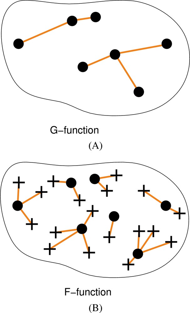
Spatial point patterns and analysis by distance functions. A. The G-function is the cumulative distribution function of point to nearest point distances. Points are shown as black dots and are joined to their nearest neighbor by line segments. B. The F-function is the cumulative distribution function of arbitrary positions to nearest point distances. Sampled positions (shown as crosses) are connected to nearest points (black dots) by line segments.

The use of this F-function variant requires an adapted segmentation method to identify and localize the “interesting” biological objects within images collected on very different types of biological material. Furthermore, the segmentation procedure needs to be automated in order to analyze a large number of images. However, few algorithms are actually available as they may vary according to the specimen. Simple software, especially the commercially available ones, used to extract and calculate 3D nuclear distributions can be used on cells grown in culture, with round shaped nuclei [27] but cannot be used for more sophisticated samples like irregularly shaped nuclei, tissue sections with clumped cells or low-contrasted fluorescent staining. For this project, we set up new automated or semi-automated segmentation algorithms adapted to the different nuclear compartments in the three dimensions of well-preserved nuclei.

4 Conclusion

The frame of the agroBI-INRA program entitled “Nuclear Architecture: spatial modeling and application to a better understanding of differentiation/dedifferentiation mechanisms” allowed a fruitful confrontation between animal and plant biologists, together with statisticians and modelers to define a common multidisciplinary approach. The first results of this joint study will be presented in a forthcoming paper. We believe that comparisons between plant and animals will be highly beneficial to understand the similarities/differences in genome organization, gene regulation and cell differentiation processes.

Acknowledgements

This work was supported by an INRA agroBI Research Program (2006–2008) (http://www.inra.fr/internet/Projets/agroBI/pageagroBI.html). We are grateful to Olivier Grandjean and Lionel Gissot, to the Laboratoire Commun de Cytologie (INRA Versailles), as well as to Pierre Adenot and the RIO MIMA2 platform (INRA Jouy-en-Josas) for technical support in confocal imaging. We thank Eric Biot, Nicole Houba-Hérin, Cathy Hue-Beauvais, Gaetan Lehman, Leila Tirichine, Liu Zichuan for biological experiments and development of image treatment and analysis tools. We thank Professor M.A. Ferguson-Smith and P. O'Brien (Cambridge Resource Centre for Comparative Genomics) for the gift of the rabbit chromosome-specific paints. We are indebted to Christophe Escudé for the LNA telomeric probe and Federico Tessadori for providing images (Figs. 1I, 1K). We thank the Région Ile-de-France for the funding of the Leica TCS SP2 confocal microscope (INRA Versailles) and the Zeiss AxioObserver imaging Apotome system (INRA, Jouy).


Bibliographie

[1] S. Gorski; T. Misteli Systems biology in the cell nucleus, J. Cell Sci., Volume 118 (2005), pp. 4083-4092

[2] T. Misteli Beyond the sequence: Cellular organization of genome function, Cell, Volume 128 (2007), pp. 787-800

[3] T. Misteli Concepts in nuclear architecture, Bioessays, Volume 27 (2005), pp. 477-487

[4] S.I. Grewal; S. Jia Heterochromatin revisited, Nat. Rev. Genet., Volume 8 (2007), pp. 35-46

[5] F. Hediger; S.M. Gasser Heterochromatin protein 1: Don't judge the book by its cover!, Curr. Opin. Genet. Dev., Volume 16 (2006), pp. 143-150

[6] P. Dimitri; R. Caizzi; E. Giordano; M. Carmela Accardo; G. Lattanzi; G. Biamonti Constitutive heterochromatin: A surprising variety of expressed sequences, Chromosoma, Volume 118 (2009), pp. 419-435

[7] H. Bouzinba-Segard; A. Guais; C. Francastel Accumulation of small murine minor satellite transcripts leads to impaired centromeric architecture and function, Proc. Natl. Acad. Sci. USA, Volume 103 (2006), pp. 8709-8714

[8] I. Djupedal; K. Ekwall Epigenetics: Heterochromatin meets RNAi, Cell Res., Volume 19 (2009), pp. 282-295

[9] M. Carchilan; M. Delgado; T. Ribeiro; P. Costa-Nunes; A. Caperta; L. Morais-Cecilio; R.N. Jones; W. Viegas; A. Houben Transcriptionally active heterochromatin in rye B chromosomes, Plant Cell, Volume 19 (2007), pp. 1738-1749

[10] N. Gilbert; S. Boyle; H. Fiegler; K. Woodfine; N.P. Carter; W.A. Bickmore Chromatin architecture of the human genome: Gene-rich domains are enriched in open chromatin fibers, Cell, Volume 118 (2004), pp. 555-566

[11] I. Alcobia; R. Dilao; L. Parreira Spatial associations of centromeres in the nuclei of hematopoietic cells: Evidence for cell-type-specific organizational patterns, Blood, Volume 95 (2000), pp. 1608-1615

[12] I. Alcobia; A.S. Quina; H. Neves; N. Clode; L. Parreira The spatial organization of centromeric heterochromatin during normal human lymphopoiesis: Evidence for ontogenically determined spatial patterns, Exp. Cell Res., Volume 290 (2003), pp. 358-369

[13] P. Fransz; J.H. De Jong; M. Lysak; M.R. Castiglione; I. Schubert Interphase chromosomes in Arabidopsis are organized as well defined chromocenters from which euchromatin loops emanate, Proc. Natl. Acad. Sci. USA, Volume 99 (2002), pp. 14584-14589

[14] S. de Nooijer; J. Wellink; B. Mulder; T. Bisseling Non-specific interactions are sufficient to explain the position of heterochromatic chromocenters and nucleoli in interphase nuclei, Nucleic Acids Res. (2009), pp. 3558-3568

[15] P.B. Talbert; R. Masuelli; A.P. Tyagi; L. Comai; S. Henikoff Centromeric localization and adaptive evolution of an Arabidopsis histone H3 variant, Plant Cell, Volume 14 (2002), pp. 1053-1066

[16] Y. Fang; D.L. Spector Centromere positioning and dynamics in living Arabidopsis plants, Mol. Biol. Cell, Volume 16 (2005), pp. 5710-5718

[17] T. Cremer; C. Cremer Chromosome territories, nuclear architecture and gene regulation in mammalian cells, Nat. Rev. Genet., Volume 2 (2001), pp. 292-301

[18] T. Cremer; M. Cremer; S. Dietzel; S. Muller; I. Solovei; S. Fakan Chromosome territories – a functional nuclear landscape, Curr. Opin. Cell Biol., Volume 18 (2006), pp. 307-316

[19] A. Berr; I. Schubert Interphase chromosome arrangement in Arabidopsis thaliana is similar in differentiated and meristematic tissues and shows a transient mirror symmetry after nuclear division, Genetics, Volume 176 (2007), pp. 853-863

[20] A. Pecinka; V. Schubert; A. Meister; G. Kreth; M. Klatte; M.A. Lysak; J. Fuchs; I. Schubert Chromosome territory arrangement and homologous pairing in nuclei of Arabidopsis thaliana are predominantly random except for NOR-bearing chromosomes, Chromosoma, Volume 113 (2004), pp. 258-269

[21] J. Ferreira; G. Paolella; C. Ramos; A.I. Lamond Spatial organization of large-scale chromatin domains in the nucleus: A magnified view of single chromosome territories, J. Cell Biol., Volume 139 (1997), pp. 1597-1610

[22] J.A. Croft; J.M. Bridger; S. Boyle; P. Perry; P. Teague; W.A. Bickmore Differences in the localization and morphology of chromosomes in the human nucleus, J. Cell Biol., Volume 145 (1999), pp. 1119-1131

[23] H. Tanabe; F.A. Habermann; I. Solovei; M. Cremer; T. Cremer Non-random radial arrangements of interphase chromosome territories: Evolutionary considerations and functional implications, Mutat. Res., Volume 504 (2002), pp. 37-45

[24] H.A. Foster; L.R. Abeydeera; D.K. Griffin; J.M. Bridger Non-random chromosome positioning in mammalian sperm nuclei, with migration of the sex chromosomes during late spermatogenesis, J. Cell Sci., Volume 118 (2005), pp. 1811-1820

[25] N. Sadoni; S. Langer; C. Fauth; G. Bernardi; T. Cremer; B.M. Turner; D. Zink Nuclear organization of mammalian genomes. Polar chromosome territories build up functionally distinct higher order compartments, J. Cell Biol., Volume 146 (1999), pp. 1211-1226

[26] R. Mayer; A. Brero; J. von Hase; T. Schroeder; T. Cremer; S. Dietzel Common themes and cell type specific variations of higher order chromatin arrangements in the mouse, BMC Cell Biol., Volume 6 (2005), p. 44

[27] A. Bolzer; G. Kreth; I. Solovei; D. Koehler; K. Saracoglu; C. Fauth; S. Muller; R. Eils; C. Cremer; M.R. Speicher; T. Cremer Three-dimensional maps of all chromosomes in human male fibroblast nuclei and prometaphase rosettes, PLoS Biol., Volume 3 (2005), p. e157

[28] F.A. Habermann; M. Cremer; J. Walter; G. Kreth; J. von Hase; K. Bauer; J. Wienberg; C. Cremer; T. Cremer; I. Solovei Arrangements of macro- and microchromosomes in chicken cells, Chromosome Res., Volume 9 (2001), pp. 569-584

[29] O. Alexandrova; I. Solovei; T. Cremer; C.N. David Replication labeling patterns and chromosome territories typical of mammalian nuclei are conserved in the early metazoan Hydra, Chromosoma, Volume 112 (2003), pp. 190-200

[30] K. Kupper; A. Kolbl; D. Biener; S. Dittrich; J. von Hase; T. Thormeyer; H. Fiegler; N.P. Carter; M.R. Speicher; T. Cremer; M. Cremer Radial chromatin positioning is shaped by local gene density, not by gene expression, Chromosoma, Volume 116 (2007), pp. 285-306

[31] D. Zink; M.D. Amaral; A. Englmann; S. Lang; L.A. Clarke; C. Rudolph; F. Alt; K. Luther; C. Braz; N. Sadoni; J. Rosenecker; D. Schindelhauer Transcription-dependent spatial arrangements of CFTR and adjacent genes in human cell nuclei, J. Cell Biol., Volume 166 (2004), pp. 815-825

[32] S.T. Kosak; J.A. Skok; K.L. Medina; R. Riblet; M.M. Le Beau; A.G. Fisher; H. Singh Subnuclear compartmentalization of immunoglobulin loci during lymphocyte development, Science, Volume 296 (2002), pp. 158-162

[33] R.R. Williams; V. Azuara; P. Perry; S. Sauer; M. Dvorkina; H. Jorgensen; J. Roix; P. McQueen; T. Misteli; M. Merkenschlager; A.G. Fisher Neural induction promotes large-scale chromatin reorganisation of the Mash1 locus, J. Cell Sci., Volume 119 (2006), pp. 132-140

[34] S. Dietzel; K. Zolghadr; C. Hepperger; A.S. Belmont Differential large-scale chromatin compaction and intranuclear positioning of transcribed versus non-transcribed transgene arrays containing beta-globin regulatory sequences, J. Cell Sci., Volume 117 (2004), pp. 4603-4614

[35] J.H. Brickner; P. Walter Gene recruitment of the activated INO1 locus to the nuclear membrane, PLoS Biol., Volume 2 (2004), p. e342

[36] J.M. Casolari; C.R. Brown; S. Komili; J. West; H. Hieronymus; P.A. Silver Genome-wide localization of the nuclear transport machinery couples transcriptional status and nuclear organization, Cell, Volume 117 (2004), pp. 427-439

[37] Y. Gruenbaum; A. Margalit; R.D. Goldman; D.K. Shumaker; K.L. Wilson The nuclear lamina comes of age, Nat. Rev. Mol. Cell Biol., Volume 6 (2005), pp. 21-31

[38] M.O. Scheuermann; J. Tajbakhsh; A. Kurz; K. Saracoglu; R. Eils; P. Lichter Topology of genes and nontranscribed sequences in human interphase nuclei, Exp. Cell Res., Volume 301 (2004), pp. 266-279

[39] A. Kurz; S. Lampel; J.E. Nickolenko; J. Bradl; A. Benner; R.M. Zirbel; T. Cremer; P. Lichter Active and inactive genes localize preferentially in the periphery of chromosome territories, J. Cell Biol., Volume 135 (1996), pp. 1195-1205

[40] I. Faro-Trindade; P.R. Cook Transcription factories: Structures conserved during differentiation and evolution, Biochem. Soc. Trans., Volume 34 (2006), pp. 1133-1137

[41] H. Albiez; M. Cremer; C. Tiberi; L. Vecchio; L. Schermelleh; S. Dittrich; K. Kupper; B. Joffe; T. Thormeyer; J. von Hase; S. Yang; K. Rohr; H. Leonhardt; I. Solovei; C. Cremer; S. Fakan; T. Cremer Chromatin domains and the interchromatin compartment form structurally defined and functionally interacting nuclear networks, Chromosome Res., Volume 14 (2006), pp. 707-733

[42] R.R. Williams; S. Broad; D. Sheer; J. Ragoussis Subchromosomal positioning of the epidermal differentiation complex (EDC) in keratinocyte and lymphoblast interphase nuclei, Exp. Cell Res., Volume 272 (2002), pp. 163-175

[43] E.V. Volpi; E. Chevret; T. Jones; R. Vatcheva; J. Williamson; S. Beck; R.D. Campbell; M. Goldsworthy; S.H. Powis; J. Ragoussis; J. Trowsdale; D. Sheer Large-scale chromatin organization of the major histocompatibility complex and other regions of human chromosome 6 and its response to interferon in interphase nuclei, J. Cell Sci., Volume 113 (2000), pp. 1565-1576 (Pt 9)

[44] N.L. Mahy; P.E. Perry; S. Gilchrist; R.A. Baldock; W.A. Bickmore Spatial organization of active and inactive genes and noncoding DNA within chromosome territories, J. Cell Biol., Volume 157 (2002), pp. 579-589

[45] P.J. Verschure; I. Van Der Kraan; J.M. Enserink; M.J. Mone; E.M. Manders; R. Van Driel Large-scale chromatin organization and the localization of proteins involved in gene expression in human cells, J. Histochem. Cytochem., Volume 50 (2002), pp. 1303-1312

[46] E. Heard; W. Bickmore The ins and outs of gene regulation and chromosome territory organisation, Curr. Opin. Cell Biol., Volume 19 (2007), pp. 311-316

[47] K.E. Brown; S.S. Guest; S.T. Smale; K. Hahm; M. Merkenschlager; A.G. Fisher Association of transcriptionally silent genes with Ikaros complexes at centromeric heterochromatin, Cell, Volume 91 (1997), pp. 845-854

[48] C. Francastel; W. Magis; M. Groudine Nuclear relocation of a transactivator subunit precedes target gene activation, Proc. Natl. Acad. Sci. USA, Volume 98 (2001), pp. 12120-12125

[49] S.T. Kosak; M. Groudine Gene order and dynamic domains, Science, Volume 306 (2004), pp. 644-647

[50] S. Chambeyron; W.A. Bickmore Does looping and clustering in the nucleus regulate gene expression?, Curr. Opin. Cell Biol., Volume 16 (2004), pp. 256-262

[51] S.H. Kim; P.G. McQueen; M.K. Lichtman; E.M. Shevach; L.A. Parada; T. Misteli Spatial genome organization during T-cell differentiation, Cytogenet. Genome Res., Volume 105 (2004), pp. 292-301

[52] R.C. Su; K.E. Brown; S. Saaber; A.G. Fisher; M. Merkenschlager; S.T. Smale Dynamic assembly of silent chromatin during thymocyte maturation, Nat. Genet., Volume 36 (2004), pp. 502-506

[53] C.S. Osborne; L. Chakalova; J.A. Mitchell; A. Horton; A.L. Wood; D.J. Bolland; A.E. Corcoran; P. Fraser Myc dynamically and preferentially relocates to a transcription factory occupied by Igh, PLoS Biol., Volume 5 (2007), p. e192

[54] J.M. Brown; J. Leach; J.E. Reittie; A. Atzberger; J. Lee-Prudhoe; W.G. Wood; D.R. Higgs; F.J. Iborra; V.J. Buckle Coregulated human globin genes are frequently in spatial proximity when active, J. Cell Biol., Volume 172 (2006), pp. 177-187

[55] I. Raska; P.J. Shaw; D. Cmarko Structure and function of the nucleolus in the spotlight, Curr. Opin. Cell Biol., Volume 18 (2006), pp. 325-334

[56] Q. Hu; Y.S. Kwon; E. Nunez; M.D. Cardamone; K.R. Hutt; K.A. Ohgi; I. Garcia-Bassets; D.W. Rose; C.K. Glass; M.G. Rosenfeld; X.D. Fu Enhancing nuclear receptor-induced transcription requires nuclear motor and LSD1-dependent gene networking in interchromatin granules, Proc. Natl. Acad. Sci. USA, Volume 105 (2008), pp. 19199-19204

[57] S.A. Lelievre; V.M. Weaver; J.A. Nickerson; C.A. Larabell; A. Bhaumik; O.W. Petersen; M.J. Bissell Tissue phenotype depends on reciprocal interactions between the extracellular matrix and the structural organization of the nucleus, Proc. Natl. Acad. Sci. USA, Volume 95 (1998), pp. 14711-14716

[58] D. Koehler; V. Zakhartchenko; L. Froenicke; G. Stone; R. Stanyon; E. Wolf; T. Cremer; A. Brero Changes of higher order chromatin arrangements during major genome activation in bovine preimplantation embryos, Exp. Cell Res. (2009), pp. 2053-2063

[59] S. Stadler; V. Schnapp; R. Mayer; S. Stein; C. Cremer; C. Bonifer; T. Cremer; S. Dietzel The architecture of chicken chromosome territories changes during differentiation, BMC Cell Biol., Volume 5 (2004), p. 44

[60] E. Meshorer; T. Misteli Chromatin in pluripotent embryonic stem cells and differentiation, Nat. Rev. Mol. Cell Biol., Volume 7 (2006), pp. 540-546

[61] A. Brero; H.P. Easwaran; D. Nowak; I. Grunewald; T. Cremer; H. Leonhardt; M.C. Cardoso Methyl CpG-binding proteins induce large-scale chromatin reorganization during terminal differentiation, J. Cell Biol., Volume 169 (2005), pp. 733-743

[62] M. Beil; D. Durschmied; S. Paschke; B. Schreiner; U. Nolte; A. Bruel; T. Irinopoulou Spatial distribution patterns of interphase centromeres during retinoic acid-induced differentiation of promyelocytic leukemia cells, Cytometry, Volume 47 (2002), pp. 217-225

[63] G. Martou; U. De Boni Nuclear topology of murine, cerebellar Purkinje neurons: Changes as a function of development, Exp. Cell Res., Volume 256 (2000), pp. 131-139

[64] N. Chaly; S.B. Munro Centromeres reposition to the nuclear periphery during L6E9 myogenesis in vitro, Exp. Cell Res., Volume 223 (1996), pp. 274-278

[65] A.V. Probst; F. Santos; W. Reik; G. Almouzni; W. Dean Structural differences in centromeric heterochromatin are spatially reconciled on fertilisation in the mouse zygote, Chromosoma, Volume 116 (2007), pp. 403-415

[66] C. Martin; N. Beaujean; V. Brochard; C. Audouard; D. Zink; P. Debey Genome restructuring in mouse embryos during reprogramming and early development, Dev. Biol., Volume 292 (2006), pp. 317-332

[67] V. Merico; J. Barbieri; M. Zuccotti; B. Joffe; T. Cremer; C.A. Redi; I. Solovei; S. Garagna Epigenomic differentiation in mouse preimplantation nuclei of biparental, parthenote and cloned embryos, Chromosome Res., Volume 15 (2007), pp. 341-360

[68] O. Mathieu; Z. Jasencakova; I. Vaillant; A.V. Gendrel; V. Colot; I. Schubert; S. Tourmente Changes in 5S rDNA chromatin organization and transcription during heterochromatin establishment in Arabidopsis, Plant Cell, Volume 15 (2003), pp. 2929-2939

[69] F. Tessadori; R. van Driel; P. Fransz Cytogenetics as a tool to study gene regulation, Trends Plant Sci., Volume 9 (2004), pp. 147-153

[70] F. Tessadori; R.K. Schulkes; R. van Driel; P. Fransz Light-regulated large-scale reorganization of chromatin during the floral transition in Arabidopsis, Plant J., Volume 50 (2007), pp. 848-857

[71] C. Baroux; A. Pecinka; J. Fuchs; I. Schubert; U. Grossniklaus The triploid endosperm genome of Arabidopsis adopts a peculiar, parental-dosage-dependent chromatin organization, Plant Cell, Volume 19 (2007), pp. 1782-1794

[72] S. Costa; P. Shaw Chromatin organization and cell fate switch respond to positional information in Arabidopsis, Nature, Volume 439 (2006), pp. 493-496

[73] F. Tessadori; M.C. Chupeau; Y. Chupeau; M. Knip; S. Germann; R. van Driel; P. Fransz; V. Gaudin Large-scale dissociation and sequential reassembly of pericentric heterochromatin in dedifferentiated Arabidopsis cells, J. Cell Sci., Volume 120 (2007), pp. 1200-1208

[74] J. Mateos-Langerak; S. Goetze; H. Leonhardt; T. Cremer; R. van Driel; C. Lanctot Nuclear architecture: Is it important for genome function and can we prove it?, J. Cell Biochem., Volume 102 (2007), pp. 1067-1075

[75] G. Chandramouly; P.C. Abad; D.W. Knowles; S.A. Lelievre The control of tissue architecture over nuclear organization is crucial for epithelial cell fate, J. Cell Sci., Volume 120 (2007), pp. 1596-1606

[76] K.J. Meaburn; T. Misteli Locus-specific and activity-independent gene repositioning during early tumorigenesis, J. Cell Biol., Volume 180 (2008), pp. 39-50

[77] P. Kaminker; C. Plachot; S.H. Kim; P. Chung; D. Crippen; O.W. Petersen; M.J. Bissell; J. Campisi; S.A. Lelievre Higher-order nuclear organization in growth arrest of human mammary epithelial cells: A novel role for telomere-associated protein TIN2, J. Cell Sci., Volume 118 (2005), pp. 1321-1330

[78] E. Chytilova; J. Macas; D.W. Galbraith Green fluorescent protein targeted to the nucleus, a transgenic phenotype useful for studies in plant biology, Annals of Botany, Volume 83 (1999), pp. 645-654

[79] N. Kato; E. Lam Chromatin of endoreduplicated pavement cells has greater range of movement than that of diploid guard cells in Arabidopsis thaliana, J. Cell Sci., Volume 116 (2003), pp. 2195-2201

[80] D. Marenduzzo; I. Faro-Trindade; P.R. Cook What are the molecular ties that maintain genomic loops?, Trends Genet., Volume 23 (2007), pp. 126-133

[81] J.M. O'Sullivan; D.M. Sontam; R. Grierson; B. Jones Repeated elements coordinate the spatial organization of the yeast genome, Yeast, Volume 26 (2009), pp. 125-138

[82] M. Dundr; U. Hoffmann-Rohrer; Q. Hu; I. Grummt; L.I. Rothblum; R.D. Phair; T. Misteli A kinetic framework for a mammalian RNA polymerase in vivo, Science, Volume 298 (2002), pp. 1623-1626

[83] R. Hancock A role for macromolecular crowding effects in the assembly and function of compartments in the nucleus, J. Struct. Biol., Volume 146 (2004), pp. 281-290

[84] D. Marenduzzo; K. Finan; P.R. Cook The depletion attraction: An underappreciated force driving cellular organization, J. Cell Biol., Volume 175 (2006), pp. 681-686

[85] S. Asakura; F. Oosawa Interactions between particles suspended in solutions of macromolecules, J. Polym. Sci. B, Volume 33 (1958), pp. 183-192

[86] M. Guenatri; D. Bailly; C. Maison; G. Almouzni Mouse centric and pericentric satellite repeats form distinct functional heterochromatin, J. Cell Biol., Volume 166 (2004), pp. 493-505

[87] A. Goncalves Dos Santos Silva; R. Sarkar; J. Harizanova; A. Guffei; M. Mowat; Y. Garini; S. Mai Centromeres in cell division, evolution, nuclear organization and disease, J. Cell Biochem., Volume 104 (2008), pp. 2040-2058

[88] A. Brunet-Simon; G. Henrion; J.P. Renard; V. Duranthon Onset of zygotic transcription and maternal transcript legacy in the rabbit embryo, Mol. Reprod. Dev., Volume 58 (2001), pp. 127-136

[89] M.J. Bissell; V.M. Weaver; S.A. Lelievre; F. Wang; O.W. Petersen; K.L. Schmeichel Tissue structure, nuclear organization, and gene expression in normal and malignant breast, Cancer Res., Volume 59 (1999), p. 1757-1763s (discussion 1763s–1764s)

[90] J.L. Salisbury The contribution of epigenetic changes to abnormal centrosomes and genomic instability in breast cancer, J. Mammary Gland Biol. Neoplasia, Volume 6 (2001), pp. 203-212

[91] M. Gue; J.S. Sun; T. Boudier Simultaneous localization of MLL, AF4 and ENL genes in interphase nuclei by 3D-FISH: MLL translocation revisited, BMC Cancer, Volume 6 (2006), p. 20

[92] N.L. Mahy; P.E. Perry; W.A. Bickmore Gene density and transcription influence the localization of chromatin outside of chromosome territories detectable by FISH, J. Cell Biol., Volume 159 (2002), pp. 753-763

[93] O. Ronneberger; D. Baddeley; F. Scheipl; P.J. Verveer; H. Burkhardt; C. Cremer; L. Fahrmeir; T. Cremer; B. Joffe Spatial quantitative analysis of fluorescently labeled nuclear structures: Problems, methods, pitfalls, Chromosome Res., Volume 16 (2008), pp. 523-562

[94] C. Shiels; N.M. Adams; S.A. Islam; D.A. Stephens; P.S. Freemont Quantitative analysis of cell nucleus organisation, PLoS Comput. Biol., Volume 3 (2007), p. e138

[95] A. Baddeley; P. Gregori; J. Mateu; R. Stoica; D. Stoyan Case Studies in Spatial Point Process Modeling, Lecture Notes in Statistics, vol. 185, Springer-Verlag, 2006

[96] P.J. Diggle Statistical Analysis of Spatial Point Patterns, A Hodder Arnold Publication, Oxford Univ. Press, London, 2003

[97] N.A.C. Cressie Statistics for Spatial Data, Series in Probability and Statistics, Wiley-Interscience, 1993

[98] K.J. McManus; D.A. Stephens; N.M. Adams; S.A. Islam; P.S. Freemont; M.J. Hendzel The transcriptional regulator CBP has defined spatial associations within interphase nuclei, PLoS Comput. Biol., Volume 2 (2006), p. e139

[99] D.W. Young; S.K. Zaidi; P.S. Furcinitti; A. Javed; A.J. van Wijnen; J.L. Stein; J.B. Lian; G.S. Stein Quantitative signature for architectural organization of regulatory factors using intranuclear informatics, J. Cell Sci., Volume 117 (2004), pp. 4889-4896

[100] H.J. Noordmans; K. van der Kraan; R. van Driel; A.W. Smeulders Randomness of spatial distributions of two proteins in the cell nucleus involved in mRNA synthesis and their relationship, Cytometry, Volume 33 (1998), pp. 297-309

[101] M. Beil; H. Braxmeier; F. Fleischer; V. Schmidt; P. Walther Quantitative analysis of keratin filament networks in scanning electron microscopy images of cancer cells, J. Microsc., Volume 220 (2005), pp. 84-95


Commentaires - Politique


Ces articles pourraient vous intéresser

Estimation of nuclear genome size of the genus Mycetophylax Emery, 1913: Evidence of no whole-genome duplication in Neoattini

Danon Clemes Cardoso; Carlos Roberto Carvalho; Maykon Passos Cristiano; ...

C. R. Biol (2012)


New principles of cell plasticity

Neil D. Theise

C. R. Biol (2002)


Fertilization and early seed formation

Christian Dumas; Peter Rogowsky

C. R. Biol (2008)