Plan
Comptes Rendus

Neurosciences/Neurosciences
The rat SCO responsiveness to prolonged water deprivation: Implication of Reissner's fiber and serotonin system
[La réactivité de l’organe sous-commissural du rat suite à une privation d’eau prolongée : implication de la fibre de Reissner et le système à sérotonine]
Comptes Rendus. Biologies, Volume 335 (2012) no. 4, pp. 253-260.

Résumés

The osmotic stress is a potent stimulus that can trigger several peripheral as well as central impairments. The brain is a vulnerable target of the osmotic stress and particularly circumventricular organs (CVOs) regarding their strategic localization as sensory organs of biochemical changes in the blood and cerebrospinal fluid circulations. The subcommissural organ (SCO) is a CVO which releases doubly in the CSF and blood circulation a glycoprotein called Reissner's fiber (RF) that has been associated to several functions including electrolyte and water balances. The present work was aimed on the assessment of the secretory activity of the SCO and its serotoninergic innervation following 2 weeks of total water restriction in Wistar rat. Using the immunohistochemistry of RF and serotonin (5HT), our data showed a significant overall reduction of RF immunoreactivity within both ependymal and hypendymal cells of the SCO of dehydrated rats compared to their corresponding controls, this decrease was concomitant with an enhancement of fibers 5HT immunoreactivity in the SCO as well as in the classical ependyma and in the dorsal raphe nucleus (DRN), constituting the origin of this innervation. The present findings support the possible involvement of the SCO in the response to prolonged water deprivation by decreasing its secretory materials which may result from either a direct peripheral hormonal control and/or the consequence of the enhanced 5HT innervation of the SCO.

Le stress osmotique est un stimulant puissant qui peut déclencher plusieurs déficiences périphériques et centrales. Le cerveau est une cible vulnérable à la pression osmotique, particulièrement les organes circumventriculaire (OCV) vu leurs localisations stratégiques comme organes sensibles aux changements biochimiques dans le sang et le LCR. L’organe sous-commissural (OSC) est un OCV qui libère doublement dans le LCR et le sang une glycoprotéine appelée fibre de Reissner (FR) qui a été associée à plusieurs fonctions y compris la balance hydrique. Ce travail avait pour objectif d’évaluer l’activité sécrétoire de l’OSC et son innervation sérotoninergique après deux semaines de restriction totale d’eau chez le rat Wistar. En utilisant l’immunohistochimie de la FR et de la 5HT, nos résultats montrent une diminution globale significative de l’immunoréactivité de la FR aussi bien dans les cellules épendymaires que dans les cellules hypendymaires de l’OSC de rats déshydratés par rapport à leurs contrôles correspondants. Cette diminution a été concomitante avec une augmentation de l’immunoréactivité des fibres à sérotonine dans cet organe ainsi que dans l’épendyme classique et dans le noyau du raphé dorsal (NRD), constituant l’origine de cette innervation. Les résultats obtenus montrent l’implication de l’OSC dans la réponse à la privation d’eau prolongée chez le rat par une diminution de sa sécrétion glycoprotéique, cette diminution peut résulter soit d’un contrôle direct de certaines hormones périphériques et/ou d’une conséquence de l’augmentation de l’innervation à 5HT au niveau de l’OSC.

Métadonnées
Reçu le :
Accepté le :
Publié le :
DOI : 10.1016/j.crvi.2012.03.011
Keywords: Rat, Dehydration, Serotonin (5HT), Reissner's fiber (RF), Immunohistochemistry
Mot clés : Rat, Déshydratation, Sérotonine (5HT), Fibre de Reissner (FR), Immunohistochimie
Hicham Chatoui 1 ; Omar El Hiba 1 ; Abdeljalil Elgot 1 ; Halima Gamrani 1

1 Equip Neurosciences, Pharmacology and Environment, Faculty of Sciences Semlalia, Cadi Ayyad University, BP 2390, Marrakesh, Morocco
@article{CRBIOL_2012__335_4_253_0,
     author = {Hicham Chatoui and Omar El Hiba and Abdeljalil Elgot and Halima Gamrani},
     title = {The rat {SCO} responsiveness to prolonged water deprivation: {Implication} of {Reissner's} fiber and serotonin system},
     journal = {Comptes Rendus. Biologies},
     pages = {253--260},
     publisher = {Elsevier},
     volume = {335},
     number = {4},
     year = {2012},
     doi = {10.1016/j.crvi.2012.03.011},
     language = {en},
}
TY  - JOUR
AU  - Hicham Chatoui
AU  - Omar El Hiba
AU  - Abdeljalil Elgot
AU  - Halima Gamrani
TI  - The rat SCO responsiveness to prolonged water deprivation: Implication of Reissner's fiber and serotonin system
JO  - Comptes Rendus. Biologies
PY  - 2012
SP  - 253
EP  - 260
VL  - 335
IS  - 4
PB  - Elsevier
DO  - 10.1016/j.crvi.2012.03.011
LA  - en
ID  - CRBIOL_2012__335_4_253_0
ER  - 
%0 Journal Article
%A Hicham Chatoui
%A Omar El Hiba
%A Abdeljalil Elgot
%A Halima Gamrani
%T The rat SCO responsiveness to prolonged water deprivation: Implication of Reissner's fiber and serotonin system
%J Comptes Rendus. Biologies
%D 2012
%P 253-260
%V 335
%N 4
%I Elsevier
%R 10.1016/j.crvi.2012.03.011
%G en
%F CRBIOL_2012__335_4_253_0
Hicham Chatoui; Omar El Hiba; Abdeljalil Elgot; Halima Gamrani. The rat SCO responsiveness to prolonged water deprivation: Implication of Reissner's fiber and serotonin system. Comptes Rendus. Biologies, Volume 335 (2012) no. 4, pp. 253-260. doi : 10.1016/j.crvi.2012.03.011. https://comptes-rendus.academie-sciences.fr/biologies/articles/10.1016/j.crvi.2012.03.011/

Version originale du texte intégral

1 Introduction

The osmotic stress is a powerful stimulus that has been associated to several peripheral and central manifestations in many species with alteration of numerous endocrine and neurotransmitter systems [1–4]. Circumventricular organs (CVO) has been meanly involved in the central response to osmotic stress such as the subfornical organ [5,6], area postrema (AP) [7], median preoptic nucleus (MnPO) [6], organum vasculosum of laminae terminalis (OVLT) [6] and the subcommissural organ (SCO) [2,8]. The latter is a derivative of the ependymal layer in the central nervous system of chordates [9,10] that releases doubly, in blood stream and cerebrospinal fluid (CSF), a glycoprotein with high molecular weight known after Reissner; Reissner's fiber (RF) [11] which has been involved in many physiological functions including its role in the clearance of monoamines [12], detoxification of the CSF [13,14], neuronal survival [15], the ontogenetic development of the spinal cord and the posterior commissure [16–19], the pathophysiology of lordosis [20], hydrocephalus [21] and its mean role in the control of water balance [2].

The SCO is a target of numerous innervations [22] namely GABAergic [23,24], noradrenergic and dopaminergic [25] and serotoninergic fibers [26,27]. One of the most abundant innervating systems within the SCO is of a serotoninergic nature [28,29]. Substantial evidences have suggested that the serotoninergic system, which originates from the dorsal raphe nucleus (DRN) [30] which influence sodium appetite, food and water intake behaviors [31,32], controls the SCO secretory activity of cat, frog, lizards, and meriones [1,29,33,34].

Otherwise, involvement of the SCO in water balance is supported by several experiments, indeed, destruction of the SCO results in lower water consumption and a decreased urine output [35,36]. Furthermore, SCO-extracts have an antidiuretic effect in cat [37,38] and influence the level of urinary sodium and sodium excretion in dog [39]. A functional relationship between SCO and adrenals has also been proposed [40–46].

The above observations suggest involvement of the SCO in the control of body homeostasis, however, little is known about its response especially the SCO secretory activity in rat submitted to prolonged dehydration.

For that we aimed by the present investigation to assess changes of SCO secretory activity of rat under prolonged water deprivation concomitantly with its serotoninergic innervation. This study was performed via an immunohistochemical analysis using antiserums against RF and serotonin (5HT) respectively.

2 Material and methods

2.1 Animals

Our animals were housed at a constant room temperature (25 °C), with a 12 h dark-light cycle and free access to food to all studied groups. Animals were treated in compliance according to guidelines of the University of Cadi Ayyad, Marrakesh (Morocco). All procedures were conducted in conformity with approved institutional protocols and in accordance with the provisions for animal care and use in the Scientific Procedures on Living Animals Act 1986 (European Council Directive: 86/609 EEC). All efforts were made to minimize animal suffering. Concerning the experimental design, we used male Wistar rats divided into two groups: Group 1 of controls (n = 5, 283.33 ± 26.40 g) with free access to food and water.

Group 2: of dehydrated rats (n = 5, 268.33 ± 23.85 g) by water deprivation during 2 weeks. To prevent excessive dehydration of the experimental animals, relative air moisture was kept at 90%.

2.2 Immunohistochemistry

At the 14th day of treatment, all animals of each group were anaesthetized with sodium pentobarbital (40 mg/kg i.p.) and perfused transcardiacally with chilled physiological saline and paraformaldehyde (4%) (Janssen Chimicas) in phosphate buffered saline (PBS, 0.1 M, pH 7.4). Brains were removed and fixed in the same fixative for 12 h at 4 °C, then dehydrated through a graded ethanol series (70–100%), passed through serial polyethylene glycol (PEG) solutions and embedded in pure PEG. 20 μm thick coronal sections were cut with a microtome according to the Bregma atlas and collected in phosphate buffered saline (PBS). Sections were taken from throughout the SCO and DRN. Free-floating sections were incubated in rabbit polyclonal anti-5HT antibody (Dakos), diluted 1/500, or in rabbit polyclonal anti-RF antibody [47], diluted 1/1000, for 48 h. Sections were then incubated for 2 h in biotinyled goat anti-rabbit immunoglobulin (Jackson Immunoresearch), diluted 1/2000, and then in avidin-biotin complex (ABC) (Vectastain ABC kit, Vecor laboratories), diluted 1/2000, for 2 h. All dilutions were performed in PBS, thorough washes between stages were performed in PBS containing 0.1% Triton X-100 and all stages occurred at room temperature unless otherwise stated. After washing with Tris buffer (Sigma) 0.5 M, pH 7.5, the peroxidase activity was revealed by incubating sections in 0.03% DAB (3,3′ diaminobenzidine, Sigma) and 0.02% H2O2 in 0.05 M Tris buffer, pH 7.5. Sections were then collected, dehydrated through a graded ethanol series and mounted in Eukit (Sigma) for light microscopy. Controls were carried out by omitting the primary antibodies during the first incubation and then incubating the SCO sections with all secondary immunoreagents. Observations were made in the same level of the SCO to prevent possible variations of the immunoreactivity between the different parts of the studied structures.

2.3 Immunolabeling quantification and statistical analysis

The quantification of immunolabeling was performed according to the protocol published by Vilaplana and Lavialle [48]. Briefly, the digitization and storage of images were performed using a Zeiss-Axioskop 40 microscope equipped by a digital Canons camera. Images were digitized into 512/512 pixels with eight bits of grey resolution and were stored in TIFF format. Image processing and quantification were performed using Adobe Photoshop v.6.0. After transformation of each image to the binary mode, the percentages of black pixels were obtained using the image histogram option of Adobe Photoshops. This percentage corresponds to the 5HT or RF immunopositive areas. For controls and dehydrated animals, five sections were randomly chosen for the quantification of each immunoassay treatment. Results are expressed as mean ± SEM. The Student's t-test was used to analyze differences between groups. Differences were considered significant for P < 0.05.

3 Results

3.1 Water deprivation reduces secretory activity of the rat SCO

After application of antiserum against the glycoprotein RF in a frontal sections through the SCO, our data show, in control rats (Fig. 1A), an heterogeneous immunostaining in all components of SCO; namely the apical and basal parts. Ependymocytes as well as the hypendymocytes of the SCO appeared strongly immunoreactive indicating an increase of the RF material ready for release doubly in blood and CSF circulations (Fig. 1A a1–a2), while in the rest of the organ, the immunostaining is weak. Dehydration for 2 weeks caused a significant overall decrease of RF immunoreactivity in both of the apical (Fig. 1Bb1) and basal (Fig. 1Bb2) parts of the SCO. Such decrease of RF immunoreactivity was confirmed by the statistical analysis (Fig. 1E).

Fig. 1

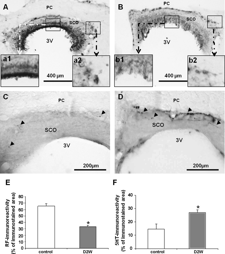
Light micrographs of frontal sections through the subcommissural organ (SCO) of adult rat, immunolabelled with antiserum against Reissner's Fiber (RF) (A and B); and against serotonin (C and D) in controls (A and C) and after 2 weeks of water deprivation (B and D). High magnification shows detail of immunoreactive ependymal (a1 and b1) and hypendymal (a2 and b2) cells. Histograms (E and F) showing the percentage of immunoreactive area to Reissner's fibers (E) and serotonin (F) in the two studied groups. Data are reported as mean ± SEM, and were subjected to Student t-test. A value of P < 0.05 was considered to indicate statistical significance between controls and dehydrated groups. PC: posterior commissure, 3V: third ventricle.

3.2 Water deprivation affects the serotoninergic innervation of the rat SCO

Substantial evidences support a serotoninergic control upon the secretory activity of the SCO in rat. Thus, to assess whether changes of RF material release by the SCO ependymocytes occurring in dehydrated rats is due to alterations of the serotoninergic outputs achieving the SCO, we undertook an immunohistochemical study of 5HT fibers within the SCO. Effectively, our findings demonstrated in control rats (Fig. 1C), many 5HT-immunoreactive fibers surrounding the ependymocytes in the basal and lateral parts and even within ependymocytes of the SCO. In dehydrated rats, an obvious enhancement in the density of 5HT-immunoreactive fibers is observed. These fibers form a dense and continuous plexus surrounding the basal and lateral parts of the SCO (Fig. 1D). Some immunopositive fibers appeared deeper within the ependymocytes of this organ. The statistical analysis supports such increase (Fig. 1F).

3.3 Water deprivation affects the 5HT immunoreactivity within the ventricular borders (VB)

To ascertain whether changes of serotoninergic innervation observed in the SCO occurring in dehydration are generalized to other brain regions, we examined in the VB of the third ventricle via immunohistochemistry the 5HT immunoreactivity, thus, in control animals; many 5HT immunopositive fibers innervated the ventricular borders and formed a highly reactive plexus (Fig. 2A). After dehydration, an obvious rise of the immunoreactivity was seen in the ependymal region with an increased density of IR fibers reaching the ependymal cells of the ventricular borders (Fig. 2B). Such increase was confirmed by the statistical analysis (Fig. 2F).

Fig. 2

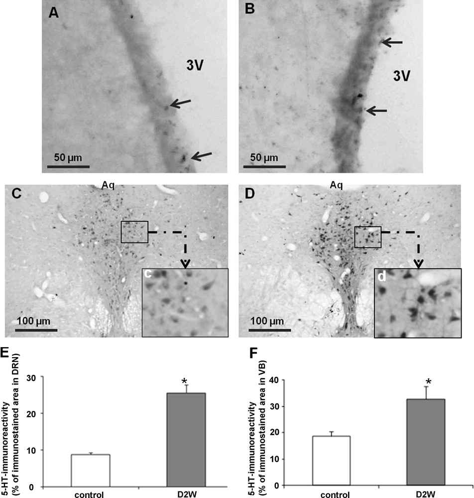
Light micrographs of frontal sections through borders of the third ventricle (VB) (A and B) and the dorsal raphe nucleus (DRN) (C and D) of adult rat, immunolabelled with antiserum against serotonin in controls (A and C) and after 2 weeks of water deprivation (B and D). High magnification shows detail of immunoreactive neurons in the DRN (c and d). Histograms (E and F) represent the percentage areas of immunoreactivity of serotonin in the DRN (E) and the VB (F) in different groups. Data are reported as mean ± SEM, and were subjected to Student t-test. A value of P < 0.05 was considered to indicate statistical significance between controls and dehydrated groups. Aq: Aqueduc de Sylvius, PC: posterior commissure, 3V: third ventricle.

3.4 Water deprivation affects serotoninergic system within the nucleus of origin: dorsal raphe nucleus (DRN)

To assess whether the increased 5HT innervation of the SCO is due to an enhancement of 5HT expression in the neurons of origin, we examined immunohistochemically the expression of 5HT in the nucleus of origin: DRN. Indeed, our finding demonstrates in dehydrated animals a significant enhancement of the immunoreactivity in all neurons forming the DRN nucleus (Fig. 2D) as compared to controls (Fig. 2C), high magnification shows details of immunoreactive neurons (Fig. 2Cc, 2Dd). The statistical analysis supports such observation (Fig. 2E). The prolonged water deprivation does not affect 5HT neurons number within the DRN between treated groups (Fig. 3).

Fig. 3

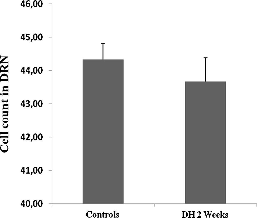
Histograms displaying the cell count in DRN of both control and dehydrated rat. Note that the prolonged water deprivation does not affect 5HT neurons number between treated groups (DH 2 weeks: dehydrated for 2 weeks).

4 Discussion

4.1 Effect of water deprivation on the secretory activity of the rat SCO

The SCO is a particular CVO that releases doubly in the CSF and the circulation [49] a glycoprotein with high molecular weight known after Reissner; Reissner's fiber [11]. This glycoprotein has been involved in many functions including neuronal survival [15], detoxification [13,14], transport of monoamines and clearance of the CSF [50], the pathophysiology of lordosis [20], hydrocephalus [21] and control of water and salt balance [2,8].

In our knowledge, to date no study has been performed concerning the effect of osmotic stress by prolonged water deprivation on the secretory activity of the SCO in the rat. The present study was aimed on the assessment of changes in the secretory activity and serotoninergic innervation of the SCO occurring during water deprivation in the rat. Indeed, our findings demonstrate after 2 weeks of total water restriction, a significant reduction of RF immunoreactivity within both of the hypendymal and the ependymal cells of the SCO. Substantial evidences have associated the changes of the SCO secretory activity with alteration of body homeostasis [1,2,8]. In the semi-desert rodent Meriones shawi, the secretory material of the SCO is significantly reduced following 1 and 3 months of total water restriction [2], whereas, in the frog Rana temporaria, submitted to osmotic stress, the growth rate of the RF and the secretory activity of the SCO have been increased [1].

According to our finding, the reduction of the RF immunoreactivity in the SCO may result from either a decreased RF biosynthesis and/or increased RF release by the SCO.

The mean role and the underlining mechanisms by which the SCO controls water and electrolytic homeostasis is not well established, some experiments are thought to involve this organ in a direct control of peripheral homeostasis. Indeed, destruction of the SCO induces a lower water consumption and a decreased urine output [35,36] and increases the affinity of the SCO-cells for Schiff's reagent (periodic acid-Schiff reaction) indicating either storage or excessive production of the secretory material [51]. Otherwise, dehydration also affects the ultrastructure of the SCO-cells; the dilated sacs and vacuoles of the endoplasmic reticulum, the membrane system of the Golgi complex and the increase in the number of ribosomes suggest a stimulation of the secretory process in the SCO of dehydrated animals [52]. Otherwise, SCO-extracts have an antidiuretic effect in cat [37,38] and influence the level of urinary sodium and sodium excretion in dog [39]. A functional relationship between SCO and adrenals has also been proposed [40,41,43–46]. The SCO may have a hormonal influence on the adrenal cortex by the production of an aldosteronotropic factor [43,53]. Vullings and Diederen in 1985 demonstrated that exposure of the SCO to aldosterone, known to be enhanced during dehydration [54], decreases the secretory activity of the SCO by means of a process of negative feedback regulation [1]. Also worth mentioning is the presence of angiotensin II (AAGII) receptors in the SCO-cells which display binding sites for angiotensin II [55] and, in some species, for atrial natriuretic peptide [56]. Indeed, some studies suggest a functional interrelation between AAGII and the secretory activity of the SCO of goat [57].

4.2 Effect of water deprivation on the serotoninergic innervation of the rat SCO

The SCO is a target of numerous innervations, namely GABAergic [23,24], noradrenergic and dopaminergic [25], and specially serotoninergic [26,27]. This latter innervation is well known to have a control on the SCO secretory activity of several species such as rat [28,58], Meriones [29], gerbil [58] and lizard [34]. Our results show a significant enhancement of the density of the 5HT-immunoreactive fibers achieving the SCO ependymocytes. Thus, the same tendency was observed in the DRN, the origin nucleus of this innervation, from which emanates the serotoninergic fibers innervating the SCO. Such increase of the 5HT innervations of the SCO seems supporting a possible decrease of RF biosynthesis rather than an increase of RF release by the SCO, since in rat, serotonin negatively controls the RF material release by the ependymocytes of the SCO [59]. In contrast to Mshawi, in which, serotonin stimulates the RF release by the SCO [29] that may explain in part, the specific differences of the SCO responsiveness to water deprivation between rat and M. shawi.

Involvement of serotonin in water intake behavior is well established [32,60]. In rat, deprivation of water for 5 days causes a selective decrease in the serotonin concentrations of the neural lobe and pars distalis [61]. In M. shawi, submitted to prolonged water deprivation, serotonin expression in the DRN, supraependymal plexus and the subcommissural organ decreased significantly [2]. While in adult soft-shelled turtles exposed to hyperosmotic stress either with chronic dehydration for 7 days or intramuscular injection of sodium chloride (3.6%, 0.5 mL daily for 7 days) or acute treatment for 0.5, 1 or 2 h with sodium chloride (3.6%, single intramuscular injection) reduced the pineal-paraphyseal serotonin [62]. Our data seem to be contradictory regarding the previous studies; such differences may result from the duration of water deprivation since the present work describes for the first time, in contrast to previous studies, the responsiveness to serotoninergic system to long period of dehydration in rat. Taken together, the above data demonstrate the implication of the serotoninergic system in water and electrolyte homeostasis. Indeed, further investigations have associated this system with the control of body homeostasis [63]. Some experiments of electrolytic or exitotoxic lesions of the DRN reduced the basal and stimulated plasma levels of the atrial natriuretic peptide [64], and increased the basal salt intake in several experimental models [32,65–67].

5 Conclusion

The present findings confirm the involvement of the SCO and the serotonin system in the response to water deprivation in the rat as demonstrated in the M. shawi [2]. The variation of the serotonin system after a prolonged dehydration induced both in the rat as in the Meriones, a decreased secretion of the RF. The mechanism for the involvement of the latter in the dehydration remains to be determined.

Disclosure of interest

The authors declare that they have no conflicts of interest concerning this article.

Acknowledgment

This work has been supported by the Moroccan CNRST (SC. Vie 05/10), le Centre national d’études et de recherches sur le Sahara (CNERS); OTAN (PDD [CP] - [LST.CLG979823]).


Bibliographie

[1] H.G.B. Vullings; J.H.B. Diederen Secretory activity of the subcommissural organ in Rana temporaria L. under osmotic stimulation, Cell Tissue Res., Volume 241 (1985), pp. 663-670

[2] A. El Got; S. Ahboucha; M.M. Bouyatas; M. Fèvre-Montange; H. Gamrani Water deprivation affects serotoninergic system and glycoprotein secretion in the sub-commissural organ of a desert rodent Meriones shawi, Neurosci. Lett., Volume 466 (2009), pp. 6-10

[3] W.D. Knight; L.L. Ji; J.T. Little; J.T. Cunningham Dehydration followed by sham rehydration contributes to reduced neuronal activation in vasopressinergic supraoptic neurons after water deprivation, Am. J. Physiol. Regul. Integr. Comp. Physiol., Volume 299 (2010), p. R1232-R1240

[4] H. Gamrani; A. Elgot; O. El Hiba; M. Fèvre-Montange Cellular plasticity in the supraoptic and paraventricular nuclei after prolonged dehydration in the desert rodent Meriones shawi: vasopressin and GFAP immunohistochemical study, Brain Res., Volume 1375 (2011), pp. 85-92

[5] D.M. Santer; W.E. Heydorn; G.J. Creed; D.C. Klein; D.M. Jacobowitz Subfornical organ: effects of salt loading and water deprivation on in vitro radioamino acid incorporation into individual proteins, Brain Res., Volume 372 (1986), pp. 107-114

[6] B.J. Oldfield; R.J. Bicknell; R.M. McAllen; R.S. Weisinger; M.J. McKinley Intravenous hypertonic saline induces Fos immunoreactivity in neurons throughout the lamina terminalis, Brain Res., Volume 561 (1991), pp. 151-156

[7] K.S. Curtis; J.G. Verbalis; E.M. Stricker Area postrema lesions in rats appear to disrupt rapid feedback inhibition of fluid intake, Brain Res., Volume 726 (1996), pp. 31-38

[8] W.B. Severs; C.D. Balaban; B.S.A.A. Morrow; C.L. Snyder; L.C. Keil The subcommissural organ: immunohistochemistry and potential relation to salt/water balance (A. Ocsche; E.M. Rodriguez; P. Fernandez-Llebrez, eds.), The Subcommissural Organ: an Ependymal Brain Gland, Springer, Berlin, Heidelberg, New York, 1993, pp. 265-277

[9] A. Oksche The subcommissural organ, J. Neurovisc. Relat., Volume 9 (1969), pp. 111-139 (Supplement)

[10] E.M. Rodriguez; A. Oksche; S. Hein; C.R. Yulis Cell biology of the subcommissural organ, Int. Rev. Cytol., Volume 135 (1992), pp. 39-121

[11] E. Reissner Beitr ge zur Kenntnis vom Bau des Rückenmarks von Petromyzon fluviatilis L, Arch. Anat. Physiol., Volume 77 (1860), pp. 545-588

[12] S. Rodríguez; K. Vio; C. Wagner; M. Barría; E.H. Navarrete; V.D. Ramírez; J.M. Pérez-Fígares; E.M. Rodríguez Changes in the cerebrospinal-fluid monoamines in rats with an immunoneutralization of the subcommissural organ-Reissner's fiber complex by maternal delivery of antibodies, Exp. Brain Res., Volume 128 (1999), pp. 278-290

[13] J.H.B. Dideren; H.G.B. Vullings; J.H.W.M. Guntschoonderwoerd The suncommissural organ-liquor fiber complex: the binding of catecholamines to the liquor fiber in frogs of the Rana esculenta complex, Acta Zool., Volume 64 (1983), pp. 47-53

[14] M.M. Bouyatas; H. Gamrani Immunohistochemical evaluation of the effect of lead exposure on subcommissural organ innervation and secretion in Shaw's Jird (Meriones shawi), Acta Histochem., Volume 109 (2007), pp. 421-427

[15] H. Monnerie; O. Boespflug-Tanguy; B. Dastugue; A. Meiniel Reissner's fibre supports the survival of chick cortical neurons in primary mixed cultures, Cell Tissue Res., Volume 282 (1995), pp. 81-91

[16] S. Gobron; I. Creveaux; R. Meiniel; R. Didier; A. Herbet; M. Bamdad; F. El Bitar; B. Dastugue; A. Meiniel Subcommissural organ/Reissner's fiber complex: characterization of SCO-spondin, a glycoprotein with potent activity on neurite outgrowth, Glia, Volume 32 (2000), pp. 177-191

[17] G. Estivill-Torrús; T. Vitalis; P. Fernández-Llebrez; D.J. Price The transcription factor Pax6 is required for development of the diencephalic dorsal midline secretory radial glia that form the subcommissural organ, Mech. Dev., Volume 109 (2001), pp. 215-224

[18] P. Fernández-Llebrez; J.M. Grondona; J. Pérez; M.F. López-Aranda; G. Estivill-Torrús; P.F. Llebrez-Zayas; E. Soriano; C. Ramos; Y. Lallemand; A. Bach; B. Robert Msx1-deficient mice fail to form prosomere 1 derivatives, subcommissural organ, and posterior commissure and develop hydrocephalus, J. Neuropathol. Exp. Neurol., Volume 63 (2004), pp. 574-586

[19] C. Hoyo-Becerra; M.D. López-Avalos; M. Cifuentes; R. Visser; P. Fernández-Llebrez; J.M. Grondona The subcommissural organ and the development of the posterior commissure in chick Embryos, Cell Tissue Res., Volume 339 (2010), pp. 383-395

[20] S. Ahboucha; H. Gamrani Differences in protein expression in the subcommissural organ of normal and lordotic lizards (Agama impalearis), Metab. Brain Dis., Volume 16 (2001), pp. 219-226

[21] A. Meiniel The secretory ependymal cells of the subcommissural organ: which role in Hydrocephalus, Int. J. Biochem. Cell Biol., Volume 39 (2007), pp. 463-468

[22] A.J. Jiménez; P. Fernández-Llebrez; J.M. Pérez-Fígares Neural input and neural control of the subcommissural organ, Microsc. Res. Tech., Volume 52 (2001), pp. 520-533

[23] H. Gamrani; M.F. Belin; M. Aguera; A. Calas; J.F. Pujol Radioautographic evidence for an innervation of the subcommissural organ by GABA-containing nerve fibres, J. Neurocytol., Volume 10 (1981), pp. 411-424

[24] D. Weissmann-Nanopoulos; M.F. Belin; M. Didier; M. Aguera; M. Partisani; M. Maitre; J.F. Pujol Immunohistochemical evidence for neuronal and non-neuronal synthesis of GABA in the rat subcommissural organ, Neurochem. Int., Volume 5 (1983), pp. 785-791

[25] C.D. Balaban; R.J. Schuerger; W.B. Severs Evidence for a noradrenergic projection to the subcommissural organ, Neurosci. Lett., Volume 180 (1994), pp. 209-213

[26] S. Ueda; N. Ihara; T. Tanabe; Y. Sano Reinnervation of serotonin fibers in the denervated rat subcommissural organ by fetal transplants. An immunohistochemical study, Brain Res., Volume 444 (1988), pp. 361-365

[27] T. Matsuura; K. Kumamoto; S. Ebara; Y. Sano Serotoninergic innervation of the mammalian subcommissural organ: qn immunohistochemical study, Biomed. Res. Suppl., Volume 3 (1989), pp. 177-186

[28] C. Bouchard; M. Arluisson Serotoninergic innervation of ependymal cells in the rat subcommissural organ. A fluorescence, electron microscopic and radioautographic study, Biol. Cell., Volume 30 (1977), pp. 61-64

[29] A. Laalaoui; S. Ahboucha; M. Didier-Bazes; M. Fèvre-Montange; A. Meiniel; H. Gamrani Postnatal secretion of the subcommissural organ of the Meriones shawi: control of serotonin innervations, Dev. Brain Res., Volume 126 (2001), pp. 75-80

[30] J.D. Mikkelsen; A. Hay-Schmidt; P.J. Larsen Central innervation of the rat ependyma and subcommissural organ with special reference to ascending serotoninergic projections from the raphe nuclei, J. Comp. Neurol., Volume 384 (1997), pp. 556-568

[31] L.C. Reis; V.R. Marinho Influence of 5-HT1A agonist on the feeding behavior of Coturnix japonica (Galliformes: Aves), Braz. J. Biol., Volume 65 (2005), pp. 675-681

[32] L.C. Reis; M.J. Ramalho; J. Antunes-Rodrigues Brain serotoninergic stimulation reduces the water intake induced by systemic and central beta-adrenergic administration, Braz. J. Med. Biol. Res., Volume 25 (1992), pp. 529-536

[33] T. Sakumoto; K. Sakai; D. Salvert; H. Sakai; H. Kimura; T. Maeda; M. Jouvet Possible role of serotonin in the secretory activity of the subcommissural organ of the cat, Neurosci. Res., Volume 1 (1984), pp. 191-197

[34] S. Ahboucha; M. Didier-Bazes; A. Meiniel; M. Fèvre-Montange; H. Gamrani Secretory activity and serotonin innervation of lizard's subcommissural organ, Neurorepor., Volume 11 (2000), pp. 2611-2615

[35] G.B. Forbes; A.M. Lewis Total sodium, potassium and chloride in adult man, J. Clin. Invest., Volume 35 (1956), pp. 596-600

[36] D.D. Brown; A.K. Afifi Histological and ablation studies on the relation of the subcommissural organ and rostral midbrain to sodium and water metabolism, Anat. Rec., Volume 153 (1965), pp. 255-263

[37] M. Palkovits; P. Földvari Über die antidiuretische Wirkung des Organon subcommissurale, Acta Biol. Hung., Volume 11 (1960), pp. 95-102

[38] G.D. Murphy; J.G. Wood Functional and microscopic studies of the subcommissural organ in the rat, Texas Rep. Biol. Med., Volume 24 (1966), pp. 729-735

[39] G.J. Gilbert Renal effect of subcommissural extract, Neurology, Volume 13 (1963), pp. 43-55

[40] M. Palkovits; G. Lukacs Karyometric examination of the system subcommissural organ-adrenal cortex in the rat, Acta Biol. Hung., Volume 13 (1963), pp. 361-369

[41] I.P. Földvari; M. Palkovits Effect of sodium and potassium restriction on the functional morphology of the subcommissural organ, Nature, Volume 202 (1964), pp. 905-906

[42] M. Palkovits Morphology and function of the subcommissural organ, Stud. Biol. Hung., Volume 41 (1965), pp. 1-105

[43] M. Palkovits; E. Monos; J. Fachet The effect of subcommissural organ lesions on aldosterone production in the rat, Acta Endocrinol., Volume 48 (1965), pp. 169-176

[44] P. Peczely; T. Muray Response of the adrenal gland to the activation of the subcommissural organ in the pigeon, Acta Morphol. Acad Sci. Hung., Volume 16 (1968), pp. 455-462

[45] U. Attila; S. Talanti Incorporation of 35 S-labelled cysteine in the subcommissural organ of the rat after adrtnalectomy and after treatment with hydrocortisone, Acta Physiol. Scand., Volume 87 (1973), pp. 422-424

[46] E.S. Gullyants; L.P. Sizyakina Histophysiological characteristics of the subcommissural organ of the brain during isolated inhibition of the adrenal zona glomerulosa, Bull. Eksp. Biol. Med., Volume 90 (1980), pp. 370-372

[47] A. Meiniel; R. Meiniel; R. Didier; I. Creveaux; S. Gobron; H. Monnerie; B. Dastugue The subcommissural organ and Reissner's fiber complex. An enigma in the central nervous system?, Prog. Histochem. Cytochem., Volume 30 (1996), pp. 1-66

[48] J. Vilaplana; M. Lavialle A method to quantify glial fibrillary acidic protein immunoreactivity on the suprachiasmatic nucleus, J. Neurosci. Methods, Volume 88 (1999), pp. 181-187

[49] E.M. Rodríguez; S. Rodríguez; S. Hein The subcommissural organ, Microsc. Res. Tech., Volume 41 (1998), pp. 98-123

[50] R. Olsson Studies on the subcommissural organ, Acta Zool., Volume 39 (1958), pp. 71-102

[51] G.J. Gilbert Subcommissural organ secretion in the dehydrated rat, Anat Rec., Volume 132 (1958), pp. 563-567

[52] J. Leonieni; L. Rechardt The effect of dehydration on the ultrastructure and cholinesterase activity of the subcommissural organ in the rat, Z. Zellforsch. Mikrosk. Anat., Volume 133 (1972), pp. 377-387

[53] H. Wetzig; J.H. Scharf; P. Krumbacher; K. Liedel; D. Marzotko Statistical studies on nuclear variation in the subcommissural organ of white rats following chronic treatment with methylthiouracil, diiodotyrosine, p-hydroxypropiophenone and alloxan in single and combination dosage, Endokrinologie, Volume 56 (1970), pp. 97-108

[54] E.A. Ezzarani; J.P. Laulin; R. Brudieux Effects of water-deprivation on aldosterone production in Brattleboro male rats congenitally lacking vasopressin, Horm. Metab. Res., Volume 17 (1985), pp. 230-233

[55] P. Ghiani; B. Uva; M. Vallarino; A. Mandich; M. Masini Angiotensin II specific receptors in subcommissural organ, Neurosci. Lett., Volume 85 (1988), pp. 212-216

[56] C.R. Mantyh; L. Kruger; N.C. Brecha; P.W. Mantyh Localization of specific binding sites for atrial natriuretic factor in the central nervous system of rat, guinea pig, cat and human, Brain Res., Volume 412 (1987), pp. 329-342

[57] M. Castañeyra-Martín; E.M. Carmona-Calero; H. Pérez-González; Y.V.I. Martínez-Peña; C. Ormazabal-Ramos; C.G. Pérez-Garcia; N. Marrero-Gordillo; A. Trujillano-Dorado; I. González-Marrero; A. Castañeyra-Perdomo Postnatal development of the secretory activity of the goat subcommissural organ. A Reissner's fibre and angiotensin II immunohistochemical study, Anat. Histol. Embryol., Volume 34 (2005), pp. 247-251

[58] L. Wiklund; J.J. Lundberg; K. Møllgård Species differences in serotoninergic in innervation and secretory activity of rat, gerbil, mouse and rabbit subcommissural organ, Acta Physiol. Scand. Suppl., Volume 452 (1977), pp. 27-30

[59] K. Møllgård; J.J. Lundberg; L. Wiklund; L. Lachenmayer; H.G. Baumgarten Morphologic consequences of serotonin neurotoxin administration: neuron-target cell interaction in the rat subcommissural organ, Ann. N. Y. Acad. Sci., Volume 305 (1978), pp. 262-288

[60] P.L. Cedraz-Mercez; A.C. Almeida; C.M. Thomaz; R.H. Costa-e-Sousa; E.L. Olivares; W.S. Côrtes; M.A. Medeiros; L.C. Reis Effect of L-5-Hydroxytryptophan on drinking behavior in Coturnix japonica (Temminck and Schlegel, 1849) (Galliformes: Aves): involvement of renin-angiotensin system, Braz. J. Biol., Volume 67 (2007), pp. 771-776

[61] R.S. Piezzi; R.J. Wurtman Pituitary serotonin content: effects of melatonin or deprivation of Water, Science, Volume 169 (1970), pp. 285-286

[62] M.S. Mahapatra; S.K. Mahata; B. Maiti Effect of stress on serotonin, norepinephrine, epinephrine and corticosterone contents in the soft-shelled turtle, Clin. Exp. Pharmacol. Physiol., Volume 18 (1991), pp. 719-724

[63] J.T. Fitzsimons Angiotensin, thirst, and sodium appetite, Physiol. Rev., Volume 78 (1998), pp. 583-686

[64] L.C. Reis; M.J. Ramalho; A.L. Favaretto; J. Gutkowska; S.M. McCann; J. Antunes-Rodrigues Participation of the ascending serotonergic system in the stimulation of atrial natriureti peptide release, Proc. Natl Acad. Sci. U. S. A., Volume 91 (1994), pp. 12022-12026

[65] E.L. Olivares; R.H. Costa-E-Sousa; H.R. Cavalcante-Lima; H.R. Lima; P.L. Cedraz-Mercez; L.C. Reis Effect of electrolytic lesion of the dorsal raphe nucleus on water intake and sodium Appetite, Braz. J. Med. Biol. Res., Volume 36 (2003), pp. 1709-1716

[66] H.R. Lima; H.R. Cavalcante-Lima; P.L. Cedraz-Mercez; R.H. Costa-E-Sousa; E.L. Olivares; D. Badauêg-Passos; M.A. Medeiros; W.S. Côrtes; L.C. Reis Brain serotonin depletion enhances the sodium appetite induced by sodium depletion or beta-adrenergic stimulation, An. Acad. Bras. Cienc., Volume 76 (2004), pp. 85-92

[67] H.R. Cavalcante-Lima; D. Badauê-Passos; W. De-Lucca; H.R. Lima; R.H. Costa-e-Sousa; E.L. Olivares; P.L. Cedraz-Mercez; R.O. Reis; M.A. Medeiros; W.S. Côrtes; L.C. Reis Chronic excitotoxic lesion of the dorsal raphe nucleus induces sodium appetite, Braz. J. Med. Biol. Res., Volume 38 (2005), pp. 1669-1675


Commentaires - Politique


Ces articles pourraient vous intéresser

The neuroactive neurosteroid Dehydroepiandrosterone Sulfate (DHEAS) modulates the serotonergic system within the dorsal Raphe nucleus and the cerebrospinal fluid release of Reissner’s fiber in rat

Omar El Hiba; Ahmed Draoui; Halima Gamrani

C. R. Biol (2020)


Alteration of dopaminergic innervation and voluntary movements after long period of thirst in a semi-desert rodent, Meriones shawi: Behavioral and immunohistochemical studies

Abdeljalil Elgot; Omar El Hiba; Halima Gamrani

C. R. Biol (2012)


Altered nigrostriatal dopaminergic and noradrenergic system prompted by systemic lead toxicity versus a treatment by curcumin-III in the desert rodent Meriones shawi

Lahcen Tamegart; Abdellatif Abbaoui; Abdelaati El Khiat; ...

C. R. Biol (2019)