Plan
Comptes Rendus

Estrogen-dependent, extrahepatic synthesis of vitellogenin in male vertebrates: A mini-review
Comptes Rendus. Biologies, Volume 340 (2017) no. 3, pp. 139-144.

Résumé

In the last years, the hormonal balance is threatened by the interferences of substances with hormone-like action (endocrine disruptor chemicals, EDCs) that may harm animal reproduction. Most EDCs are resistant to environmental degradation and are considered ubiquitous contaminants. EDCs may have synthetic or natural origins. Pesticides used in intensive agriculture contain large amounts of chemicals with estrogenic properties, such as the alkylphenol nonylphenol (NP). Besides, animal feeding operations are important sources of natural estrogen metabolites introduced into the environment through manure application in organic farming. In both cases, EDCs can reach animals, including humans particularly at risk due to their position in the food chain. This is the reason for which it is important to use terrestrial vertebrates as sentinels in soil biomonitoring programmes. Today, the most validated biomarker of estrogenic exposure is the expression in male liver of the vitellogenin (VTG), an estrogen-dependent glycolipophosphoprotein naturally expressed only in the liver of oviparous females during the reproductive season. This report summarizes the data available on the EDC-dependent expression and the synthesis of VTG in male vertebrates, highlighting our latest studies that demonstrate the ability of testis and epididymis of the lacertid Podarcis sicula to synthesize VTG following estrogenic exposure. These findings provide, for the first time, evidence on an extrahepatic expression and synthesis of VTG in a terrestrial vertebrate and lay the groundwork for a new value of the VTG as a biomarker of environmental contamination. In addition, the results open a new scenario on the role of VTG in cells other than oocytes.

Métadonnées
Reçu le :
Accepté le :
Publié le :
DOI : 10.1016/j.crvi.2017.01.005
Mots clés : Ectopic synthesis of vitellogenin, Endocrine disruptor chemicals, Estrogen, Manure, Nonylphenol, Podarcis sicula, Vertebrates, Testis
Mariailaria Verderame 1 ; Rosaria Scudiero 1

1 Department of Biology, University of Naples Federico II, Via Mezzocannone 8, 80134 Napoli, Italy
@article{CRBIOL_2017__340_3_139_0,
     author = {Mariailaria Verderame and Rosaria Scudiero},
     title = {Estrogen-dependent, extrahepatic synthesis of vitellogenin in male vertebrates: {A} mini-review},
     journal = {Comptes Rendus. Biologies},
     pages = {139--144},
     publisher = {Elsevier},
     volume = {340},
     number = {3},
     year = {2017},
     doi = {10.1016/j.crvi.2017.01.005},
     language = {en},
}
TY  - JOUR
AU  - Mariailaria Verderame
AU  - Rosaria Scudiero
TI  - Estrogen-dependent, extrahepatic synthesis of vitellogenin in male vertebrates: A mini-review
JO  - Comptes Rendus. Biologies
PY  - 2017
SP  - 139
EP  - 144
VL  - 340
IS  - 3
PB  - Elsevier
DO  - 10.1016/j.crvi.2017.01.005
LA  - en
ID  - CRBIOL_2017__340_3_139_0
ER  - 
%0 Journal Article
%A Mariailaria Verderame
%A Rosaria Scudiero
%T Estrogen-dependent, extrahepatic synthesis of vitellogenin in male vertebrates: A mini-review
%J Comptes Rendus. Biologies
%D 2017
%P 139-144
%V 340
%N 3
%I Elsevier
%R 10.1016/j.crvi.2017.01.005
%G en
%F CRBIOL_2017__340_3_139_0
Mariailaria Verderame; Rosaria Scudiero. Estrogen-dependent, extrahepatic synthesis of vitellogenin in male vertebrates: A mini-review. Comptes Rendus. Biologies, Volume 340 (2017) no. 3, pp. 139-144. doi : 10.1016/j.crvi.2017.01.005. https://comptes-rendus.academie-sciences.fr/biologies/articles/10.1016/j.crvi.2017.01.005/

Version originale du texte intégral

1 The environmental soil pollution by xenoestrogens: the case of manure and nonylphenol

Many substances poured in the environment mimic the effects of animal endogenous hormones interfering with the endocrine system [1], in particular with the male reproductive physiology [2,3]. These substances, known as endocrine disruptor chemicals (EDCs), originate from a variety of sources. Today, a natural and plentiful source of EDCs is given by the metabolites of steroid hormones in the manure used for the fertilization of the soils dedicated to organic farming [1,4,5], whereas an artificial source is the presence in pesticides of substances such as the alkylphenol polyethoxylates.

To date, little attention has been paid to the steroid hormones and to the products of their metabolism in manure. The organic farming is exponentially growing and, along with it, the use of manure as the principal choice for soil fertility. Indeed, manure ensures a proper recycling of biological by-products. However, it must be not overlooked that organic manure of animal origin contributes to a significant load of estrogen hormones in the natural environment [6], since farm animals excrete conjugated steroid hormones that persist in the manure for several months [7]. It has been estimated that pregnant cows can excrete from 700 to 17,000 mg/day of estrone in urine; in the dairy cattle, a combined (faecal + urinary) excretion of 384 mg of 17ß-estradiol was calculated [8]. In addition, the conjugated and biologically inactive forms of excreted hormones can be easily converted into free steroids by soil microorganisms as Escherichia coli [9–11]. Bioassays of estrogenicity on water samples from streams across the United States revealed that higher estrogenic activity was frequently associated with manure application to the cultivated fields in the neighbourhood [12].

As regards EDCs of synthetic origin, nonylphenol (NP) is the estrogen-like compound produced in larger quantities. NP is persistent in sediments and in aquatic environment, bioaccumulable, extremely toxic and it is used primarily to produce surfactants for a wide variety of applications and consumer products [1]. NP is commonly used as a co-formulant in pesticides.

NP is a clear-to-pale yellow viscous liquid at room temperature [13–15]. The air concentrations of NP are generally expected to be low [14], but a research has shown that in some circumstances there may be water-to-air volatilization that results in significant atmospheric concentrations of NP substances [16]. NP has been detected in many different habitats, such as groundwater, sediment, soil, freshwater, saltwater.

The human exposure is a result of the presence of NP in detergents, cleaners, agricultural and indoor pesticides, food packaging and cosmetics and it has been confirmed by biomonitoring data from breast milk [17], umbilical cord blood [18] and urine [19]. The maximum level of NP found in the breast milk was 56.3 μg/L, leading to an estimated maximum dose for an infant of 3.9 μg/kg/day [17].

Due to its structural similarity with estradiol-17β, NP is able to bind the estrogen receptors stimulating the transcription of the downstream genes (as the estrogen receptors itself or vitellogenin) [20] interfering with the reproductive and developmental events, in particular.

Most of the research on NP in the environment was carried out in ecosystems where the human impact is predominant, as in rivers in which the contribution of wastewater effluents to flow is significant [21]. Nevertheless, agricultural ecosystems can potentially be contaminated by spraying of pesticides containing NP as co-formulates, landfilling of sludge or by the application of sewage sludge or pulp and paper mill sludge [22,23]. In addition, NP remains in the soil, thus accumulating moderately [24].

2 Vitellogenin in extrahepatic tissues of vertebrates

Vitellogenin (VTG), the major precursor of the yolk proteins, is an estrogen-dependent and sex-specific protein naturally synthesized in the liver of the females of oviparous vertebrates during the reproductive period [25,26]. In males, VTG gene is silent, but it may be activated by estrogenic exposure [27–29]. For this reason, the finding of VTG in the liver and/or in plasma of males is considered a good biomarker of xeno-estrogenic pollution.

A huge amount of studies was performed on males of aquatic organisms, naturally or experimentally exposed to estrogen-like substances and were mainly aimed to highlight the hepatic induction of VTG [27–35].

A possible extrahepatic synthesis of VTG was suggested by Wallace [26], according to which in females 5–10% of the total VTG could be produced by the oocyte itself; this process was referred to as endogenous or ectopic vitellogenesis.

Over the years, many studies resumed the possibility of an extrahepatic origin for VTG protein by using biochemical and molecular approaches (Table 1). Investigations carried out on oviparous fish suggest that estrogenic hormones and estrogen-like compounds are able to induce the expression of the VTG gene in both hepatic and extrahepatic tissues [36–38]. It has been demonstrated that in male zebrafish 17α-ethinylestradiol induces VTG expression and synthesis in dose- and time-dependent pattern in skin and eye [39] and estradiol-17β or EDCs are able to determine the VTG synthesis in the heart, cerebral tissue [40], epidermis [41], gills [42,43], white adipose tissue [44], intestine and muscle tissue [38].

Table 1

Estrogen-induced expression of extrahepatic VTG in male vertebrates.

Table 1
Species Treatment Tissue References
Danio rerio 17α-ethinylestradiol Skin Zhang et al., 2014 [39]
Eye
Estradiol 17-β/EDC Heart Yin et al., 2009 [66]
Cerebral tissue
Epidermis Jin et al., 2008 [67]
Gills Islinger et al., 2003 [42]
Wang et al., 2010 [43]
White adipose tissue Tingaud et al., 2012 [44]
Intestine Wang et al., 2005 [38]
Muscle tissue
Testis
Acipenser transmontanus Estradiol 17-β Testis Bidwell et al., 1995 [36]
Oryzias latipes Estradiol 17-β/NP Spermatocytes Kobayashi et al., 2005 [37]
Koger et al., 1999 [45]
Melanotaenia fluviatilis Estradiol 17-β Seminiferous epithelium Shanthanagouda et al., 2013 [46]
Woods et al., 2009 [47]
Tanichthys albonube Estradiol 17-β Testis Wang et al., 2010 [43]
Torpedo marmorata Estradiol 17-β/NP Testis Del Giudice et al., 2011; 2012 [48,49]
Kidney
Podarcis sicula Estradiol 17-β/NP/manure Testis Verderame et al., 2016 [55]
Verderame, 2016 [63]

Many studies focused on gonads of male fish experimentally exposed to E2 or estrogen-like substances. In Acipenser transmontanus, VTG transcripts were detected in the testis of the E2-treated samples [36]; a same result was obtained also in zebrafish [38]. In the testis of Oryzias latipes treated with E2 or NP, spermatocytes are able to transcribe the VTG gene [37,45]. In specimens of Melanotaenia fluviatilis experimentally exposed to E2, VTG-mRNA was found in the seminiferous epithelium [46], whereas the VTG protein was localized in the cellular spaces surrounding the spermatids [47]. Gene expression analysis demonstrated the presence of VTG-mRNA in the testes of Tanichthys albonube treated with E2 [43]. Again, the male germ cells of medaka incorporate and accumulate VTG after treatment with E2 or estrogen-like substances [37].

Among the cartilaginous fish, experiments carried out on Torpedo marmorata demonstrated the VTG expression and synthesis in testis and kidney following intraperitoneal injection of E2 or NP [48,49]. Interestingly enough, in this species the VTG synthesis has been found to occur also within the ovarian follicle cells in both previtellogenic and vitellogenic phases [50].

3 Expression and synthesis of VTG in the terrestrial oviparous vertebrate Podarcis sicula

In the literature, until 2015 no data are present on the extrahepatic expression and/or synthesis of VTG in terrestrial oviparous vertebrates. In addition, studies on the reproductive status of wildlife exposed to manure-fertilized cropland are very limited, so we decided to investigate the presence of endogenous VTG in testis of the lizard Podarcis sicula caught in areas devoted to organic farming and in males experimentally fed with NP-polluted food.

The Italian wall lizard Podarcis sicula is a small lacertid, considered an excellent pollution bioindicator model for ecotoxicological studies [51–56]. It is widespread in the countryside and lives in narrow ranges sheltering under the ground and the rocks [57]. As prey for many small mammals and birds, it fills an important role also as soil top predator contributing, in several areas, to the control of agricultural pests.

To make this animal a good model organism for studies on vitellogenesis mechanisms is also the in-depth knowledge of its reproductive cycle in both male and female [58–62]. In this oviparous species, VTG synthesis occurs in female liver during the breeding season, under the control of the estrogen receptor alpha (ERα) [59]. The protein is also detected in the liver and the plasma of males experimentally treated with E2 [59] or exposed to NP-contaminated food [51,54]. VTG expression in the P. sicula male liver is also accompanied by the expression of the silent ERα gene [51,54]. These estrogen-contaminated animals show also a sharp slowdown of spermatogenesis, a condition that seriously threatens the continuity of this species. More recently, it has been demonstrated expression and synthesis of VTG also in the liver of P. sicula collected in cultivated fields devoted to organic farming, fertilized only with manure [56]. These animals, however, did not show any impairment of spermatogenesis [56].

We decided to verify the ability of P. sicula testis to express and synthesize VTG following exposure to estradiol-17β. Sexually mature males in the mating period were intraperitoneally injected with E2 at the same concentration able to stimulate in these males the hepatic synthesis of VTG [59]. Immunohistochemical and in situ hybridization analyses demonstrate the presence of both VTG transcript and protein in all the germ cells forming the seminiferous tubule (Fig. 1) [55].

Fig. 1

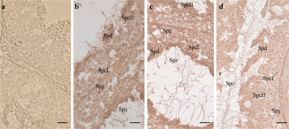
In situ hybridization on Podarcis sicula testis sections, incubated with DIG-probe to detect VTG-mRNA. The brown hybridization signal was absent in the testis of wildlife males collected on uncultivated, rural areas [a]. In the samples treated with E2 [b], fed with NP-polluted food [c] or housing in manure treated soil [d], the signal was evident in the cytoplasm of all the germinal cells of the seminiferous epithelium, i.e. spermatogonia (Spg), primary spermatocytes (SpcI), secondary spermatocytes (SpcII), spermatids (Spt), and spermatozoa (Spz). See [55] for details on methods. Bar: 30 μm.

Once experimentally assessed the estrogen-induced synthesis of VTG in the testis, we investigate the induction of testicular VTG in two possible environmentally estrogenic exposures derived from:

  • • estrogen metabolites naturally present in animal manure;
  • • NP-polluted food and water.

Biochemical and biomolecular investigations demonstrated that both estrogenic exposures, i.e. animals caught in a field where the only fertilizer used was manure or in the animals experimentally exposed to NP-polluted diet, are able to switch on the VTG gene in the germ cells of the seminiferous epithelium (Fig. 1) [63].

While there is no doubt on the endogenous origin of VTG transcripts, the protein observed in lizard testis could have a dual origin. It is conceivable that VTG protein could be synthesized from the transcripts present in the same cells, but it could not be excluded that, as occurs in the ovary during the oocyte vitellogenesis [64], the hepatic VTG protein, synthesized under estrogenic stimulation and released into the bloodstream, could be taken up by receptor-mediated endocytosis in the testis. The failure to detect VTGR transcripts in testis of lizards environmentally or experimentally contaminated with EDCs [56] strongly suggests that the VTG protein detected in this tissue derives entirely from the biosynthetic process locally activated by the estrogenic contamination (Fig. 2).

Fig. 2

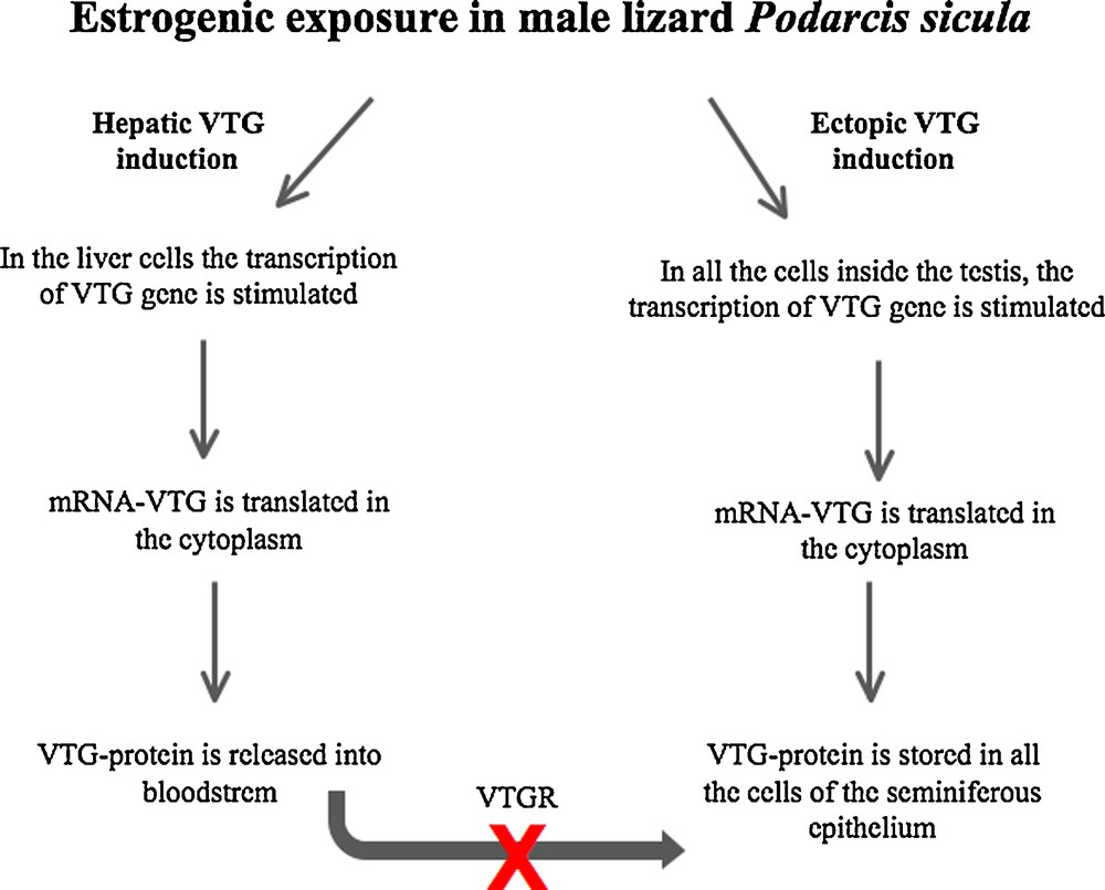
Schematic illustration of hepatic and extrahepatic synthesis of VTG in male lizard Podarcis sicula exposed to estrogenic environment.

4 Concluding remarks

Taken together, these data demonstrate the ability of both aquatic and terrestrial oviparous vertebrates to synthesize VTG in the male gonad. The presence of VTG in the gonads opens a new scenario on the possibility of using this protein as a biomarker of environmental estrogen pollution, as well as on its role in the impairment of reproduction observed in estrogen contaminated males. Our data also demonstrate the estrogenic nature of manure and this sheds new light on the use of animal manure as a fertilizer. Indeed, the excessive and uncontrolled use of manure as a fertilizer in organic farming could endanger the reproduction and survival of soil organisms. Considering that in the last past years there has been a huge increase in organic farming, estimating to grow by 8.9% per year [65], soil contamination by estrogen metabolites could soon represent a serious risk for wildlife.

Finally, the result opens new scenarios and questions on the role of VTG in cells other than oocytes.

Disclosure of interest

The authors declare that they have no competing interest.

Acknowledgement

The authors gratefully acknowledge Prof. Ermelinda Limatola for her critical review of the manuscript.


Bibliographie

[1] A. Priac; N. Morin-Crini; C. Druart; S. Gavoille; C. Bradu; C. Lagarrigue; G. Torri; P. Winterton; G. Crini Alkylphenol and alkylphenol polyethoxylates in water and wastewater: a review of options for their elimination, Arabian. J. Chem. (2014) http://dx.doi.org/10.1016/j.arabjc.2014.05.011​ (In press)

[2] S. Carreau; R.A. Hess Oestrogens and spermatogenesis, Philos. Trans. R. Soc. Lond. B. Biol. Sci., Volume 365 (2010), pp. 1517-1535

[3] A. Joseph; B.D. Shur; R.A. Hess Estrogen, efferent ductules and the epididymis, Biol. Reprod., Volume 84 (2011), pp. 207-217

[4] S.L. Bartelt-Hunt; S. Devivo; L. Johnson; W.L. Kranz; T.L. Mader; C.A. Shapiro; S.J. van Donk; D.P. Shelton; D.D. Tarkalson; T.C. Zhang Effect of composting on the fate of steroids in beef cattle manure, J. Environ. Qual., Volume 42 (2013), pp. 1159-1166

[5] A. Valdehita; A. Quesada-Garcı́a; M.M. Delgado; J.V. Martı́n; M.C. Garcıa-Gonzalez; M.L. Fernandez-Cruz; J.M. Navas In vitro assessment of thyroidal and estrogenic activities in poultry and broiler manure, Sci. Total. Environ., Volume 472 (2014), pp. 630-641

[6] G. Andaluri; R.P.S. Suri; K. Kumar, Environmental Monitoring and Assessment (2012), pp. 1197-1205

[7] B. Schiffer; A. Daxenberger; K. Meyer; H.H. Meyer The fate of trenbolone acetate and melengestrol acetate after application as growth promoters in cattle: environmental studies, Environ. Health. Perspect., Volume 109 (2001), pp. 1145-1151

[8] A.C. Johnson; R.J. Williams; P. Matthiessen The potential steroid hormone contribution of farms animals to freshwaters the united kingdom as a case study, Sci. Total. Environ., Volume 362 (2006), pp. 166-178

[9] C. Desbrow; E.J. Routledge; G.C. Brighty; J.P. Sumpter; M. Waldock Identification of estrogenic chemicals in STW effluent: chemical fractionation and in vitro biological screening, Environ. Sci. Technol., Volume 32 (1998), pp. 1549-1558

[10] T.A. Ternes; P. Kreckel; J. Mueller Behaviour and occurrence of estrogens in municipal sewage treatment plants-II. Aerobic batch experiments with activated sludge, Sci. Total. Environ., Volume 225 (1999), pp. 91-99

[11] C. Baronti; R. Curini; G. D’Ascenzo; A. Di Corcia; A. Gentili; R. Samperi Monitoring natural and synthetic estrogens at activated sludge sewage treatment plants and in a receiving river water, Environ. Sci. Technol., Volume 34 (2000), pp. 5059-5066

[12] D.A. Alvarez; N.W. Shappell; L.O. Billey; D.S. Bermudez; V.S. Wilson; D.W. Kolpin; S.D. Perkins; N. Evans; W.T. Foreman; J.L. Gray; M.J. Shipitalo; M.T. Meyer Bioassay of estrogenicity and chemical analyses of estrogens in streams across the United States associated with livestock operations, Water. Res., Volume 47 (2013), pp. 3347-3363

[13] M. Ahel; J. McEvoy; W. Giger Bioaccumulation of the lipophilic metabolites of non-ionic surfactants in freshwater organisms, Environ. Pollut., Volume 79 (1993), pp. 243-248

[14] EU, European Union Risk Assessment Report. 4-nonylphenol [branched] and nonylphenol. 2nd Priority List, Vol. 10, 2002

[15] A. Seidel Kirk-Othmer encyclopedia of chemical technology, John Wiley & Sons, Inc, Hoboken, NJ, 2004 (148 pp.)

[16] Environment Canada. Canadian environmental quality guidelines for nonylphenol and its ethoxylates [water, sediment and soil] scientific supporting document. ecosystem health: science-based solutions report No. 1-3. National guidelines and standards office environmental quality branch, environment Canada, 2002

[17] N. Ademollo; F. Ferrara; M. Delise; F. Fabietti; E. Funari Nonylphenol and octylphenol in human breast milk, Environ. Int., Volume 34 (2008), pp. 984-987

[18] M. Chen; C.C. Chang; Y.J. Shen; J.H. Hung; B.R. Guo; H.Y. Chuang; I.F. Mao Quantification of prenatal exposure and maternal-fetal transfer of nonylphenol, Chemosphere, Volume 73 (2008), pp. 5245-5349

[19] A. Calafat; Z. Kuklenyik; J. Reidy; S. Caudill; J. Ekong; L. Needham Urinary concentration of bisphenol A and 4-nonylphenol in a human reference population, Environ. Health. Perspect., Volume 113 (2008), pp. 391-395

[20] T. Celius; T.B. Haugen; T. Grotmol; B.T. Walther A sensitive zonagenetic assayfor rapid in vitro assessment of estrogenic potency of xenobiotics and mycotoxins, Environ. Health. Perspect., Volume 107 (1999), pp. 63-68

[21] S. Jobling; J.P. Sumpter Detergent components in sewage effluent are weakly estrogenic to fish: an in vitro study using rainbow trout [Oncorhynchus mykiss] hepatocytes, Aquat. Toxicol., Volume 27 (1993), pp. 361-372

[22] S. Jobling; D. Sheahan; J.A. Osborne; P. Matthiessen; J.P. Sumpter Inhibition of testicular growth in rainbow trout [Oncorhynchus mykiss] exposed to estrogenic alkylphenolic chemicals, Environ. Toxicol. Chem., Volume 15 (1996), pp. 194-202

[23] S. Brown; D. Devin-Clarke; M. Doubrava; G. O’Connor Fate of 4-nonylphenol in a biosolids amended soil, Chemosphere, Volume 75 (2009), pp. 549-555

[24] Office of environmental health hazard assessment [OEHHA], 2009

[25] J.R. Tata; D.F. Smith Vitellogenesis: a versatile model for hormonal regulation of gene expression, Recent Prog. Hormone Res., Volume 35 (1979), pp. 47-90

[26] R.A. Wallace Vitellogenesis and oocyte growth in non-mammalian vertebrates, Dev. Biol., Volume 1 (1985), pp. 127-177

[27] B. Ruggeri; M. Ubaldi; A. Lourdusamy; L. Soverchia; R. Ciccocioppo; G. Hardiman; M.E. Baker; F. Palermo; A.M. Polzonetti-Magni Variation of the genetic expression pattern after exposure to estradiol-17b and 4-nonylphenol in male zebrafish (Danio rerio), Gen. Comp. Endocrinol., Volume 158 (2008), pp. 138-144

[28] T. El-Sayed Ali; S.H. Abdel-Aziz; A.F. El-Sayed; S. Zeid Structural and functional effects of early exposure to 4-nonylphenol on gonadal development of Nile tilapia [Oreochromis niloticus]: a histological alterations in ovaries, Fish Physiol. Biochem., Volume 40 (2014), pp. 1509-1519

[29] M. Staniszewska; L. Falkowska; P. Grabowski; J. Kwaśniak; S. Mudrak-Cegiołka; A.R. Reindl; A. Sokołowski; E. Szumiło; A. Zgrundo Bisphenol A, 4-tertoctylphenol, and 4-nonylphenol in the gulf of Gdañsk (Southern Baltic), Arch. Environ. Contam. Toxicol., Volume 67 (2014), pp. 335-347

[30] M. Ahel; W. Giger Aqueous solubility of alkylphenols and alkylphenol polyethoxylates, Chemosphere, Volume 26 (1993), pp. 1461-1470

[31] T. Christiansen; B. Korsgaard; A. Jespersen Effects of nonylphenol and 17 boestradiol on vitellogenin synthesis, testicular structure and cytology in male eelpout Zoarces viviparus, J. Exp. Biol., Volume 201 (1998), pp. 179-192

[32] A. Arukwe; A. Goksyr; R. Thibaut; J. Cravedi Metabolism and organ distribution of nonylphenol in Atlantic salmon (Salmo salar), Mar. Environ. Res., Volume 50 (2000), pp. 141-145

[33] K. Kinnberg; B. Korsgaard; P. Bjerregaard; A. Jespersen Effects of nonylphenol and 17 beta-estradiol on vitellogenin synthesis and testis morphology in male platyfish Xiphophorus maculatus, J. Exp. Biol., Volume 203 (2000), pp. 171-181

[34] K. Kinnberg; B. Korsgaard; P. Bjerregaard Concentration-dependent effects of nonylphenol on testis structure in adult platyfish Xiphophorus maculatus, Mar. Environ. Res., Volume 50 (2000), pp. 169-173

[35] J.S. Seo; Y.M. Lee; S.O. Jung; I.C. Kim; Y.D. Yoon; J.S. Lee Nonylphenol modulates expression of androgen receptor and estrogen receptor genes differently in gender types of the hermaphroditic fish Rivulus marmoratus, Biochem. Biophys. Res. Commun., Volume 346 (2006), pp. 213-223

[36] C.A. Bidwell; D.M. Carlson Characterization of vitellogenin from white sturgeon, Acipenser transmontanus, J. Mol. Evol., Volume 41 (1995), pp. 104-112

[37] K. Kobayashi; S. Tamotsu; K. Yasuda; T. Oishi Vitellogenin immunohistochemistry in the liver and the testis of the Medaka Oryzias latipes, exposed to 17β-estradiol and p-nonylphenol, Zool. Sci., Volume 22 (2005), pp. 453-461

[38] H. Wang; J.T.T. Tan; A. Emelyanov; V. Korzh; Z.Y. Gong Hepatic and extrahepatic expression of vitellogenin genes in the zebrafish, Danio rerio. Gene, Volume 356 (2005), pp. 91-100

[39] Z. Zhang; N. Ren; K. Kannan; J. Nan Occurrence of endocrine-disrupting phenols and estrogens in water and sediment of the Songhua River Northeastern China, Arch. Environ. Contam. Toxicol., Volume 66 (2014), pp. 361-369

[40] G.G. Ying; B. Williams; R. Kookana Environmental fate of alkylphenols and alkylphenol ethoxylates-a review, Environ. Int., Volume 28 (2002), pp. 215-226

[41] X. Jin; Y. Wang; W. Jin; K. Rao; J.P. Giesy; H. Hollert; K.L. Richardson; Z. Wang Ecological risk of nonylphenol in China surface waters based on reproductive fitness, Environ. Sci. Technol, Volume 48 (2014), pp. 1256-1262

[42] M. Islinger; D. Willimski; A. Volkl; T. Braunbeck Effects of 17α-ethinylestradiol on the expression of three estrogen-responsive genes andcellular ultrastructure of liver and testes in male zebrafish, Aquat. Toxicol., Volume 62 (2003), pp. 85-103

[43] R.L. Wang; Y. Gao; L.H. Zhang; Y.K. Zhang; Z.Q. Fang; J.G. He; W.M. Zhang; G.Z. Ma Cloning, expression, and induction by 17-beta-estradiol [E-2] of a vitellogenin gene in the white cloud mountain minnow Tanichthys albonubes, Fish Physiol. Biochem., Volume 36 (2010), pp. 157-164

[44] A. Tingaud-Sequeira; A. Knoll-Gellida; M. Andre; P.J. Babin Vitellogenin expression in white adipose tissue in female teleost fish, Biol. Reprod., Volume 86 (2012), p. 38

[45] C.S. Koger; S.J. Teh; D.E. Hinton Variations of light and temperature regimes and resulting effects on reproductive parameters in medaka [Oryzias latipes], Biol. Reprod., Volume 61 (1999), pp. 1287-1293

[46] A.H. Shanthanagouda; D. Nugegoda; K.L. Hassell; J.G. Patil Exposure to estrogenic chemicals induces ectopic expression of VTG in the testis of rainbowfish Melanotaenia fluviatilis, Bull. Environ. Contam. Toxicol., Volume 91 (2013), pp. 438-443

[47] M. Woods; A. Kumar; M. Barton; A. Woods; R. Kookana Localisation of estrogen responsive genes in the liver and testis of Murray rainbowfish Melanotaenia fluviatilis exposed to 17beta-estradiol, Mol. Cell. Endocrinol., Volume 303 (2009), pp. 57-66

[48] G. Del Giudice; M. Prisco; M. Agnese; M. Verderame; E. Limatola; P. Andreuccetti Expression of vitellogenin in the testis and kidney of the spotted ray Torpedo marmorata exposed to 17b-estradiol, Gen. Comp. Endocrinol., Volume 174 (2011), pp. 318-325

[49] G. Del Giudice; M. Prisco; M. Agnese; M. Verderame; L. Rosati; E. Limatola; P. Andreuccetti Effects of nonylphenol on vitellogenin synthesis in adult males of the spotted ray Torpedo marmorata, J. Fish Biol., Volume 80 (2012), pp. 2112-2121

[50] M. Prisco; S. Valiante; M. Romano; L. Ricchiari; A. Liguoro; V. Laforgia; E. Limatola; P. Andreuccetti Ovarian follicle cells in Torpedo marmorata synthesize vitellogenin, Mol. Reprod. Dev., Volume 67 (2004), pp. 424-429

[51] M. Verderame; M. Prisco; P. Andreuccetti; F. Aniello; E. Limatola Experimentally nonylphenol-polluted diet induces the expression of silent genes VTG and ERα in the liver of male lizard Podarcis sicula, Environ. Pollut., Volume 159 (2011), pp. 1101-1107

[52] A. Cardone Testicular toxicity of methyl thiophanate in the Italian wall lizard (Podarcis sicula): morphological and molecular evaluation, Ecotoxicology, Volume 21 (2012), pp. 512-523

[53] A. Cardone Imidacloprid induces morphological and molecular damages on testis of lizard [Podarcis sicula], Ecotoxicology, Volume 24 (2015), pp. 94-105

[54] M. Verderame; E. Limatola Interferences of an environmental pollutant with estrogen-like action in the male reproductive system of the terrestrial vertebrate Podarcis sicula, Gen. Comp. Endocrinol., Volume 213 (2015), pp. 9-15

[55] M. Verderame; E. Limatola; R. Scudiero Ectopic synthesis of vitellogenin in testis and epididymis of estrogen-treated lizard Podarcis sicula, Gen. Comp. Endocrinol., Volume 235 (2016), pp. 57-63

[56] M. Verderame; E. Limatola; R. Scudiero Estrogenic contamination by manure fertilizer in organic farming: a case study with the lizard Podarcis sicula, Ecotoxicology, Volume 25 (2016), pp. 105-114

[57] L. Marsili; S. Casini; G. Mori; S. Ancora; N. Bianchi; A. D’Agostino; M. Ferraro; M.C. Fossi The Italian wall lizard [Podarcis sicula] as a bioindicator of oil field activity, Sci. Total. Environ., Volume 407 (2009), pp. 3597-3604

[58] S. Filosa Biological and cytological aspects of the ovarian cycle in Lacertas, Raf. Monitore Zoologico Italiano, Volume 7 (1973), pp. 151-165

[59] M. Verderame; E. Limatola Molecular identification of estrogen receptors (ERα and ERβ) and their differential expression during VTG synthesis in the liver of lizard Podarcis sicula, Gen. Comp. Endocrinol., Volume 168 (2010), pp. 231-238

[60] M. Verderame; F. Angelini; E. Limatola Expression of the estrogen receptor alpha switches off the secretory activity in the epididymal channel of the lizard Podarcis sicula, Mol. Reprod. Dev., Volume 79 (2012), pp. 107-117

[61] M. Verderame; F. Angelini; E. Limatola Spermatogenic waves and expression of AR and ERs in germ cells of Podarcis sicula, Int. J. Zool., Volume 2014 (2014), p. 965617

[62] M. Verderame The involvement of the androgen receptor in the secretion of the epididymal corpus in the lizard Podarcis sicula Int, J. Zool., Volume 2014 (2014), p. 457830

[63] M. Verderame Manure as nonylphenol and estradiol promotes the extrahepatic synthesis of vitellogenin in the testis of a terrestrial vertebrate, the lizard Podarcis sicula, Austin. Biol., Volume 1 (2016), p. 1006

[64] E. Limatola; S. Filosa Exogenous vitellogenesis and micropinocytosis in the lizard Podarcis sicula treated with follicle-stimulating hormone, Gen. Comp. Endocrinol., Volume 75 (1989), pp. 165-176

[65] J. Paull The uptake of organic agriculture: a decade of worldwide development, J. Social. Dev. Sci., Volume 2 (2011), pp. 111-120

[66] N. Yin; X. Jin; J. He; Z. Yin Effects of adrenergic agents on the expression of zebrafish (Danio rerio) vitellogenin Ao1, Toxicol. Appl. Pharmacol., Volume 238 (2009), pp. 20-26

[67] Y.X. Jin; W.Y. Wang; G.D. Sheng; W.P. Liu; Z.W. Fu Hepatic and extrahepatic expression of estrogen-responsive genes in male adult zebrafish (Danio rerio) as biomarkers of short-term exposure to 17-beta-estradiol, Environ. Monit. Assess., Volume 146 (2008), pp. 105-111


Commentaires - Politique


Ces articles pourraient vous intéresser

Health status of the lizard Podarcis siculus (Rafinesque-Schmaltz, 1810) subject to different anthropogenic pressures

Mariailaria Verderame; Rosaria Scudiero

C. R. Biol (2019)


Role of estrogen receptors, P450 aromatase, PCNA and p53 in high-fat-induced impairment of spermatogenesis in rats

Mariailaria Verderame; Vincenzo Migliaccio; Rosaria Scudiero

C. R. Biol (2018)


Immunolocalization of hepatic estrogen and progesterone receptors in the female lizard Uromastyx acanthinura

Sadjia Benmansour Hammouche; Soumia Remana; Jean-Marie Exbrayat

C. R. Biol (2012)