

1 Introduction
In NMR experiments, a single sequence of two r.f. pulses separated by a time τ is capable of producing a train of echoes (multiple spin echoes, MSE) occurring at multiples of τ (Fig. 1a). This has been interpreted as a non-linear effect due to interaction between spins and the dipolar field generated by the bulk nuclear magnetization. This effect, originally observed in solid and liquid 3He [1,2] has also been observed in liquids, at room temperature [3,4]. Although this ‘classical’ mean-field approach provides a correct explanation of the experimental observations [5], an alternative quantum-mechanical interpretation has been proposed [6–9]. In the ‘quantum’ framework, the signal arises from intermolecular multiple quantum coherences (iMQC) involving the (usually neglected) interaction between distant spins belonging to different molecules. iMQC can be detected as unexpected cross-peaks in 2D-spectroscopy based on the CRAZED (COSY Revamped by Asymmetric Z-gradient Echo Detection) sequence (Fig. 1b). The dipolar interaction distance (so called ‘correlation distance’) can be set by appropriate experimental parameters. Typically, for highly diffusive liquids such as water, the correlation distance ranges between 10 μm and a few millimetres. The equivalence between the ‘classical’ and the ‘quantum-mechanical’ approaches has been demonstrated by different authors [8,10–12]. MSE experiments can be thought of as the superimposition of CRAZED experiments: the nth-order MSE echo corresponds to the signal of intermolecular n-quantum coherence in the CRAZED experiment.

Pulse sequences used to generate (a) Multiple Spin Echoes (MSE) and (b) intermolecular n-quantum coherences (CRAZED sequence). As discussed in the text, MSE experiments can be thought of as the superimposition of CRAZED experiments. In all the experiments described in this paper the second r.f. pulse (β) = 120°.
NMR based on the distant dipolar-field effects has been proposed as a structure-sensitive method to investigate heterogeneous samples [13–17]. It has been demonstrated that if the nuclear magnetization is spatially modulated, the dipolar-field results mainly from magnetization found within a distance less than half the wavelength of the modulation (which corresponds to the correlation distance in the ‘quantum’ framework) [13,17]. This is a remarkable consequence of the non-locality of the dipolar field. As a result, in MSE-based images, the pixel intensity depends on the magnetization distribution within a volume bounded by the modulation wavelength, even if this volume is larger (or smaller) than a voxel [13,17]. By tuning the modulation wavelength, it might be possible to obtain a contrast dependent on the magnetization distribution within sub-voxel structures.
It has been suggested that signal arising from the long-range dipolar-field effect also depends on magnetic susceptibility variations over the correlation distance [6,18]. This selected distance-sensitive mechanism presents great potentials in medical magnetic resonance imaging (MRI). Recent studies have suggested that the sensitivity of iMQC to the local blood oxygenation level could be advantageous (compared to the traditional MRI techniques) to determine more specifically activation areas in the human brain [19–22]. The localization of micro-tumours and micro-vessels has also been considered [18]. Long-range dipolar-field MRI has been used to enhance diffusion-weighted MRI [23], to map the absolute value of nuclear magnetization at equilibrium [24] and for the characterization of trabecular bone quality [25,26]. It has been ascertained that MSE contrast does not depend on typical experimental parameters (T1, T2, T2*, magnetization density, diffusion coefficient and flow) in the same way as traditional SE (spin-echo) or GE (gradient-echo) contrasts [18,19,27–29]. Despite such stimulating results, no experimental corroboration of a specific length-scale contrast, sensitive to local magnetic field variations at the sub-voxel scale, has been provided.
MSE experiments in porous media, where the inter-pulse delay time varies systematically, have shown discrete drops in signal intensity for certain values of the modulation wavelength [25,30,31]. It has been argued that these ‘dips’ occur when the pitch of modulation matches the size of pores. These ‘dips’ have been interpreted as evidence of a diffraction-like phenomenon [25] associated with sensitivity to local variations in magnetic susceptibility in the solid-liquid porous interface [31].
NMR diffraction in structured liquid samples has been originally demonstrated by Mansfield using a single sequence constituted by a r.f. pulse, followed by a steady field gradient [32]. Robyr and Bowtell have demonstrated theoretically that diffraction-like effects also are expected in MSE and CRAZED experiments involving samples with periodical structures if the modulation wavelength is a multiple of the reciprocal lattice vector [15].
Strongly ordered samples (such as arrays of packed glass beads, for example) have been frequently used to demonstrate the local-field sensitive mechanism acting in the porous interface [24,31]. The fact that a diffraction phenomenon in structured samples can generate additional multiple echoes interfering with dipolar echoes has not always been fully appreciated. MSE measurements can therefore be misevaluated if no adequate pulse sequence is used to discriminate dipolar echoes from diffraction ones.
The aims of this paper are: to determine the conditions allowing diffraction-like effects to be observed in MSE experiments, to explain how diffraction in MSE can modify signal intensities and to discuss the use of phase cycling as a method to eliminate interference from diffraction echoes.
2 Theory
In an isotropic fluid an interspin vector samples all directions during an NMR time scale experiment (typically from milliseconds to seconds), through molecular diffusion, and therefore the dipolar coupling averages to zero. For long-range distances between interacting spins, dipolar averaging through diffusion becomes less effective and the net effect of the dipolar interaction in a spherical sample vanishes by symmetry. It has been demonstrated that the long-range inter-molecular interaction could be reintroduced by applying an external field gradient, which breaks the spherical symmetry [1]. This long-range interaction between spins can be described as the resulting mean ‘distant dipolar field’ (Bd) [1]:
| (1) |
The non-linear effects of this mean dipolar field can be understood by inspection of the Bloch Equation. If, for the sake of simplicity, relaxation and diffusion are neglected in the rotating frame, the Bloch equation in the presence of a steady field gradient G and of the dipolar field Bd is:
| (2) |
| (3) |
Let us consider an MSE sequence (Fig. 1a) applied to a homogeneous sample. Just after the second r.f. pulse, the transverse and longitudinal magnetization are spatially modulated by the steady field gradient G, applied continuously throughout the time interval τ:
| (4a) |
| (4b) |
| (5a) |
| (5b) |
| (6) |
It should be noted that for structured samples (i.e. in which M0 depends on r), Eq. (6) shows that an(t) = an(r, t), for n > 1.
The relative amplitude of the second-order echo (A2) compared to the first-order echo (A1) can be evaluated by assuming that the second r.f. pulse = 90° (the angle value is not critical to observe MSE and diffraction effects, except if the r.f. angle is a multiple of 180°) and that G is parallel to B0:
| (7) |
Eq. (7) was obtained for an ideal experiment. In real experiments, transverse relaxation and diffusion restrict the choice of the delay time τ [3,4]. Relaxation, diffusion and flow modify the dipolar field, and consequently the amplitude of the non-linear echoes. In heterogeneous samples, the signal depends on magnetic susceptibility variations and on the magnetization distribution at the correlation distance scale. Thus, the dependence of MSE signal on the correlation-distance scale suggests that this technique could be used as a method to study structured samples.
In order to understand the effects of magnetization modulation by the lattice in the MSE experiments involving structured samples, let us write the total transverse magnetization MΣ in the Fourier space (the k-space).
| (8) |
Let us examine the case where dipolar-field effects can be neglected (which is the most usual case). In this case the only relevant term in Eq. (8) is n = 1. Then
| (9) |
If the dipolar field is taken in account, for n = 2:
| (10) |
In order to gain insight into the diffraction effects on the dipolar-field echoes in periodic samples, let us examine an example of MSE in a single regular structure. For the sake of simplicity, we will consider a one-dimensional array of N closed boxes (of size d) containing uniformly distributed spins (magnetization density = 1) equally spaced (lattice constant = Δs) along an arbitrary direction given by the unitary vector
| (11) |

One-dimensional model of a structured sample, and corresponding (simulated) MSE experiment results. (a) Magnetization distribution in the one-dimensional lattice constituted from N closed boxes (of size d) containing uniformly distributed spins (magnetization density = 1) equally spaced (lattice constant = Δs) along an arbitrary direction given by the unitary vector
One-dimensional model of a structured sample, and corresponding (simulated) MSE experiment results. (a) Magnetization distribution in the one-dimensional lattice constituted from N closed boxes (of size d) containing uniformly distributed spins (magnetization density = 1) equally spaced (lattice constant = Δs) ... Lire la suite
Inspection of Eq. (11) indicates that (for N >> 1)? only k values multiples of the lattice wave vector (2π/Δs) will produce a nonzero M0(k). The time evolution of MSE signal can be evaluated by substituting for M0(k) from Eq. (11) into Eqs. (9) and (10) – terms of order higher than 2 in the Fourier series have been neglected, for the sake of simplicity). Figs. 2b and 2c display a simulation of MSE in the one-dimensional array described above. Multiple diffraction peaks are visible around both the first- and second-order MSE peaks. In real experiments, diffraction peaks originating from the first-order MSE peak can be more intense than the second-order MSE peak.
3 Materials and methods
In order to study diffraction-like effects in echoes generated by the long-range dipolar field, we performed both MSE and CRAZED experiments on a structured sample, constituted from a packed array of parallel hollow cylinders (i.d. ~0.9 mm, o.d. ~1.4 mm, length, 200 mm) filled with doped water (T2 ~ 100 ms, T1 ~ 200 ms). All cylinders were sealed and the array was inserted in a 180-mm long glass tube (i.d. ~14.5 mm, o.d. ~18.0 mm). The alignment of the tubes was verified through NMR imaging. Another sample, constituted from packed glass beads (diameter ~ 2.8 mm) surrounded by water, was occasionally used to verify the results obtained with the cylinders.
Experiments were carried out in a 4.7-T SMIS small animal imager, with a 200-mm-diameter horizontal bore magnet (MR Research Systems, UK), using a 10-cm-long, 72-mm-diameter birdcage r.f. coil. A long repetition time (TR = 5 s) was used in order to ensure complete magnetization recovery.
Two sets of MSE experiments were performed: one with a steady field gradient oriented along the main axis of the tubes (z-axis) and one with a gradient perpendicular to the tubes (x-axis). For both experiments, the gradient amplitude (G) and delay time τ were maintained unchanged. The r.f. pulse β was set to 120° in all experiments in order to maximize the second-order spin echo [2]. Simple phase cycling was used to retain only the desired nth order of a MSE experiment. The phase cycling used to select the first-order echo was (90°)(x,–x) – (β°)(x,x) – ACQ(x,–x), and (90°)(x,–x) – (β°)(x,x) – ACQ(x,x) to select the second-order echo. Another set of MSE experiments was made to study the effect of the delay time duration and of the gradient amplitude over the temporal position of the diffraction peaks.
CRAZED experiments were carried out, by setting the pulsed field gradient along and perpendicular to the main axis of the tubes. In each case, the first pulsed gradient (G1) was maintained constant, while the second gradient (G2) was systematically made to vary.
4 Results
Fig. 3 displays the results of MSE experiments in which the field gradient was oriented along and perpendicular to the tubes, respectively. In both experiments, three echoes were observed (at t = 30, 60 and 90 ms), corresponding to the first, second and third orders of MSE. In Fig. 3b, ‘satellite’ peaks are observed corresponding to the diffraction echoes. If it is assumed that the tubes were arranged into a hexagonal array (this was confirmed by imaging), with a lattice parameter (in the direction x) dx ≈ 1.4 mm (outer diameter of the cylinders), then the (first-order) diffraction peaks are expected at the time Δt = |τ – t| = 2 π/γ G dx ≈ 6.8 ms relative to the position of the MSE peaks. This agrees with our experimental results.
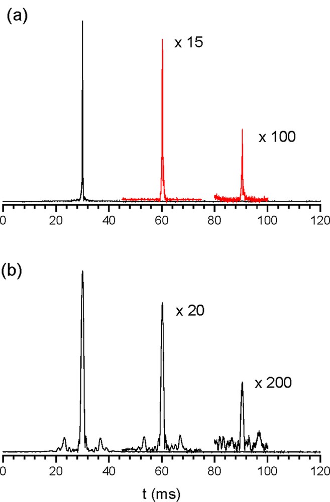
MSE observed in an array of hollow tubes filled with doped water. The steady field gradient was applied (a) along the main axis of the tubes and (b) perpendicular to the tubes. In both experiments, G (amplitude of modulation gradient) was ~2.5 mT m–1 and τ (delay between the r.f. pulses) was 30 ms. The diffraction peaks are clearly observed in MSE only when the field gradient is perpendicular to the tubes. Magnification relative to the first-order echo has been indicated in (a) and (b). Masquer
MSE observed in an array of hollow tubes filled with doped water. The steady field gradient was applied (a) along the main axis of the tubes and (b) perpendicular to the tubes. In both experiments, G (amplitude of modulation ... Lire la suite
Figs. 4a–c display the results of the MSE experiment where the delay time τ was made to vary, while the steady gradient was kept constant. When τ is incremented by Δτ, MSE echoes and their respective ‘satellite’ echoes are all shifted by n Δτ, where n is the MSE order. In Figs. 4d and 4f, the gradient amplitude G was made to vary, while the delay time τ was kept constant. If G is changed to α G (where α is an arbitrary positive number), the relative positions of the diffraction echoes changes to Δt/α. An interesting situation happens when a suitable combination of gradient amplitude and delay time are set and a satellite echo is focused at a time t multiple of τ. Fig. 5 displays two MSE experiments in which this condition is met. If no phase cycling is used, a diffraction echo arising from first-order MS echo overlaps the second-order MSE.
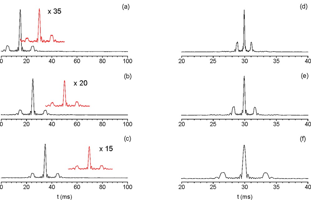
Effect of the delay time duration and of the gradient amplitude over the temporal position of the diffraction peaks. From (a) through (c): the gradient amplitude G was 1.7 mT m–1 and the delay time τ was (a) 15 ms, (b) 25 ms and (c) 35 ms. Magnification relative to the first-order echo has been indicated in (a) through (c). Phase cycling was used to select only the first-order echo (main line) or the second-order echo (insert). Second-order echoes have been shifted vertically to prevent the two displays from overlapping. Diffraction peaks are visible in both first- and second-order MSE experiments. From (d) through (f): the delay time τ was 30 ms and gradient amplitude G was (d) 14.6 mT m–1, (e) 9.7 mT m–1 and (f) 4.9 mT m–1. First-order echoes have been normalized in all sub-figures. Masquer
Effect of the delay time duration and of the gradient amplitude over the temporal position of the diffraction peaks. From (a) through (c): the gradient amplitude G was 1.7 mT m–1 and the delay time τ was (a) 15 ms, ... Lire la suite

Overlapping second-order MSE and diffraction echoes. In (a) and (b), phase cycling was used to select the first-order MSE (line down) or the second-order MSE (line up). (a) Diffraction NMR pattern observed in an array of parallel hollow cylinders. The gradient amplitude G was 1.7 mT m–1 and the delay time τ was 9.6 ms. Magnification relative to the first-order echo has been indicated. (b) Diffraction NMR pattern observed in a packed glass bead sample (diameter ~ 2.8 ms). G was 1.9 mT m–1 and the delay time τ was 15 ms. A second-order MSE was refocused at t = 30 ms (inset). Multiple diffraction peaks are visible around the first-order MSE (main line). Masquer
Overlapping second-order MSE and diffraction echoes. In (a) and (b), phase cycling was used to select the first-order MSE (line down) or the second-order MSE (line up). (a) Diffraction NMR pattern observed in an array of parallel hollow cylinders. The ... Lire la suite
Diffraction-like effects can also be observed in pulsed-field-gradient experiments (Fig. 6). In an n-CRAZED sequence (Fig. 1b), an echo is focused at the time t = n τ after the second r.f. pulse, when the ratio between gradient amplitudes (G2/G1) is n (where n = 0,1,2, ...); otherwise, no echo is refocused. While in the MSE experiments described here, the k-space was sampled in the time domain (kn = γ G (n τ – t), where t was made to vary during the acquisition), in CRAZED experiments the k-space was sampled in the gradient amplitude domain (kn = γ δ (n G1 – G2), where δ was maintained constant and G2 was made to vary). Thus, Fig. 6 is analogous to Fig. 3. As in the MSE experiments, we detected echoes in periodical samples even when the condition kn = 0 was not met (in pulsed-field-gradient experiments, this condition corresponds to G2 = n G1).
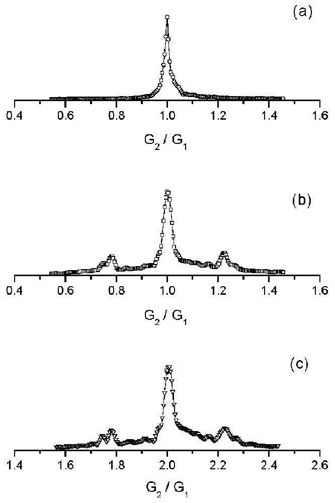
Signal amplitude of CRAZED experiments in an array of hollow tubes filled with doped water: the pulsed field gradient was applied (a) along the main axis of the tubes and (b–c) perpendicularly to the tubes. In all experiments, the amplitude of the first pulsed gradient G1 was 24.7 mT m–1, the gradient pulse duration δ 3 ms and the delay time τ 30 ms. Figs. 6a and 6b display the signal when G2/G1 varies around 1 and Fig. 6c when G2/G1 varies around 2. Satellite peaks due to the sample structure are present in both Figs. 6b and c. The symbols correspond to the experimental data (echo amplitude). The solid line is only used as a guide. The data have been normalized. Masquer
Signal amplitude of CRAZED experiments in an array of hollow tubes filled with doped water: the pulsed field gradient was applied (a) along the main axis of the tubes and (b–c) perpendicularly to the tubes. In all experiments, the amplitude ... Lire la suite
Figs. 6a and 6b display the echo amplitude at t = τ, in an experiment in which the field gradient pulses were parallel and perpendicular to the tubes, respectively. The maximum intensities in Figs. 6a and 6b correspond to the case n = 1 (Eq. (5)), i.e., when dipolar-field effects are neglected. The two ‘satellite’ peaks visible in Fig. 6b correspond to the diffraction condition for k1 = γ δ (G1 – G2).
In Fig. 6c, pulsed gradients were oriented as in Fig. 6b, but a different amplitude range was used for G2. The maximum intensity corresponds to the case n = 2 (Eq. (5)), in which dipolar-field effects are taken into consideration. As in Fig. 6b, two ‘satellite’ peaks are visible, corresponding to the diffraction condition for k2=γ δ|2G1−G2|.
5 Discussion
Over the past few years, it was suggested that long-range dipolar-field effects in NMR could be used as a new method to study structured samples, including living systems. It was also suggested that MSE was sensitive to local-field variations in heterogeneous samples. Local-field variations at different length scales could be probed by ‘tuning’ an experimental parameter: the correlation distance. This unique feature of MSE presents potentially important applications in medical imaging. Calibrated porous samples have frequently been used to demonstrate this mechanism. Simple Fourier transform arguments show that diffraction-like effects are expected in samples with a strong periodicity. In such samples, without careful setting of timings and pulse cycling, multiple echoes originating from NMR diffraction could interfere with the (commonly weak) second-order MSE signal. Indiscriminate choice of the acquisition window can result in the detection of pseudo-modulations of the MSE signal in the k-space, arising from the periodical pattern of diffraction peaks.
Vous devez vous connecter pour continuer.
S'authentifier