Plan
Comptes Rendus

Biological modelling / Biomodélisation
Biological processes in organised media
[Processus biologiques en milieux structurés]
Comptes Rendus. Biologies, Volume 326 (2003) no. 2, pp. 149-159.

Résumés

Embedding a simple Michaelis–Menten enzyme in a gel slice may allow the catalysis of not only scalar processes but also vectorial ones, including uphill transport of a substrate between two compartments, and may make it seem as if two enzymes or transporters are present or as if an allosterically controlled enzyme/transporter is operating. The values of kinetic parameters of an enzyme in a partially hydrophobic environment are usually different from those actually measured in a homogeneous aqueous solution. This implies that fitting kinetic data (expressed in reciprocal co-ordinates) from in vivo studies of enzymes or transporters to two straight lines or a sigmoidal curve does not prove the existence of two different membrane mechanisms or allosteric control. In the artificial transport systems described here, a functional asymmetry was sufficient to induce uphill transport, therefore, although the active transport systems characterised so far correspond to proteins asymmetrically anchored in a membrane, the past or present existence of structurally symmetrical systems of transport in vivo cannot be excluded. The fact that oscillations can be induced in studies of the maintenance of the electrical potential of frog skin by addition of lithium allowed evaluation of several parameters fundamental to the functioning of the system in vivo (e.g., relative volumes of internal compartments, characteristic times of ionic exchanges between compartments). Hence, under conditions that approach real biological complexity, increasing the complexity of the behaviour of the system may provide information that cannot be obtained by a conventional, reductionist approach.

La fixation d'enzymes michaéliennes dans une lame de gel peut suffire à les rendre aptes à catalyser des processus, non seulement scalaires, mais également vectoriels, y compris le transport actif d'un substrat entre deux compartiments, et à les faire se comporter comme le feraient un double mécanisme enzymatique ou de transport ou un processus allostérique. Dans un environnement partiellement hydrophobe, les paramètres cinétiques apparents n'ont en général rien à voir avec les véritables paramètres caractéristiques du comportement de la même protéine en solution aqueuse. Réciproquement, lorsque l'on observe (en coordonnées inverses) que des systèmes enzymatiques ou de transport in vivo s'ajustent mieux à deux approximations linéaires ou à une courbe sigmoı̈de qu'à une seule droite, ceci ne prouve pas qu'interviennent deux systèmes enzymatiques ou de transport différents ou un processus allostérique. Avec les systèmes de transport étudiés ici, il est apparu qu'une asymétrie fonctionnelle pouvait suffire à induire un transport à contre-gradient en l'absence de toute asymétrie structurale ; aussi, bien que tous les systèmes de transport actif isolés jusqu'ici correspondent à des protéines à structure asymétrique par rapport à la membrane où elles sont insérées, on ne peut pas exclure que des systèmes de transport à structure symétrique existent ou aient existé au cours de l'évolution. Avec des systèmes réels, tel que celui qui maintient le potentiel électrique de la peau de grenouille, augmenter la complexité du comportement du système par l'induction d'oscillations électriques par addition de lithium a permis d'atteindre des données sur ce système qu'il n'aurait pas été possible d'obtenir par l'approche réductionniste traditionnelle.

Métadonnées
Reçu le :
Accepté le :
Publié le :
DOI : 10.1016/S1631-0691(03)00062-3
Keywords: immobilised enzymes, enzyme kinetics, aqueous/hydrophobic reaction media, artificial transport systems, electric oscillations
Mot clés : enzymes immobilisées, cinétique enzymatique, réaction en milieu partiellement aqueux/partiellement hydrophobe, systèmes artificiels de transport, oscillations électriques
Michel Thellier 1 ; Jean-Claude Vincent 2 ; Stéphane Alexandre 2 ; Jean-Paul Lassalles 3 ; Brigitte Deschrevel 2 ; Victor Norris 1 ; Camille Ripoll 1

1 Laboratoire des « Processus intégratifs cellulaires », faculté des sciences, université de Rouen, 76821 Mont-Saint-Aignan cedex, France
2 Laboratoire « Polymères, Biopolymères et Membranes », faculté des sciences, université de Rouen, 76821 Mont-Saint-Aignan cedex, France
3 Laboratoire des « Processus ioniques membranaires », faculté des sciences, université de Rouen, 76821 Mont-Saint-Aignan cedex, France
@article{CRBIOL_2003__326_2_149_0,
     author = {Michel Thellier and Jean-Claude Vincent and St\'ephane Alexandre and Jean-Paul Lassalles and Brigitte Deschrevel and Victor Norris and Camille Ripoll},
     title = {Biological processes in organised media},
     journal = {Comptes Rendus. Biologies},
     pages = {149--159},
     publisher = {Elsevier},
     volume = {326},
     number = {2},
     year = {2003},
     doi = {10.1016/S1631-0691(03)00062-3},
     language = {en},
}
TY  - JOUR
AU  - Michel Thellier
AU  - Jean-Claude Vincent
AU  - Stéphane Alexandre
AU  - Jean-Paul Lassalles
AU  - Brigitte Deschrevel
AU  - Victor Norris
AU  - Camille Ripoll
TI  - Biological processes in organised media
JO  - Comptes Rendus. Biologies
PY  - 2003
SP  - 149
EP  - 159
VL  - 326
IS  - 2
PB  - Elsevier
DO  - 10.1016/S1631-0691(03)00062-3
LA  - en
ID  - CRBIOL_2003__326_2_149_0
ER  - 
%0 Journal Article
%A Michel Thellier
%A Jean-Claude Vincent
%A Stéphane Alexandre
%A Jean-Paul Lassalles
%A Brigitte Deschrevel
%A Victor Norris
%A Camille Ripoll
%T Biological processes in organised media
%J Comptes Rendus. Biologies
%D 2003
%P 149-159
%V 326
%N 2
%I Elsevier
%R 10.1016/S1631-0691(03)00062-3
%G en
%F CRBIOL_2003__326_2_149_0
Michel Thellier; Jean-Claude Vincent; Stéphane Alexandre; Jean-Paul Lassalles; Brigitte Deschrevel; Victor Norris; Camille Ripoll. Biological processes in organised media. Comptes Rendus. Biologies, Volume 326 (2003) no. 2, pp. 149-159. doi : 10.1016/S1631-0691(03)00062-3. https://comptes-rendus.academie-sciences.fr/biologies/articles/10.1016/S1631-0691(03)00062-3/

Version originale du texte intégral

1 Introduction

In the last century, considerable progress has been made in separating, identifying and purifying the molecules that constitute living cells. In particular, purification of information-bearing molecules such as nucleic acids, proteins and polysaccharides has elucidated the relationship between structure and function under rigorously defined, controlled in vitro conditions. Despite the continuing importance of this reductionist approach to both fundamental biology and biotechnology, it is clear that macromolecules are in completely different conditions in vitro from in vivo. In the latter, they are located in compartments or at compartment boundaries and are surrounded by a variety of small mobile solutes that may be ionised; this heterogeneity means that the important parameters are not only concentrations of reactants and products but also electric fields, gradients of solute activities (including pH gradients), and transport and reaction rates.

In the present paper, we approach the problem of biological complexity by studying the behaviour of two different types of systems: (i) simple, in vitro enzymatic systems in which the media is either of a controllable structural complexity or of an aqueous-hydrophobic nature and (ii) a complex, in vivo system subjected to an extra constraint that in the case presented here is the frog skin after a specific alteration of its ionic environment.

2 Simple, in vitro enzymatic systems

This section concerns simple, in vitro enzymatic systems in which the media is either of a controllable structural complexity or of an aqueous-hydrophobic nature.

A number of studies have dealt with the in vitro behaviour of immobilised enzymes [1–3]. Studies in our group, as explained in the examples given below, illustrate the emergence of a complex behaviour in cases in which simple, ‘monosteric’ (as opposed to ‘allosteric’) enzymes were inserted in various types of artificial structures.

2.1 The problem

The simplest possible behaviour for a monosteric enzyme, E, catalysing the reaction between substrate, S, and product, P, according to:

SEP(1)
in a homogeneous, aqueous medium, is that described (when P=0) by Michaelis and Menten's kinetic equation [4]
V=VmS/(Km+S)(2)
in which the italic symbol (e.g., S) characterises the concentration of the corresponding substance (e.g., S), and Vm and Km are the maximum rate and the Michaelis constant of the reaction. The corresponding curve in the direct system of co-ordinates is a hyperbola (Fig. 1a), but it is customary to use the reciprocal system of co-ordinates {1/S,1/V} (equation (3)):
1V=KmVm1S+1Vm(3)
which gives a straight line, whose intercepts with the co-ordinate axes are −1/Km and 1/Vm (Fig. 1b) [5]. In such a case, the reaction kinetics are said to be ‘monophasic’.

Fig. 1

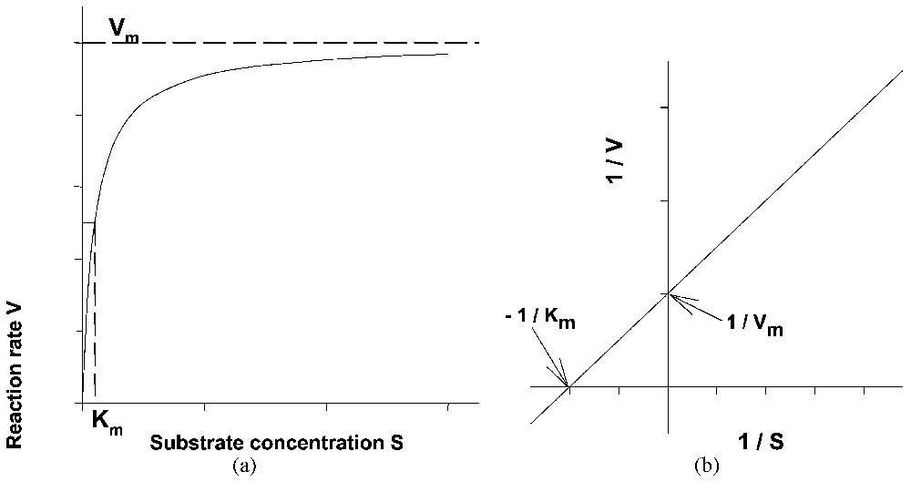
Representative curve of the kinetics of a Michaelis and Menten enzyme reaction using either (a) the direct or (b) the reciprocal system of coordinates.

Now, let us consider model systems in which monosteric enzymes, Ej, are inserted at random in a gel slice, with thickness l, separating two aqueous solutions, e and i (Fig. 2) [6]. Locally, the rate, Vj, of the catalysed reaction(s) of substrate(s) Sj and product(s) Pj (4):

SjEjPj(4)
is again a hyperbolic function of Sj (for Pj=0), which may be written as:
Vj=Vmjγjλj(5)
λj=Sj/(Kmj+Sj)(6)
In these equations Vmj, γj, λj and Kmj (assumed to be constant with respect to pH) are the maximum rate, the possible pH dependence, the substrate dependence and the Michaelis constant of enzyme Ej in solution, respectively. Within the gel slice, concentration Sj obeys the differential equation:
Sj/t=Dj(2Sj/x2)-Vj(x,t)(7)
where t is time, Dj the diffusion coefficient of Sj and x the distance along l with x=0 and x=l at the boundaries of e and i. The flux, Jj of Sj at time t and at point x in the gel slice is written as:
Jj(x,t)=-Dj(Sj/x)x,t(8)
while the rate of disappearance, Vj¯, of Sj from one of the aqueous solutions (e.g., e) is written
Vje¯=-Jj(0,t)A/ve(9)
where A is the surface area of the gel slice and ve the volume of compartment e.

Fig. 2

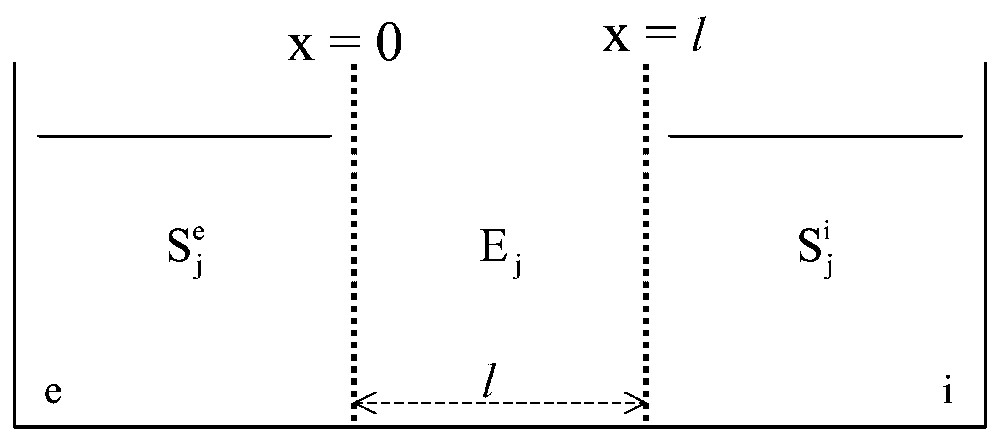
General scheme of the in vitro systems studied. A gel slice, l in thickness and containing one or several enzymes, Ej, distributed at random, is used to separate two aqueous media, e and i, containing one or several solutes Sj.

2.2 Kinetic behaviour of a monosteric enzyme inserted in a gel slice separating aqueous compartments

In the simplest possible case, i.e. when the system is monoenzymatic (index j thus may be deleted), the concentration of S is the same in e and i (Se=Si), the system is without any pH effect (γ=constant) and the stationary state has been established everywhere in the gel slice (∂S/∂t=0), one may consider the dimensionless, diffusion–reaction parameter:

αl=Vmγl2/KmD=τ diff /τ react (10)
In equation (10), τdiff and τreact are the characteristic times for the diffusion of S and for the reaction, respectively, with:
τ diff =l2/D and τ react =Km/Vmγ(11)
The larger the contribution of the reaction and the smaller that of diffusion, the larger is the value of αl and vice versa. Computing the dependence of -1/Ve¯ as a function of Km/Se (reciprocal system of co-ordinates), the case when αl⪡1 gives a linear plot (i.e. there are Michaelis–Menten kinetics in the gel slice at all values of substrate concentration and the system behaviour thus remains monophasic) with the apparent Km being equal to the actual Km of the enzyme in solution and Vm¯ being proportional to the Vm of the enzyme in solution. However, as αl increases, the plot of {Km/Se,-1/Ve¯} ceases to be linear. The curve has a monotonous negative curvature and its extremities (low and high S values) can be approximated by two different straight lines (dual-phasic behaviour). In this case, although there is a single monosteric enzyme present in the system, one could be tempted to wrongly interpret the kinetic data as corresponding to the presence of two different monosteric enzymes (one detectable at low and the other at high substrate concentrations). With pH-dependent systems (i.e. when the reaction produces or consumes protons or hydroxyl groups), situations are encountered in which the plot in the reciprocal system of co-ordinates {Km/Se,-1/Ve¯} ceases to be linear and possibly becomes sigmoidal. Therefore, even with an enzyme of the Michaelis–Menten type, it is possible to obtain in the reciprocal system of co-ordinates (Fig. 3) a complex, non-linear (e.g., dual-phasic or sigmoidal) kinetic behaviour by imposing appropriate (and possibly rather simple) structural conditions.

Fig. 3

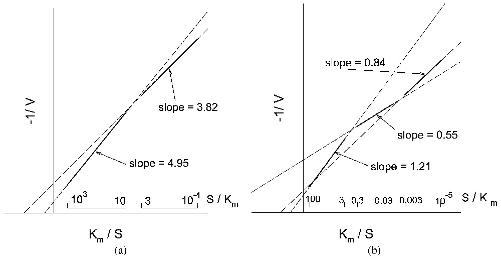
Kinetic behaviour (reciprocal system of coordinates) of a monosteric enzyme system, similar to that described in Fig. 2, for increasing values of αl: (a) dual-phasic behaviour and (b) sigmoidal behaviour.

2.3 Two-enzyme artificial first-order transport systems

Let us consider again [6] a model system made of a gel slice separating two aqueous compartments, e and i, but now let us insert two monosteric enzymes, E1 and E2, differing in their optimum pH value and catalysing the associated reactions

S+ XY E1 PX +Y(12)
PX E2S+X(13)
at random in the gel slice. The overall reaction
XY X+Y(14)
is assumed to be exergonic in the direction of the splitting of XY into X and Y. Different pH values, pHe and pHi, are imposed on compartments e and i to create a pH gradient in the gel slice, and the pHe and pHi values are chosen so as to make E1 active only in a layer, l1, of the gel slice close to compartment e and E2 active only in a layer, l2, close to compartment i (Fig. 4). Under such conditions, substrate S diffusing from compartment e is transformed into PX by enzyme E1 in layer l1; the part of PX which diffuses towards l2 is transformed back into S by enzyme E2; this newly-formed S diffuses partially towards compartment i and, depending on the concentration profiles in the gel slice, the overall process can produce an uphill transport of S at the expense of the energy provided by the exergonic splitting of XY. The system under consideration thus is a model of a first-order [7] process of transport.

Fig. 4

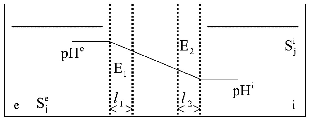
Two-enzyme systems. In a system structurally similar to that described in Fig. 2, two enzymes, E1 and E2, are distributed at random in the gel slice. Two different pH values, pHe and pHi, are imposed on compartments e and i, in order that only enzyme E1 is active in layer l1 facing e and only E2 is active in layer l2 facing i.

As an example, let us consider the simple case when the two enzymes have the same Vm and the same Km in solution (Vm1=Vm2=Vm and Km1=Km2=Km) and the two layers, l1 and l2, have the same thickness (l1=l2=l¯). Within each layer, the enzyme activity is treated as constant with x, while the intermediate layer (thickness nl¯) is considered as purely diffusive, without any significant enzyme activity. The experiment starts with identical concentrations of S in compartments e and i (i.e. at time 0, Se=Si=S). Under these conditions, a dimensionless, diffusion–reaction parameter:

αl¯=Vmγl¯2/KmD(15)
can again be defined and the initial rate of transport, V'¯, can be computed as in section 2.1. When plotting these data in reciprocal co-ordinates (1/S,1/V'¯), three cases may be considered.
  • • When αl¯1, whether or not there is a pH feedback, the plot is linear; the transport process thus behaves as a single Michaelis–Menten type process; moreover, in this case, the apparent Km of the transport process is found to be equal to the actual Km of the enzymes in solution.
  • • When αl¯1, again whether or not there is a pH feedback, the plot in reciprocal co-ordinates appears to be made of two asymptotic straight lines, corresponding to SKm and SKm (dual-phasic plot), connected by a monotonous curve. Although there are only two types of enzymes with identical values for Km and for Vm, it might seem that there are two transport processes, one with low apparent Km and Vm and the other with high apparent Km and Vm playing the major role at low and high substrate concentrations, respectively. Moreover the apparent Km and Vm of transport no longer bear a simple relationship to the actual Km and Vm of the enzymes in solution.
  • • When αl¯1 and when a pH feedback exists (e.g., consumption or production of protons in reactions (12) and (13)), the curve connecting the two asymptotic straight lines may become sigmoidal.

2.4 Monoenzymatic artificial second-order transport systems

In the preceding sections 2.2 and 2.3, we discussed purely theoretical systems. Let us now consider from both the theoretical and experimental points of view a soluble enzyme constrained to work as a transporter by particular conditions of structure and concentrations [8,9].

Yeast alcohol deshydrogenase (ADH) catalyses the reversible reaction:

(16)
A gel slice, G, containing a homogeneous distribution of ADH was inserted between two barriers, B, that prevented enzyme leakage and were practically impermeable to NAD+ while being permeable to NADH and OH. Gel slice and barriers were then used to separate two aqueous compartments, e and i, containing identical mixtures of alcohol, acetaldehyde, NADH and phosphate buffer, but no NAD+ (Fig. 5). Constant pH values, more alkaline in i than in e, were imposed in the aqueous compartments, thus creating a pH gradient in the gel slice. After an initial decrease of the NADH concentration in both compartments, corresponding to NADH equilibration of the gel slice with the bathing solutions, NADH concentration began to increase in i, while it continued to decrease in e. After a few days, the NADH concentration in i was higher than the initial NADH concentrations in the aqueous compartments and almost twice as high as the final concentration in e (Fig. 6). The qualitative interpretation is as follows. At the outset of the experiment, NADH diffuses from e and i into the gel slice. In the part of the gel close to e, where the concentration of H+ is the highest, NADH is transformed into NAD+ as a consequence of the pH-dependence of the enzyme, then NAD+ diffuses into the gel but cannot diffuse into e and i because of the barriers B. In the part of the gel close to i, where the concentration of H+ is lowest, NAD+ tends to be transformed back into NADH, thus increasing the NADH concentration in this part of the gel. As a consequence, NADH tends to diffuse toward compartment i. This corresponds to an uphill transport of NADH driven by the pH gradient (a transport of OH ions from i to e rather than a transport of H+ ions from e to i), while electric neutrality in compartments e and i is ensured via exchanges of saline ions provided by the buffer. Such a system is therefore a model of second-order active transport [7].

Fig. 5

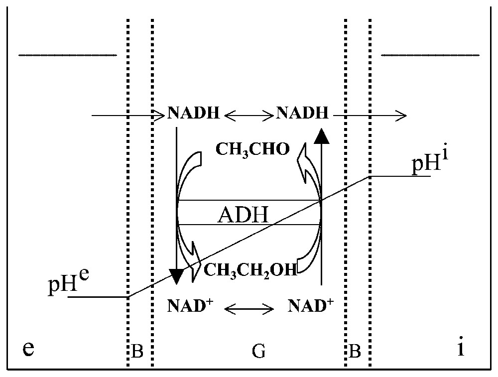
A gel slice, G, with both sides covered with a barrier, B, is used to separate two aqueous compartments, e and i. The gel slice contains a random distribution of the enzyme ADH which catalyses the oxido-reduction reaction CH3CHO/CH3CH2OH with the cofactor NAD. Two different pH values, pHe and pHi, are imposed on e and i in order that the reaction is in favour of the alcohol on the e side of the gel and in favour of the aldehyde on the i side. NADH, but not NAD+, can diffuse freely through barrier B.

Fig. 6

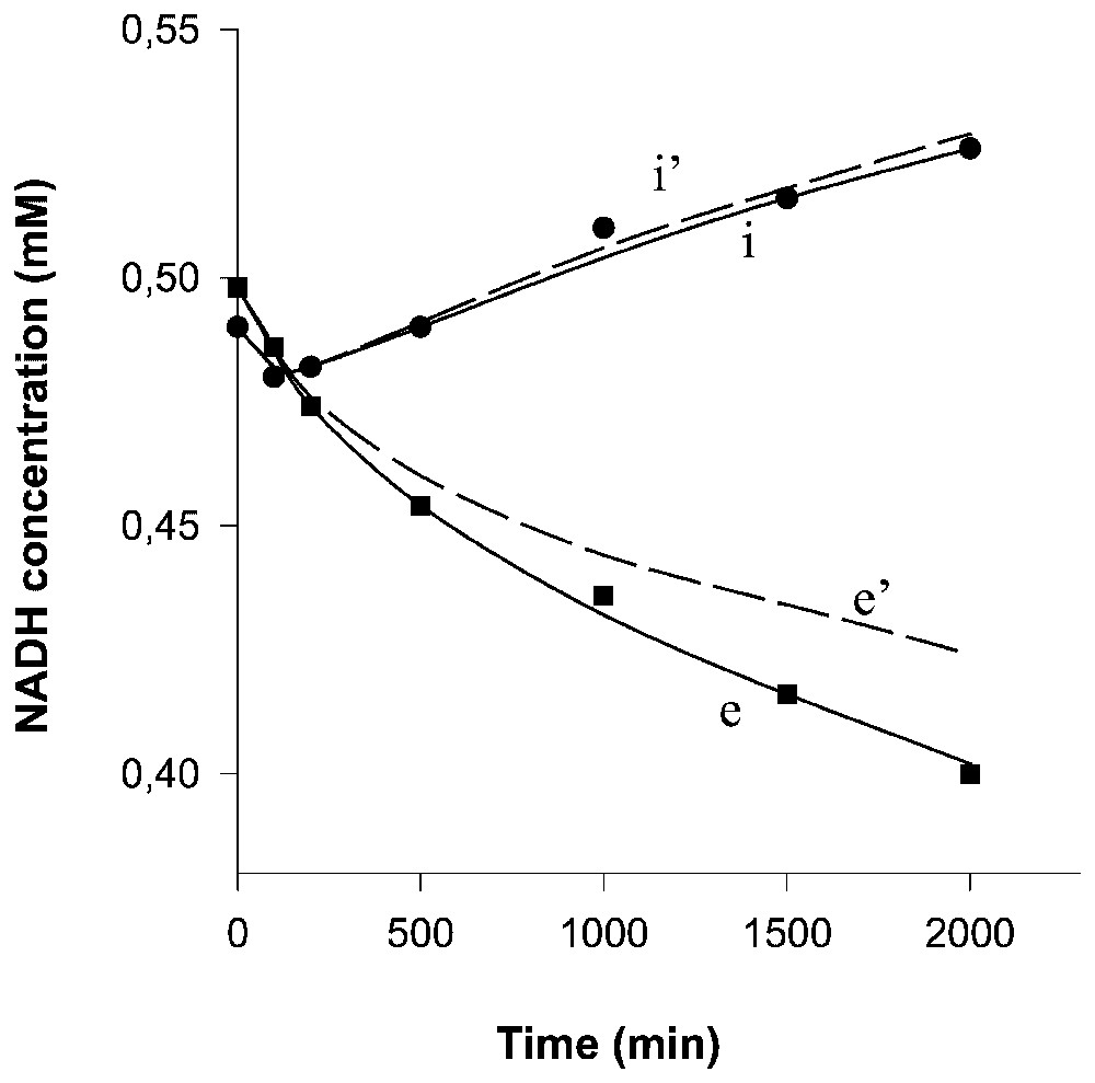
Computed time-courses of the NADH concentrations in compartments e (curve e) and i (curve i). Curves e' and i' correspond to the concentration changes due only to the transport (i.e. after subtracting the spontaneous, non-enzymatic NADH degradation). Experimental points in compartments e (■) and i (●).

The analysis and the numerical calculations [9] were performed essentially as above (sections 2.1 and 2.2) using diffusion–reaction equations of the type:

cj/t=Dj(2cj/x2)+Vj(cj,ck)(17)
where cj and ck represent the concentrations of species j and k, t the time, Dj the diffusion coefficient of j, x the space co-ordinate in the gel and Vj the enzymatic rate of the reaction involving j. Initially, the experimental data did not seem to be in accord with the time-courses of NADH concentrations predicted from the numerical simulations. However, when a non-enzymatic, pH-dependent degradation of NADH (as determined in a complementary experiment) was taken into account, the fit between the experimental data and the predictions of the model became extremely good (Fig. 6).

2.5 Enzyme-catalysed reactions in an aqueous/hydrophobic reaction medium

The kinetics of the hydrolysis and synthesis reactions of the peptide bond of the dipeptide, N-Cbz-l-tryptophanyl-glycineamide, catalysed by α-chymotrypsin, have been studied in mixtures of water and 1,4-butanediol [10]. Although the polarity of 1,4-butanediol is not very high, it is miscible with water in all proportions. The initial reaction rates decreased exponentially with decreasing water content in the solvent mixture. The study of the substrate dependencies have revealed that both the apparent and the actual kinetic parameters were dependent on the water content, and thus on the polarity, of the solvent mixture. However, the exponential decrease in the initial rate of hydrolysis was due mainly to the Km increase and only slightly to the modification of the Vm.

A reduction of the water content from 100 to 20% (v/v) did not alter Vm by more than a factor of 4. Such variation in Vm is due to changes in the conformation of the enzyme. One of the main causes of such changes in mixtures of water and organic solvent is the substitution of essential water molecules in the vicinity of the protein surface by organic solvent molecules. However, with organic solvents like 1,4-butanediol, the interactions between organic solvent molecules and the enzyme are similar to those between water molecules and enzyme; these organic solvents thus do not alter the enzyme conformation very much. Another cause is the possible modification of the ionisation/neutralisation constant of ionisable groups of the protein, in particular in the active site, as a consequence of the variation of the polarity of the reaction medium.

Decreasing the polarity of the reaction medium by decreasing its water content led to an exponential increase in the apparent Km for the hydrolysis of the dipeptide (see above) and in the solubility of this dipeptide (Fig. 7). Since the interactions between substrate and the active site of α-chymotrypsine are mainly hydrophobic, this means that decreasing the water content of the reaction medium tends to favour the interactions of the dipeptide with the solvent mixture and thus to weaken the interactions of the dipeptide with the active site of the enzyme. Moreover, it has been established that the ratio of the actual rate constants for the formation/dissociation of the enzyme–substrate complex in the solvent mixture is given by the corresponding ratio in aqueous medium divided by the equilibrium constant for the transfer of the dipeptide from water to the solvent mixture (which is equal to the ratio of the dipeptide solubility in the solvent mixture to that in water).
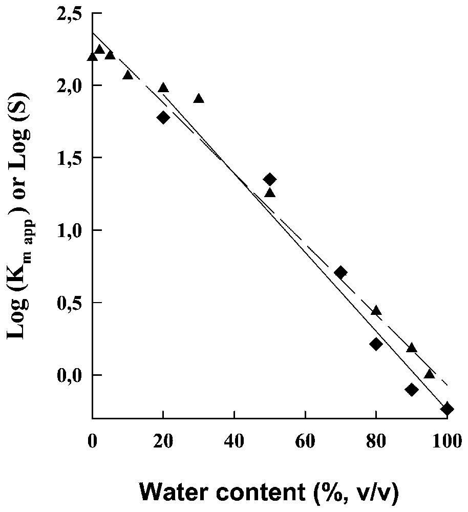
Fig. 7

Logarithms of the apparent Km, Km app , for the hydrolysis of the dipeptide ( ) and for the solubility limit, S, of this dipeptide (▴) as a function of the water content of the solvent mixture.

3 Effect of lithium on the electrical potential of frog skin

Frogs maintain the saline concentration of their internal medium, even when immersed in freshwater ponds, due to the activity of a Mg-dependent Na-K-ATPase that creates a steady transepithelial potential difference in the range of a few tens to more than a hundred millivolts (positive inside). This active ion-pumping system has been studied in great detail, especially by Ussing's group [11,12], using the ventral skin of frogs mounted between two aqueous compartments filled with appropriate saline solutions. Maintaining the electrical potential of frog skin in these conditions requires sodium in the external medium. Lithium is the only ion that can be used instead [13]. However, when all or part of the external sodium is replaced by lithium, the electric potential frequently oscillates [14–16], which it never does in the absence of lithium (Fig. 8).

Fig. 8

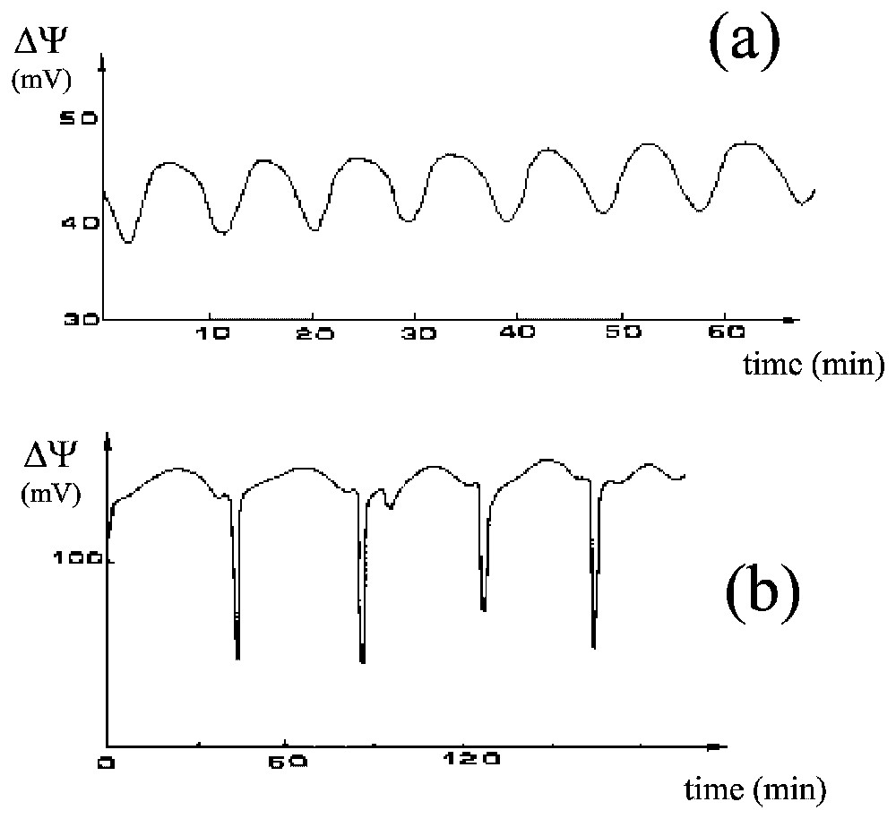
Two cases of oscillations of the electric potential difference, ΔΨ (mV), of the frog skin, (a) almost sinusoidal and (b) much more complex.

A body of evidence obtained by others, cited in Lassalles et al. [17], demonstrates that all the ionic processes are controlled at the level of the skin epithelium, which comprises a few layers of cells separating the external medium e from the internal medium i. The epithelium may be modelled as follows (Fig. 9): it is made of two main compartments, C1 and C2; the membrane, a, at the external face of the epithelium is relatively permeable to Na+ and Li+ whilst it is almost impermeable to K+; the membrane, b, between compartments C1 and C2 is permeable to K+ but not to Na+ and contains the Na-K-ATPase pumping sodium and lithium from C1 to C2 and potassium from C2 to C1; the face, c, between epithelium and internal medium is not an actual membrane, but may be considered rather as a non-selective diffusive barrier.

Fig. 9

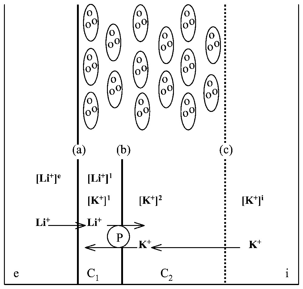
Modelling frog skin epithelium. Top: schematic representation of the cellular structure of frog skin epithelium, mounted between two aqueous, saline solutions, e and i. Bottom: the epithelium model for ionic exchange. The frog epithelium is modelled as corresponding to two compartments C1 and C2 with three barriers, a between e and C1, b between C1 and C2 and c between C2 and i. Symbol [x]j stands for concentration of cation x in compartment j. The Na-K-ATPase, P, pumps actively Li+ from C1 to C2 and K+ from C2 to C1.

In studies of the oscillatory process, our group [17–20] has shown experimentally that (i) when no transepithelial potential is imposed, sustained oscillations with a period of about 10 min are maintained for several hours, (ii) an oscillation of the Na+ influx accompanies the electric oscillation (the two oscillations have approximately the same period but are not in phase), (iii) under conditions of imposed potential the transepithelial electric current has damped oscillations, (iv) the shape of the oscillations in potential is quite variable (from almost sinusoidal to very complex), (v) theophyllin (which induces an accumulation of cyclic AMP within the cells) promotes a significant decrease in the mean electric potential of the skin but does not affect the characteristics of the oscillation very much, (vi) important factors influencing the oscillations include temperature, the permeability of the external membrane to lithium and the potassium concentration in the internal medium, (vii) no evident correlation exists between skin area and the characteristics of the oscillations, which may therefore have a local origin (possibly in local oscillators at the level of the cell) and the coupling of these oscillators would generate the macroscopic oscillations and (viii) synchronisation of local oscillators can be controlled by varying the coupling resistance in the absence of diffusion, which is consistent with electrical coupling rather than diffusion being responsible for the synchronisation.

After extensive numerical simulation of the oscillatory process, it proved impossible to obtain all the above characteristics unless the model had a few well-defined properties, the following ones in particular.

  • • If £1 and £2 represent the volumes of compartments C1 and C2 per unit surface of epithelium, they obey the relation £12⪡1, i.e. compartment C2 has to be much larger than compartment C1. This makes it possible to choose one of the various possible modes of transport of Na+ through the epithelium proposed in the literature and cited in Lassalles et al. [17]. Indeed, £2⪢£1 does not support the idea that compartment C1 merely corresponds to most of the cytoplasmic volume and C2 is restricted to a few endoplasmic cisternae, whereas it strongly supports the idea that compartment C1 corresponds to a few cytoplasmic vacuoles transporting Na+ (and Li+) and compartment C2 is part or all of the intercellular spaces and possibly some endoplasmic cisternae (in this case, most of the cell cytoplasm would remain inactive in Na+ (or Li+) transport).
  • • The main variables are the concentration of Li+ in compartment C1 and that of K+ in compartment C2. When the characteristic times τ1 and τ2 are defined as corresponding to Li+ modifications in C1 and K+ modifications in C2, respectively, the ratio of the characteristic times obeys the relation 0<τ1/τ2<1 with 1 min⩽τ1⩽5 min.
  • • Two parameters, ρ and u, represent the relative importance of the active to the passive fluxes of Li+ and K+, respectively, such that ρ⪢1 and u⪢1, with ρu⩽2ρ (i.e., ρ and u are of the same order of magnitude).
  • P represents the permeability coefficients of interfaces a, b and c for ions K+ and Li+, such that PKb/PKc⪡1 and PLia/PKc⪡1.

4 Discussion and conclusions

Embedding simple Michaelis–Menten enzymes in a gel slice allows them to catalyse not only scalar processes but also vectorial ones, including the uphill transport of a substrate between two compartments. Moreover, a system containing a single species of a simple enzyme can be made to behave as if it contained two different enzymes (or transporters) or an allosterically controlled enzyme (or transporter). These effects are more pronounced the larger the term characteristic of the reaction (Vmγ/Km) compared with that of substrate diffusion (D/l2). The existence of a pH feedback also tends to increase these effects. In the presence of a partially hydrophobic environment (which is likely to be the case with membrane-bound enzymes or transport systems in vivo), the measured kinetic parameters are usually different, sometimes very different from those characteristic of the protein behaviour in homogeneous, aqueous solutions. Another group [21,22] has also shown that it is possible to induce oscillatory phenomena in immobilised enzyme systems or spatio-temporal pattern formation in immobilised bienzymatic systems.

These results are of general relevance to the interpretation of kinetic data on membrane-bound enzymes or transport systems. In reciprocal co-ordinates, such experimental data are often better fitted by two straight lines (one for the high and the other for the low concentrations) than by a single line. In line with the original proposal of Epstein and co-workers [23–25], most authors interpret such data as revealing the presence of two different membrane mechanisms of reaction or transport, with the apparent Km and Vm of the membrane processes being equated to the actual Km and Vm of the catalytic proteins involved. Similarly, sigmoidal experimental curves have been taken as indicative of an allosteric character of the membrane-bound active proteins [26]. Our results clearly show that such interpretations can be wrong, since the complexity of the kinetic behaviour may as well correspond to the partially hydrophobic nature and other features of the cellular structures in which the enzyme or transport systems are inserted (for a more detailed discussion, see Thellier et al. [27]). Indeed, an alternative interpretation to allosteric interaction has been given for sigmoidal kinetic behaviour in an actual biological system in vivo [28].

A real cell membrane is obviously not the same as a thick homogenous gel slice or a water/hydrophobic mixture. Nevertheless, the values of the parameters used in the experiments reviewed here cover the range of biological possibilities [6,8,9]. Moreover, since real membranes are far more complex than our gel slices or aqueous/hydrophobic mixtures, it is likely that the effects of membrane structure and/or hydrophobicity in vivo are even more numerous and diverse than they are in vitro.

In the artificial transport systems discussed here (sections 2.3 and 2.4), a functional asymmetry such as a proton gradient sufficed to induce an active uphill transport even under symmetrical structural conditions. Therefore, although all the active-transport systems isolated until now correspond to proteins asymmetrically anchored in a membrane, one cannot exclude the possibility that structurally symmetrical systems of transport do exist in as yet unstudied organisms or did once exist earlier in evolution.

Situations exist in biology (see Vincent et al. [9]) where the same enzymatic activity is present on each side of a membrane and where the ionic conditions are different in the media on each side. Such a situation, which closely resembles the artificial ones studied here, is important in the homeostasis of inorganic ions in cellular compartments or in the operation of relay and amplification mechanisms (in which the direction of transfer depends on differences in ion concentrations in the compartments). Moreover, the frog epithelium in vivo resembles some of the in vitro set-ups described here (sections 2.3 and 2.4), since it can be modelled as a thick two-compartment slice with both passive ion diffusion and active ion transport by an enzyme.

It is remarkable that increasing the complexity of the behaviour of the living frog-skin system by inducing electric oscillations via lithium addition to the external solution provided information on this system (such as the relative size of compartments and the values of other parameters) that could not have been provided by a conventional, reductionist approach. This may be the basis for the introduction of ‘complexification’, as opposed to reductionism, as a method for studying those properties of a complex system that are destroyed when adopting the conventional, reductive, biochemical approaches.

Further study of the model systems discussed above would benefit from knowledge of the distribution of ions. This may be provided by secondary ion mass spectrometry (SIMS), a method largely used by physicists for materials studies (see, e.g., [29,30]), now available for application to biological specimens [31–33] after appropriate preparation of dehydrated tissue sections [34,35] or using frozen hydrated samples [36].


Bibliographie

[1] E. Sélégny; S. Avrameas; G. Broun; D. Thomas Membranes à activité enzymatique : synthèse de membranes à enzymes liées par covalence, caractérisation de l'activité catalytique par diffusion-réaction, C. R. Acad. Sci. Paris, Ser. C, Volume 266 (1968), pp. 1431-1434

[2] E. Sélégny; G. Broun; D. Thomas Enzymatically active model membranes: experimental illustrations and calculations on the basis of diffusion-reaction kinetics of their functioning, of regulatory effects, of facilitated, retarded and active transport, Physiol. Vég., Volume 9 (1971), pp. 25-50

[3] J.M. Engasser; C. Horvath Inhibition of bound enzymes. I. Antienergistic interaction of chemicals and diffusional inhibition, Biochemistry, Volume 13 (1974), pp. 3845-3849

[4] L. Michaelis; M.L. Menten Die Kinetic der Invertinwirkung, Biochem. Z., Volume 49 (1913), pp. 333-369

[5] H. Lineweaver; D. Burk The determination of enzyme dissociation constants, J. Am. Chem. Soc., Volume 56 (1934), pp. 658-666

[6] J.-C. Vincent; M. Thellier Theoretical analysis of the significance of whether or not enzymes or transport systems in structured media follow Michaelis–Menten kinetics, Biophys. J., Volume 41 (1983), pp. 23-28

[7] P. Mitchell Active transport and ion accumulation (M. Florkin; E.H. Stotz, eds.), Comprehensive Biochemistry, Vol. 22, Elsevier, Amsterdam, 1967, pp. 167-197

[8] J.-C. Vincent; S. Alexandre; M. Thellier How a soluble enzyme can be forced to work as a transport system: description of an experimental design, Arch. Biochem. Biophys., Volume 261 (1988), pp. 405-408

[9] J.-C. Vincent; S. Alexandre; M. Thellier How a soluble enzyme can be forced to work as a transport system: theoretical interpretation, Bioelectrochem. Bioenerg., Volume 20 (1988), pp. 215-222

[10] B. Deschrevel; J.-C. Vincent; M. Thellier Kinetic study of the α-chymotrypsin-catalyzed hydrolysis and synthesis of a peptide bond in a monophasic aqueous/organic reaction medium, Arch. Biochem. Biophys., Volume 304 (1993), pp. 45-52

[11] H.H. Ussing Active and passive transport of the alkali metal ions (O. Eichler; A. Farah, eds.), Handbuch der experimentellen Pharmakologie, Chapter VII, Springer Verlag, Berlin, 1960, pp. 45-143

[12] C.L. Voute; H.H. Ussing Some morphological aspects of active sodium transport: the epithelium of the frog skin, J. Cell Biol., Volume 36 (1968), pp. 625-638

[13] W. Nagel Influence of lithium upon the intracellular potential of frog skin epithelium, J. Membr. Biol., Volume 37 (1977), pp. 347-359

[14] S. Takenaka Studies on the quasiperiodic oscillations of the electric potential of the frog skin (part I), Jpn J. Med. Sci. (III Biophys.), Volume 4 (1936), pp. 143-197

[15] S. Takenaka Studies on the quasiperiodic oscillations of the electric potential of the frog skin (part II), Jpn J. Med. Sci. (III Biophys.), Volume 4 (1936), pp. 198-293

[16] T. Teorell Rhythmical potential impedance variations in isolated frog skin induced by lithium ions, Acta Physiol. Scand., Volume 31 (1954), pp. 268-284

[17] J.-P. Lassalles; C. Hyver; M. Thellier Oscillation of the electrical potential of frog skin under the effect of Li+: theoretical formulation, Biophys. Chem., Volume 14 (1981), pp. 65-80

[18] M. Thellier; J.-P. Lassalles; T. Stelz; A. Hartmann; A. Ayadi Oscillations de potentiel et de courant électriques transépithéliaux sous l'effet du lithium, C. R. Acad. Sci. Paris, Ser. D, Volume 282 (1976), pp. 2111-2114

[19] J.-P. Lassalles; M. Thellier; C. Hyver Oscillations de potentiel électrique membranaire sous l'effet du lithium (P. Delattre; M. Thellier, eds.), Elaboration et justification des modèles : applications en biologie, Part II, Maloine, Paris, 1979, pp. 461-495

[20] J.-P. Lassalles; A. Hartmann; M. Thellier Oscillations of the electrical potential of frog skin under the effect of Li+: experimental approach, J. Membr. Biol., Volume 56 (1980), pp. 107-119

[21] J.-F. Hervagault; D. Thomas Oscillatory phenomena in immobilized enzyme systems, Methods Enzymol., Volume 135 (1987), pp. 554-569

[22] S. Cortassa; H. Sun; J.P. Kernevez; D. Thomas Pattern formation in an immobilized bienzyme system. A morphogenetic model, Biochem. J., Volume 269 (1990), pp. 115-122

[23] E. Epstein; C.E. Hagen A kinetic study of the absorption of alkali cations by barley roots, Plant Physiol., Volume 27 (1952), pp. 457-474

[24] E. Epstein; J.E. Legett The absorption of alkaline earth cations by barley roots: kinetics and mechanisms, Am. J. Bot., Volume 41 (1954), pp. 785-791

[25] E. Epstein Dual pattern of ion absorption by plant cells and by plants, Nature, Volume 212 (1966), pp. 1324-1327

[26] A.D.M. Glass Regulation of potassium absorption by barley roots: an allosteric model, Plant Physiol., Volume 58 (1976), pp. 33-37

[27] M. Thellier; C. Ripoll; J.-C. Vincent; D. Mikulecky Interpretation of the fluxes of substances exchanged by cellular systems with their external medium (H. Greppin; M. Bonzon; R. Degli Agosti, eds.), Some Physicochemical and Mathematical Tools for Understanding of Living Systems, Publications of the University, Geneva, Switzerland, 1993, pp. 221-277

[28] J.A. Desimone; S. Price An alternative to allosteric interactions as causes of sigmoidal dose versus response curves: application to glucose-induced secretion of insulin, Biochim. Biophys. Acta, Volume 538 (1978), pp. 120-126

[29] K. Gammer; M. Gritsch; A. Peeva; R. Kögler; H. Hutter SIMS investigation of gettering centers in ion-implanted and annealed silicon, J. Trace Microprobe Tech., Volume 20 (2002), pp. 47-55

[30] M. Rosner; C. Kleber; M. Schreiner; H. Hutter SIMS and TM-AFM studies on weathered Cu, Zn and brass (CuZn10, CuZn30) surfaces, J. Trace Microprobe Tech., Volume 21 (2003), pp. 49-62

[31] M. Tafforeau; M.C. Verdus; V. Norris; G. White; M. Demarty; M. Thellier; C. Ripoll SIMS study of the calcium-deprivation step related to epidermal meristem production induced in flax by cold shock or radiation from a GSM telephone, J. Trace Microprobe Tech., Volume 20 (2002), pp. 611-623

[32] G.S. Groenewold; G.L. Gresham; A.K. Gianotto; R. Avci Direct characterization of surface chemicals on hair samples using imaging SIMS, J. Trace Microprobe Tech., Volume 18 (2000), pp. 107-119

[33] M. Thellier; C. Dérue; M. Tafforeau; L. Le Sceller; M.-C. Verdus; P. Massiot; C. Ripoll Physical methods for in vitro analytical imaging in the microscopic range in biology, using radioactive or stable isotopes, J. Trace Microprobe Tech., Volume 19 (2001), pp. 143-162

[34] N. Grignon; J. Jeusset; E. Lebeau; C. Moro; A. Gojon; P. Fragu SIMS localization of nitrogen in the leaf of soybean: basis of quantitative procedures by localized measurements of isotopic ratios, J. Trace Microprobe Tech., Volume 17 (1999), pp. 477-490

[35] F. Lhuissier; F. Lefèbvre; D. Gibouin; M. Demarty; M. Thellier; C. Ripoll Secondary ion mass spectrometry imaging of the fixation of 15N-labelled NO in pollen grains, J. Microscopy, Volume 198 (2000), pp. 108-115

[36] C. Dérue; D. Gibouin; F. Lefèbvre; B. Rasser; A. Robin; L. Le Sceller; M.-C. Verdus; M. Demarty; M. Thellier; C. Ripoll A new cold stage for SIMS analysis and imaging of frozen-hydrated biological samples, J. Trace Microprobe Tech., Volume 17 (1999), pp. 451-460


Commentaires - Politique


Ces articles pourraient vous intéresser

Quasi-periodic behaviour in a model for the lithium-induced, electrical oscillations of frog skin

Christophe Letellier; Jean-Paul Lassalles; Vic Norris; ...

C. R. Biol (2002)


Recognition and transport of amphiphilic molecules by a new class of inorganic ditopic receptors. The synthesis of M-t Bu4-salphen-3n-cr-n complexes and their use (M = Mn,Fe, n = 6) in the transport of tryptophan and serotonin across bulk liquid membranes

Dimitri Coucouvanis; Dell Rosa; Jay Pike

C. R. Chim (2003)


Spatio-temporal variations of functional groups in a Populus nigra L. entomocenosis in the Mitidja plain (Algeria)

Zahr-Eddine Djazouli; Bahia Doumandji-Mitiche; Daniel Petit

C. R. Biol (2009)