Plan
Comptes Rendus

Plant biology and physiology / Biologie et physiologie végétales
Helicoidal pattern in secondary cell walls and possible role of xylans in their construction
[Texture hélicoïdale des parois secondaires et rôle des xylanes dans leur construction.]
Comptes Rendus. Biologies, Volume 327 (2004) no. 9-10, pp. 785-790.

Résumés

The helicoidal organization of secondary cell walls is overviewed from several examples. Both the plywood texture and the occurrence of characteristic defects strongly suggest that the wall ordering is relevant of a cholesteric liquid-crystal assembly that is rapidly and strongly consolidated by lignification. A preferential localization of glucuronoxylans, major matrix components, and in vitro re-association experiments emphasize their preeminent role: (1) during the construction of the composite as directing the cellulose microfibrils in a helicoidal array; (2) during the lignification of the composite as a host structure for lignin precursors.

L'organisation hélicoïdale des parois secondaires est illustrée à partir de quelques exemples. La texture en contreplaqué et l'existence de défauts caractéristiques suggèrent que la mise en place de l'ordre pariétal passe par un stade cristal liquide cholestérique, qui est rapidement et fortement consolidé par la lignification. La localisation préférentielle des glucuronoxylanes, composés majeurs de la matrice, et les résultats d'expériences de réassociation in vitro révèlent leur rôle prééminent : (1) pendant la construction du composite comme agents de rotation permettant l'agencement hélicoïdal des microfibrilles de cellulose ; (2) pendant la lignification du composite comme structure hôte pour les précurseurs de lignine.

Métadonnées
Reçu le :
Accepté le :
Publié le :
DOI : 10.1016/j.crvi.2004.04.008
Keywords: cell wall, xylans, helicoidal texture, biological plywood
Mots-clés : parois cellulaires, xylanes, texture hélicoïdale, contreplaqué biologique

Danièle Reis 1 ; Brigitte Vian 1

1 Laboratoire de pathologie végétale, UMR 217, INA P-G, 16, rue Claude-Bernard, 75231 Paris cedex 05, France
@article{CRBIOL_2004__327_9-10_785_0,
     author = {Dani\`ele Reis and Brigitte Vian},
     title = {Helicoidal pattern in secondary cell walls and possible role of xylans in their construction},
     journal = {Comptes Rendus. Biologies},
     pages = {785--790},
     publisher = {Elsevier},
     volume = {327},
     number = {9-10},
     year = {2004},
     doi = {10.1016/j.crvi.2004.04.008},
     language = {en},
}
TY  - JOUR
AU  - Danièle Reis
AU  - Brigitte Vian
TI  - Helicoidal pattern in secondary cell walls and possible role of xylans in their construction
JO  - Comptes Rendus. Biologies
PY  - 2004
SP  - 785
EP  - 790
VL  - 327
IS  - 9-10
PB  - Elsevier
DO  - 10.1016/j.crvi.2004.04.008
LA  - en
ID  - CRBIOL_2004__327_9-10_785_0
ER  - 
%0 Journal Article
%A Danièle Reis
%A Brigitte Vian
%T Helicoidal pattern in secondary cell walls and possible role of xylans in their construction
%J Comptes Rendus. Biologies
%D 2004
%P 785-790
%V 327
%N 9-10
%I Elsevier
%R 10.1016/j.crvi.2004.04.008
%G en
%F CRBIOL_2004__327_9-10_785_0
Danièle Reis; Brigitte Vian. Helicoidal pattern in secondary cell walls and possible role of xylans in their construction. Comptes Rendus. Biologies, Volume 327 (2004) no. 9-10, pp. 785-790. doi : 10.1016/j.crvi.2004.04.008. https://comptes-rendus.academie-sciences.fr/biologies/articles/10.1016/j.crvi.2004.04.008/

Version originale du texte intégral

Le texte intégral ci-dessous peut contenir quelques erreurs de conversion par rapport à la version officielle de l'article publié.

1 Introduction

Secondary walls of dicotyledons are composites made of cellulose and glucuronoxylans (GX) often associated with a reinforcement of lignins [1]. When consolidated, they represent permanent constructions that are hardly destructible. They are found in stone cells, sclereids and wood components in which they ensure a role of support, protection and conduction, all properties characteristic of land plants. The actual wall texture is impossible to visualize by a direct observation with the transmission electron microscope (TEM). Since the pioneer studies of N. Parameswaran and his group [2,3], the development of techniques of wall decrustation [4–9] has allowed us to expose the actual cellulose framework of the secondary walls and at the same time to decipher some modalities of their construction.

The present paper is devoted to the helicoidal organization of secondary walls. We show that the construction of consolidated cell walls implies a phase, if a brief one, of cholesteric liquid-crystal-like ordering. A simple hypothesis proposes that GX could be implied both in the assembly of the cholesteric composite and in its hardening by lignin incrustation.

2 The basic helicoidal pattern of consolidated walls is liquid-crystal-like

It is known that numerous cell walls are constructed according to a helicoidal pattern, the so-called helicoidal plywood [4]. Basically, it is made of regular series of planes in which the direction of the cellulose microfibrils is changed by a regular angle, as stairs of a spiral staircase, with an axis perpendicular to the cell surface (Fig. 1A). In fact, a great flexibility exists and alterations of the basic model are often relevant to differentiation [4,8,9] (Fig. 1B).

Fig. 1

Schematic organization of helicoidal structures. A: Regular plywood; B: irregular plywood with transient helicoidal domain.

We illustrate several examples of stony cells: the endocarp of cherry and walnut forming a coherent tissue protecting the seed (Figs. 2–5) and stone cells of pear that constitute groups of cells within the fruit pulp (Fig. 4). Whatever the example, the thickness of the wall is very important, reaching 10 μm, and the cell lumen is dramatically reduced at maturity. The cells are spherical close structures, being either regularly polyhedral (Fig. 2) or polylobate resembling a jigsaw puzzle (Fig. 3). Moreover, numerous pit canals, the role of which is to maintain the symplastic transport, open gaps and produce a recess during wall construction, all factors that are evidently the origin of constraints during wall assembly.

Fig. 2

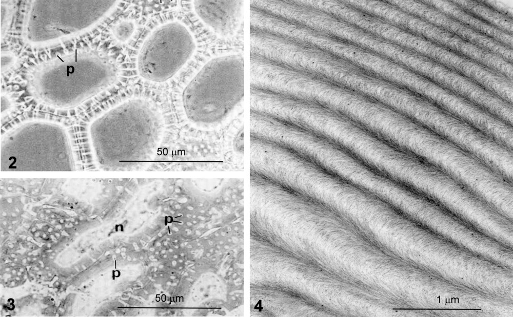
Cherry endocarp in the course of secondary-wall thickening and lignification. Freehand section. Phase contrast. Polygonal cells forming a coherent tissue. The walls are thick with numerous pit canals, p, front or profile view. From [6].

Fig. 3

Walnut endocarp in the course of secondary wall thickening and lignification. Polylobed shaped cells, bulged and associated like the pieces of a puzzle; p, pit canal; n, nucleus. From [7].

Fig. 4

Helicoidal pattern in stone cells of pear. PATAg test of polysaccharides following extraction of lignin and hemicellulose matrix. Bow-shaped arcs that are regular and monotonous.

Fig. 5

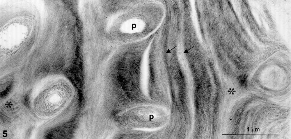
Helicoidal pattern in cherry endocarp. Zone rich in pit canals, p. Defects are visible: distortion (arrows), vortices in pits, saddle-like disclination (stars).

Fig. 4 provides the aspect of a typical helicoidal pattern where regular and monotonous nested arcs are seen, the thickness of the arc depending on the sectioning plane. The final number of arcs is variable, the maximum being up to 80–100 in stone cells. Such construction is relevant to a regular helicoidal structure (Fig. 1A) [4,9,10]. Besides these regular plywoods, apparent imperfect helicoidal arrays are also often encountered, especially in zones where pit canals are abundant. In these areas, one can recognize characteristic figures of distortions, vortices and disclinations such as saddle-like figures (Fig. 5). All these defects are diagnostic of a liquid-crystal behaviour submitted to strains. As it has been discussed from other biological models, the defects that develop here provide information on the liquid-crystal phase that could operate during wall construction [11,12].

On the whole, both the helicoidal texture and the occurrence of characteristic defects strongly suggest that the wall ordering is relevant to a cholesteric liquid-crystal assembly, if only briefly, that is rapidly and strongly consolidated by lignification [6,7]. It is clear that the constraints imposed by the spherical shape and the occurrence of pits are not an obstacle to the construction of an actual cellulosic helicoidal system.

3 Localization of glucuronoxylans reveals their possible role in helicoidal construction

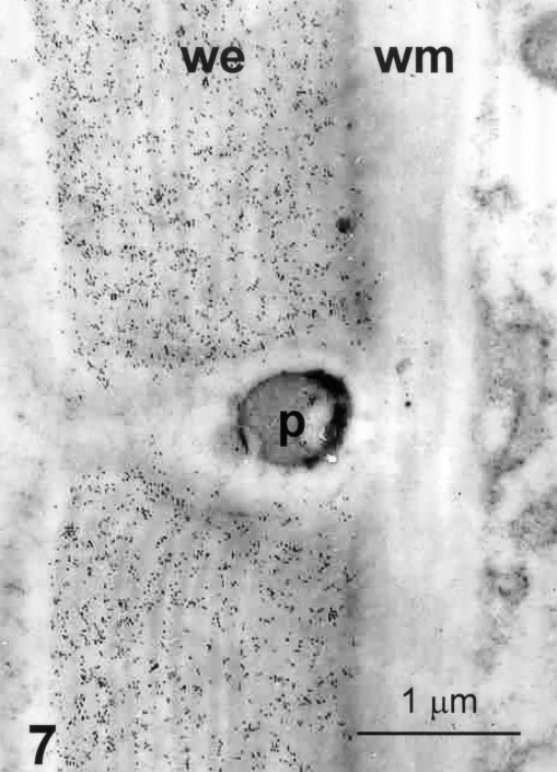
At maturity, the cell wall of stony cells contains about 30% of cellulose, 30% of GX and 40% of lignins. An immunodetection of GX was performed in the early stages, before lignification. An overview at the light microscope of cherry endocarp clearly shows an intense labelling in endocarp cells, whereas the neighbouring mesocarp cells, the cell walls of which are thin and not helicoidal in organization, are unlabelled (Fig. 6). Examination at higher magnification with the TEM of a contact endocarp/mesocarp confirms that the GX are present only in the endocarp (Fig. 7). They are regularly distributed within the whole thickness of the secondary helicoidal wall and are absent in the compound ‘middle lamella–primary wall’ and in pit canals (Fig. 8).

Fig. 6

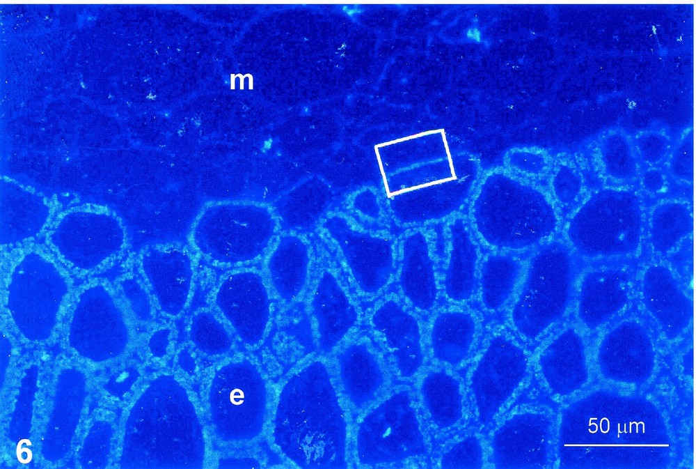
Immunodetection of GX. Cherry endocarp, 30 days post anthesis. Semithin section, anti-GX antibody, colloidal gold–silver intensification, UV epipolarisation. Cell walls in the course of secondary thickening, not yet lignified. Intense labelling of GX in secondary walls of endocarp, e. No labelling in mesocarp, m.

Fig. 7

Immunodetection of GX. Same material. Ultrathin section. Anti-GX antibody, colloidal gold. Detail of the square area of Fig. 6. Homogenous labelling within the whole thickness of the endocarp secondary wall, we. The wall of adjacent mesocarp cell, wm, and the middle lamella are not labelled; p, pit canal.

Fig. 8

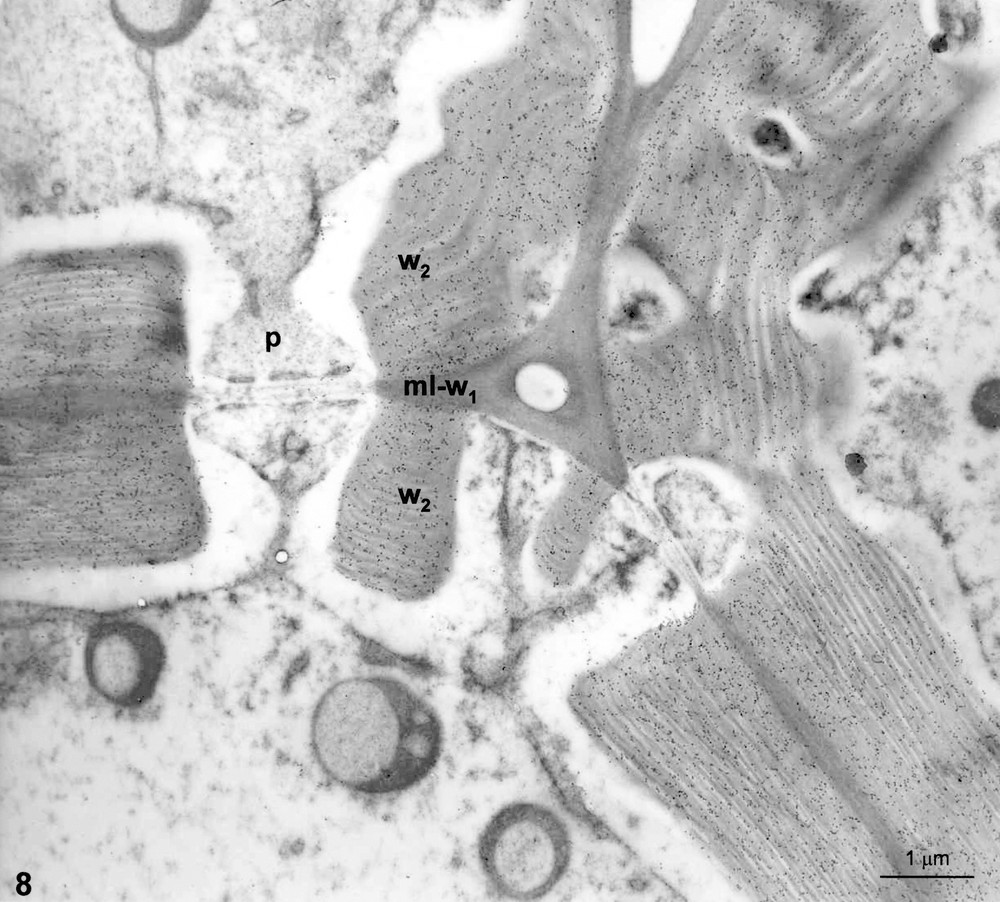
Immunodetection of GX. Same technique as for Fig. 7. Walls in the course of thickening and lignification. Intense and regular labelling on secondary walls, w2, on helicoidal structures, both in regular and distorted regions. Note that no labelling occurs on middle lamella–primary walls, mlw1, and pit canals, p.

Therefore, in secondary cell walls, a narrow correlation exists between the occurrence of GX and the occurrence of helicoidal organization. Cellulose and GX are clearly synchronically deposited when the helicoid is built, before lignification.

The case of cell walls of wood fibres provides another interesting example of secondary wall organization. Classically, it is known that the wall structure of fibres is typically three-layered (S1, S2 and S3 layers). Fine observations revealed the occurrence of helicoidal transition domains giving rise to a V-shape (Fig. 9). Such constructions correspond to an irregular plywood (Fig. 1B) [5,6,13]. An affinity labelling of GX by means of xylanase–gold complexes [5] revealed a preferential localization of GX in the transition zone between S1 et S2 layers, precisely in the zone where the transient helicoid occurs (Fig. 10). Moreover, a surface labelling of extracted cellulose microfibrils revealed the existence of an acid coat bonded onto the microfibrils, thus indicating a tight coating of the cellulose microfibrils by the GX [7,13] (Fig. 11). The acidic surfactant is likely responsible for the spacing pattern of the carbohydrate components by preventing the aggregation of the parallel-aligned microfibrils (Fig. 12A).

Fig. 9

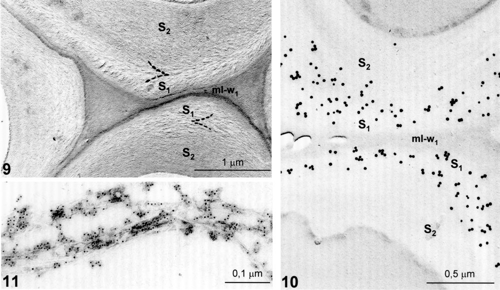
Cell wall of linden tree fibre. Chlorite delignification. DMSO extraction. Shadowing on thin section. Transitory helicoid between two blocked zones S1 and S2 of the secondary wall with a V-shaped appearance (dotted lines); mlw1, middle lamella–primary wall. From [5].

Fig. 10

Localization of GX by means of xylanase–gold labelling. Platinum shadowing. Preferential localization of GX at the boundary between S1 and S2; mlw1, middle lamella–primary wall. From [5].

Fig. 11

Decoration of cellulose microfibrils with cationic gold particles revealing GX surfactant. Stripped microfibrils dissociated by ultrasonic treatment.

Fig. 12

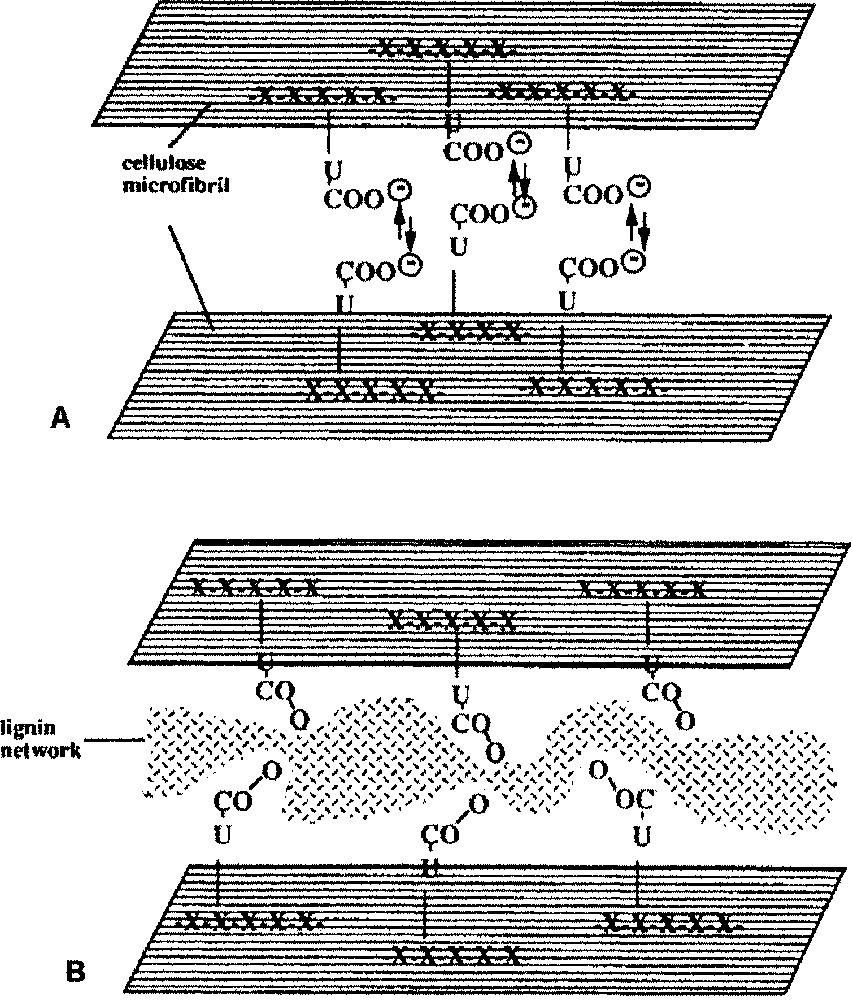
The glucuronoxylan–cellulose composite: possible successive steps of assembly and consolidation. A, Positioning and spacing pattern of the coated microfibrils, due to electrostatic repulsion of the acid coat; B, further consolidation by lignin intercalation.

Such co-crystallization of cellulose and GX led us to hypothesize that GX could direct the cellulose microfibrils in a helicoidal array [13]. This was a point that had to be explored.

4 Glucuronoxylans as twisting agents in helicoidal array

Experiments were tempted with quince slime, i.e. a helicoidal non-consolidated cell wall. This constitutes another paradigm model, since the walls remain completely fluid and cholesteric [6,14–16]. Attempts of in vitro reassociation of isolated cellulose/GX components from aqueous solutions were performed [14]. A spontaneous reassociation into cholesteric-like order was obtained from totally dispersed suspensions. Its structure was similar to the in situ native organization. When the system was deprived of GX, the cellulose flocculated, thus indicating the antifloc role of GX during assembly [15]. Glucuronoxylans possess surface charges and molecular structure (flexible side chains) that regulate fibrillogenesis as a selective moderator [17] and favour the formation of a cholesteric assembly. They could play a role of ‘twisting agent’, ‘helper molecules’, ‘molecular organizers’ or ‘lubricating agents’ driving the cellulose microfibrils into helicoidal rotation [16,18,19]. Fig. 12A shows how, in the early construction of cell wall, the face to face carboxylic groups of the GX could generate electrostatic repelling forces that prevent the aggregation of the microfibrils and favour their parallel alignment.

5 Concluding remarks

The foregoing data have focussed on the role of GX in the early assembly of helicoidal walls. It was tempting to consider their involvement in the following events, i.e. when lignins are progressively inserted in the pre-existing network at cell maturity. The cellulose/GX composite is a charged and highly anisotropic construction forming a sort of ‘host structure’ for lignin precursors. The incrustation of lignin can be compared to a form of intercalation chemistry where exogenous monomers can be intercalated, taking advantage of the gaps due to the internal electrostatic repelling, as documented for advanced industrial materials [20]. Here, lignin precursors are inserted and polymerised in the gaps extended between microfibrils, thus stiffening the xylan cellulose composite and safeguarding its initial geometry (Fig. 12B). In such a model, the GXs seem to play a key role in the successive steps of the morphogenesis of lignified secondary cell walls. Other plant systems have evidently cell walls constructed with other dominant hemicelluloses (arabinoxylans of grasses, glucomannans). The question remains open to understand whether they also could play a preeminent role in wall construction.


Bibliographie

[1] B. Monties Plant cell walls as fibrous lignocellulosic composites: relations with lignin structure and function, Anim. Feed Sci. Technol., Volume 32 (1991), pp. 159-179

[2] N. Parameswaran; M. Sinner Topological study of the wall of beech bark sclereids by enzymatic and acidic degradation, Protoplasma, Volume 101 (1979), pp. 197-215

[3] N. Parameswaran; W. Liese Ultrastructural localization of cell wall components in wood cells, Holz als Roh. Werkstoff., Volume 40 (1982), pp. 145-155

[4] J.-C. Roland; D. Reis; B. Vian; B. Satiat-Jeunemaitre; M. Mosiniak Morphogenesis of plant cell wall at the supramolecular level. Internal geometry and versatility of helicoidal expression, Protoplasma, Volume 140 (1987), pp. 75-91

[5] B. Vian; D. Reis; M. Mosiniak; J.-C. Roland The glucuronoxylans and the helicoidal shift in cellulose microfibrils in linden wood: cytochemistry in muro and on isolated molecules, Protoplasma, Volume 131 (1986), pp. 185-189

[6] D. Reis; B. Vian; J.-C. Roland Cellulose–glucuronoxylans and plant cell wall structure, Micron, Volume 25 (1994), pp. 171-187

[7] D. Reis; J.-C. Roland; M. Mosiniak; D. Darzens; B. Vian The sustained and warped pattern of a xylan–cellulose composite: the stony endocarp model, Protoplasma, Volume 166 (1992), pp. 21-34

[8] B. Satiat-Jeunemaître; M. Mosiniak Variations in morphogenetic rhythm of secondary cell wall deposition in Prunus (sp.) sclerocytes, Giorn. Bot. Ital., Volume 126 (1992), pp. 615-629

[9] A.C. Neville Biology of Fibrous Composites. Development Beyond the Cell Membrane, Cambridge University Press, Cambridge, 1993

[10] A.C. Neville; S. Levy The helicoidal concept in plant cell wall ultrastructure and morphogenesis (C.T. Brett; J.R. Hilman, eds.), Biochemistry of Plant Cell Wall, Cambridge University Press, Cambridge, 1985, pp. 99-124

[11] Y. Bouligand Cholesteric order in biopolymers (B. Blumstein, ed.), Mesomorphic order in Polymer and Polymerization in Liquid Crystalline Media, American Chemical Society, Washington, DC, 1978, pp. 261-297

[12] G.D. Mazur; J.C. Regier; F.C. Kalafatos Morphogenesis of silk chorion: sequential modification in an early helicoidal framework through expansion and densification, Tissue Cell, Volume 21 (1989), pp. 227-242

[13] B. Vian; J.-C. Roland; D. Reis; M. Mosiniak Distribution and possible morphogenetic role of the xylans within the secondary vessel wall of linden wood, IAWA Bull., Volume 13 (1992), pp. 269-282

[14] D. Reis; B. Vian; H. Chanzy; J.-C. Roland Liquid-crystal-type assembly of native cellulose–glucuronoxylans extracted from plant cell wall, Biol. Cell, Volume 67 (1991), pp. 173-178

[15] B. Vian; D. Reis; D. Darzens; J.-C. Roland Cholesteric-like crystal analogs in glucuronoxylan-rich cell wall composites: experimental approach of acellular re-assembly from native cellulosic suspension, Protoplasma, Volume 180 (1994), pp. 70-81

[16] R.M. Abeysekera; J.H.M. Willison Development of helicoidal texture in the prerelease mucilage of quince (Cydonia oblonga) seed epidermis, Can. J. Bot., Volume 66 (1988), pp. 460-467

[17] R.H. Atalla States of aggregation in native celluloses, Cellulose'91, 11th Syracuse Cellulose Conference, New Orleans, LA, USA, 1991, p. 124

[18] A.C. Neville A pipe-cleaner molecular model for morphogenesis of helicoidal cell walls based on cellulose complexity, J. Theor. Biol., Volume 131 (1988), pp. 343-354

[19] I.A. Nieduzinski; R.H. Marchessault Structure of d-(1–4)-xylan hydrate, Biopolymers, Volume 11 (1972), pp. 1344-1355

[20] J. Rouxel; A. Meerschaut; P. Gressier The design of low-dimension solids, Synth. Mat., Volume 34 (1989), pp. 597-607


Cité par

  • Yu Guo; Lichao Jiao; Jie Wang; Lingyu Ma; Yang Lu; Yonggang Zhang; Juan Guo; Yafang Yin Analyses of high spatial resolution datasets identify genes associated with multi-layered secondary cell wall thickening in Pinus bungeana, Annals of Botany, Volume 133 (2024) no. 7, p. 953 | DOI:10.1093/aob/mcae023
  • Hailin Li; Rong Xu; Cai Wang; Xiaolin Zhang; Peiling Li; Zhiyang Wu; Dan Zong Determination and Analysis of Endogenous Hormones and Cell Wall Composition between the Straight and Twisted Trunk Types of Pinus yunnanensis Franch, Forests, Volume 15 (2024) no. 9, p. 1626 | DOI:10.3390/f15091626
  • Charlotte Permann; Andreas Holzinger Zygospore formation in Zygnematophyceae predates several land plant traits, Philosophical Transactions of the Royal Society B: Biological Sciences, Volume 379 (2024) no. 1914 | DOI:10.1098/rstb.2023.0356
  • Justin G. A. Whitehill; Macaire M. S. Yuen; Angela Chiang; Carol E. Ritland; Jörg Bohlmann Transcriptome features of stone cell development in weevil‐resistant and susceptible Sitka spruce, New Phytologist, Volume 239 (2023) no. 6, p. 2138 | DOI:10.1111/nph.19103
  • Meredith Rose Barr; Luke Forster; Carmine D'Agostino; Roberto Volpe Alkaline pretreatment of walnut shells increases pore surface hydrophilicity of derived biochars, Applied Surface Science, Volume 571 (2022), p. 151253 | DOI:10.1016/j.apsusc.2021.151253
  • Charlotte Permann; Notburga Gierlinger; Andreas Holzinger Zygospores of the green alga Spirogyra: new insights from structural and chemical imaging, Frontiers in Plant Science, Volume 13 (2022) | DOI:10.3389/fpls.2022.1080111
  • Mohamed Abdel-Raheem Insect Pest Control in Orchards, Handbook of Research on Principles and Practices for Orchards Management (2022), p. 277 | DOI:10.4018/978-1-6684-2423-0.ch015
  • Sebastian J. Antreich; Jessica C. Huss; Nannan Xiao; Adya Singh; Notburga Gierlinger The walnut shell network: 3D visualisation of symplastic and apoplastic transport routes in sclerenchyma tissue, Planta, Volume 256 (2022) no. 3 | DOI:10.1007/s00425-022-03960-w
  • Yuanyuan Zhang; Jiajun Mao; Jingsong Peng; Antoni P. Tomsia; Lei Jiang; Qunfeng Cheng Ginkgo seed shell provides a unique model for bioinspired design, Proceedings of the National Academy of Sciences, Volume 119 (2022) no. 49 | DOI:10.1073/pnas.2211458119
  • Jacob D. Crowe; Pengchao Hao; Sivakumar Pattathil; Henry Pan; Shi-You Ding; David B. Hodge; Jacob Krüger Jensen Xylan Is Critical for Proper Bundling and Alignment of Cellulose Microfibrils in Plant Secondary Cell Walls, Frontiers in Plant Science, Volume 12 (2021) | DOI:10.3389/fpls.2021.737690
  • Ayano Higaki; Yui Kadowaki; Arata Yoshinaga; Keiji Takabe Xylan deposition and lignification in differentiating tension wood fibers in Mallotus japonicus (Euphorbiaceae) with multi-layered structure, Holzforschung, Volume 75 (2021) no. 1, p. 1 | DOI:10.1515/hf-2020-0001
  • Yin Chang; Rox Middleton; Yu Ogawa; Tom Gregory; Lisa M. Steiner; Alexander Kovalev; Rebecca H. N. Karanja; Paula J. Rudall; Beverley J. Glover; Stanislav N. Gorb; Silvia Vignolini Cell wall composition determines handedness reversal in helicoidal cellulose architectures of Pollia condensata fruits, Proceedings of the National Academy of Sciences, Volume 118 (2021) no. 51 | DOI:10.1073/pnas.2111723118
  • Nannan Xiao; Martin Felhofer; Sebastian J. Antreich; Jessica C. Huss; Konrad Mayer; Adya Singh; Peter Bock; Notburga Gierlinger Twist and lock: nutshell structures for high strength and energy absorption, Royal Society Open Science, Volume 8 (2021) no. 8, p. 210399 | DOI:10.1098/rsos.210399
  • Peter N. Ciesielski; M. Brennan Pecha; Aaron M. Lattanzi; Vivek S. Bharadwaj; Meagan F. Crowley; Lintao Bu; Josh V. Vermaas; K. Xerxes Steirer; Michael F. Crowley Advances in Multiscale Modeling of Lignocellulosic Biomass, ACS Sustainable Chemistry Engineering, Volume 8 (2020) no. 9, p. 3512 | DOI:10.1021/acssuschemeng.9b07415
  • F. Robert Gleuwitz; Christian Friedrich; Marie-Pierre G. Laborie Lignin-Assisted Stabilization of an Oriented Liquid Crystalline Cellulosic Mesophase, Part A: Observation of Microstructural and Mechanical Behavior, Biomacromolecules, Volume 21 (2020) no. 3, p. 1069 | DOI:10.1021/acs.biomac.9b01352
  • Jiawei Wang; Eji Minami; Haruo Kawamoto Thermal reactivity of hemicellulose and cellulose in cedar and beech wood cell walls, Journal of Wood Science, Volume 66 (2020) no. 1 | DOI:10.1186/s10086-020-01888-x
  • Pengyun Li; Qilin Zhang; Xun Zhang; Xueming Zhang; Xuejun Pan; Feng Xu Subcellular dissolution of xylan and lignin for enhancing enzymatic hydrolysis of microwave assisted deep eutectic solvent pretreated Pinus bungeana Zucc, Bioresource Technology, Volume 288 (2019), p. 121475 | DOI:10.1016/j.biortech.2019.121475
  • Martin P. Wierzbicki; Victoria Maloney; Eshchar Mizrachi; Alexander A. Myburg Xylan in the Middle: Understanding Xylan Biosynthesis and Its Metabolic Dependencies Toward Improving Wood Fiber for Industrial Processing, Frontiers in Plant Science, Volume 10 (2019) | DOI:10.3389/fpls.2019.00176
  • Hongmei Yuan; Lijuan Zhao; Wendong Guo; Ying Yu; Lei Tao; Liguo Zhang; Xixia Song; Wengong Huang; Lili Cheng; Jing Chen; Fengzhi Guan; Guangwen Wu; Huiyu Li Exogenous Application of Phytohormones Promotes Growth and Regulates Expression of Wood Formation-Related Genes in Populus simonii × P. nigra, International Journal of Molecular Sciences, Volume 20 (2019) no. 3, p. 792 | DOI:10.3390/ijms20030792
  • Martin P. Wierzbicki; Nanette Christie; Desré Pinard; Shawn D. Mansfield; Eshchar Mizrachi; Alexander A. Myburg A systems genetics analysis in Eucalyptus reveals coordination of metabolic pathways associated with xylan modification in wood‐forming tissues, New Phytologist, Volume 223 (2019) no. 4, p. 1952 | DOI:10.1111/nph.15972
  • Junlan Gao; Min Yu; Shiliu Zhu; Liang Zhou; Shengquan Liu Effects of exogenous 24-epibrassinolide and brassinazole on negative gravitropism and tension wood formation in hybrid poplar (Populus deltoids × Populus nigra), Planta, Volume 249 (2019) no. 5, p. 1449 | DOI:10.1007/s00425-018-03074-2
  • Hui Wei; Xiaowen Chen; Joseph Shekiro; Erik Kuhn; Wei Wang; Yun Ji; Evguenii Kozliak; Michael Himmel; Melvin Tucker Kinetic Modelling and Experimental Studies for the Effects of Fe2+ Ions on Xylan Hydrolysis with Dilute-Acid Pretreatment and Subsequent Enzymatic Hydrolysis, Catalysts, Volume 8 (2018) no. 1, p. 39 | DOI:10.3390/catal8010039
  • Xia Zhou; Dayong Ding; Tingting You; Xun Zhang; Keiji Takabe; Feng Xu Synergetic Dissolution of Branched Xylan and Lignin Opens the Way for Enzymatic Hydrolysis of Poplar Cell Wall, Journal of Agricultural and Food Chemistry, Volume 66 (2018) no. 13, p. 3449 | DOI:10.1021/acs.jafc.8b00320
  • Shingo Kiyoto; Arata Yoshinaga; Eva Fernandez-Tendero; Arnaud Day; Brigitte Chabbert; Keiji Takabe Distribution of Lignin, Hemicellulose, and Arabinogalactan Protein in Hemp Phloem Fibers, Microscopy and Microanalysis, Volume 24 (2018) no. 4, p. 442 | DOI:10.1017/s1431927618012448
  • María Graciela Aguayo; William Gacitúa; Paulina Valenzuela; Regis Teixeira Mendonça; Pablo Reyes Chemical characteristics of woods from several Chilean native forest species and their relationship with nanomechanical cell properties, Southern Forests: a Journal of Forest Science, Volume 80 (2018) no. 4, p. 293 | DOI:10.2989/20702620.2017.1376269
  • Jan J. Lyczakowski; Krzysztof B. Wicher; Oliver M. Terrett; Nuno Faria-Blanc; Xiaolan Yu; David Brown; Kristian B. R. M. Krogh; Paul Dupree; Marta Busse-Wicher Removal of glucuronic acid from xylan is a strategy to improve the conversion of plant biomass to sugars for bioenergy, Biotechnology for Biofuels, Volume 10 (2017) no. 1 | DOI:10.1186/s13068-017-0902-1
  • Akira Yamakawa; Shiho Suzuki; Takeshi Oku; Kenta Enomoto; Motohide Ikeda; Joseph Rodrigue; Keita Tateiwa; Yoshinobu Terada; Hiroyuki Yano; Shinichi Kitamura Nanostructure and physical properties of cellulose nanofiber-carbon nanotube composite films, Carbohydrate Polymers, Volume 171 (2017), p. 129 | DOI:10.1016/j.carbpol.2017.05.012
  • Jasna Simonović Radosavljević; Jelena Bogdanović Pristov; Aleksandra Lj. Mitrović; Gabor Steinbach; Gregory Mouille; Srđan Tufegdžić; Vuk Maksimović; Dragosav Mutavdžić; Dušica Janošević; Marina Vuković; Győző Garab; Ksenija Radotić Parenchyma cell wall structure in twining stem of Dioscorea balcanica, Cellulose, Volume 24 (2017) no. 11, p. 4653 | DOI:10.1007/s10570-017-1460-1
  • Noppadon Sathitsuksanoh; Scott Renneckar Characterization Methods and Techniques, Introduction to Renewable Biomaterials (2017), p. 107 | DOI:10.1002/9781118698600.ch4
  • Prashant Mohan‐Anupama Pawar; Christine Ratke; Vimal K. Balasubramanian; Sun‐Li Chong; Madhavi Latha Gandla; Mathilda Adriasola; Tobias Sparrman; Mattias Hedenström; Klaudia Szwaj; Marta Derba‐Maceluch; Cyril Gaertner; Gregory Mouille; Ines Ezcurra; Maija Tenkanen; Leif J. Jönsson; Ewa J. Mellerowicz Downregulation of RWA genes in hybrid aspen affects xylan acetylation and wood saccharification, New Phytologist, Volume 214 (2017) no. 4, p. 1491 | DOI:10.1111/nph.14489
  • Ayano Higaki; Arata Yoshinaga; Keiji Takabe Heterogeneous distribution of xylan and lignin in tension wood G-layers of the S1+G type in several Japanese hardwoods, Tree Physiology, Volume 37 (2017) no. 12, p. 1767 | DOI:10.1093/treephys/tpx144
  • Richard M. Parker; Bruno Frka-Petesic; Giulia Guidetti; Gen Kamita; Gioele Consani; Chris Abell; Silvia Vignolini Hierarchical Self-Assembly of Cellulose Nanocrystals in a Confined Geometry, ACS Nano, Volume 10 (2016) no. 9, p. 8443 | DOI:10.1021/acsnano.6b03355
  • Melanie Blumentritt; Douglas J. Gardner; Barbara J.W. Cole; Stephen M. Shaler Influence of hot-water extraction on ultrastructure and distribution of glucomannans and xylans in poplar xylem as detected by gold immunolabeling, Holzforschung, Volume 70 (2016) no. 3, p. 243 | DOI:10.1515/hf-2015-0030
  • Silvia Vignolini; Thomas Gregory; Mathias Kolle; Alfie Lethbridge; Edwige Moyroud; Ullrich Steiner; Beverley J. Glover; Peter Vukusic; Paula J. Rudall Structural colour from helicoidal cell-wall architecture in fruits ofMargaritaria nobilis, Journal of The Royal Society Interface, Volume 13 (2016) no. 124, p. 20160645 | DOI:10.1098/rsif.2016.0645
  • Brian Ribbans; Yujie Li; Ting Tan A bioinspired study on the interlaminar shear resistance of helicoidal fiber structures, Journal of the Mechanical Behavior of Biomedical Materials, Volume 56 (2016), p. 57 | DOI:10.1016/j.jmbbm.2015.11.004
  • Mathias Sorieul; Alan Dickson; Stefan Hill; Hamish Pearson Plant Fibre: Molecular Structure and Biomechanical Properties, of a Complex Living Material, Influencing Its Deconstruction towards a Biobased Composite, Materials, Volume 9 (2016) no. 8, p. 618 | DOI:10.3390/ma9080618
  • Justin G. A. Whitehill; Hannah Henderson; Mathias Schuetz; Oleksandr Skyba; Macaire Man Saint Yuen; John King; A. Lacey Samuels; Shawn D. Mansfield; Jörg Bohlmann Histology and cell wall biochemistry of stone cells in the physical defence of conifers against insects, Plant, Cell Environment, Volume 39 (2016) no. 8, p. 1646 | DOI:10.1111/pce.12654
  • Hoang-Linh Nguyen; Yun Jo; Minkyu Cha; Yun Cha; Dong Yoon; Naresh Sanandiya; Ekavianty Prajatelistia; Dongyeop Oh; Dong Hwang Mussel-Inspired Anisotropic Nanocellulose and Silver Nanoparticle Composite with Improved Mechanical Properties, Electrical Conductivity and Antibacterial Activity, Polymers, Volume 8 (2016) no. 3, p. 102 | DOI:10.3390/polym8030102
  • Geoffrey Daniel Microscope Techniques for Understanding Wood Cell Structure and Biodegradation, Secondary Xylem Biology (2016), p. 309 | DOI:10.1016/b978-0-12-802185-9.00015-2
  • Qiang Li; Keiichi Koda; Arata Yoshinaga; Keiji Takabe; Masatsugu Shimomura; Yuji Hirai; Yutaka Tamai; Yasumitsu Uraki Dehydrogenative Polymerization of Coniferyl Alcohol in Artificial Polysaccharides Matrices: Effects of Xylan on the Polymerization, Journal of Agricultural and Food Chemistry, Volume 63 (2015) no. 18, p. 4613 | DOI:10.1021/acs.jafc.5b01070
  • Melanie Horbens; Dominik Branke; Roland Gärtner; Axel Voigt; Florian Stenger; Christoph Neinhuis Multi-scale simulation of plant stem reinforcement by brachysclereids: A case study in apple fruit peduncles, Journal of Structural Biology, Volume 192 (2015) no. 1, p. 116 | DOI:10.1016/j.jsb.2015.08.002
  • Marta Derba‐Maceluch; Tatsuya Awano; Junko Takahashi; Jessica Lucenius; Christine Ratke; Inkeri Kontro; Marta Busse‐Wicher; Ondrej Kosik; Ryo Tanaka; Anders Winzéll; Åsa Kallas; Joanna Leśniewska; Fredrik Berthold; Peter Immerzeel; Tuula T. Teeri; Ines Ezcurra; Paul Dupree; Ritva Serimaa; Ewa J. Mellerowicz Suppression of xylan endotransglycosylase PtxtXyn10A affects cellulose microfibril angle in secondary wall in aspen wood, New Phytologist, Volume 205 (2015) no. 2, p. 666 | DOI:10.1111/nph.13099
  • Eveline Q.P. Tavares; Marcos S. Buckeridge Do plant cell walls have a code?, Plant Science, Volume 241 (2015), p. 286 | DOI:10.1016/j.plantsci.2015.10.016
  • Shingo Kiyoto; Arata Yoshinaga; Keiji Takabe Relative deposition of xylan and 8-5′-linked lignin structure in Chamaecyparis obtusa, as revealed by double immunolabeling by using monoclonal antibodies, Planta, Volume 241 (2015) no. 1, p. 243 | DOI:10.1007/s00425-014-2181-4
  • Melanie Horbens; Alexander Feldner; Monika Höfer; Christoph Neinhuis Ontogenetic tissue modification in Malus fruit peduncles: the role of sclereids, Annals of Botany, Volume 113 (2014) no. 1, p. 105 | DOI:10.1093/aob/mct262
  • Ohkyung Kwon Investigation of Layered Structure of Fiber Cell Wall in Korean Red Pine by Confocal Reflection Microscopy, Applied Microscopy, Volume 44 (2014) no. 2, p. 61 | DOI:10.9729/am.2014.44.2.61
  • Ohkyung Kwon Investigation of Bordered Pit Ultrastructure in Tracheid of Korean Red Pine (Pinus densiflora) by Confocal Reflection Microscopy, Journal of the Korean Wood Science and Technology, Volume 42 (2014) no. 3, p. 346 | DOI:10.5658/wood.2014.42.3.346
  • Hyunjung Jin; Jihye Do; Soo-Jeong Shin; Joon Weon Choi; Young Im Choi; Wook Kim; Mi Kwon Exogenously applied 24-epi brassinolide reduces lignification and alters cell wall carbohydrate biosynthesis in the secondary xylem of Liriodendron tulipifera, Phytochemistry, Volume 101 (2014), p. 40 | DOI:10.1016/j.phytochem.2014.02.003
  • Kurt V. Fagerstedt; Ewa Mellerowicz; Tatyana Gorshkova; Katia Ruel; Jean-Paul Joseleau Cell Wall Polymers in Reaction Wood, The Biology of Reaction Wood (2014), p. 37 | DOI:10.1007/978-3-642-10814-3_3
  • Marta Busse‐Wicher; Thiago C. F. Gomes; Theodora Tryfona; Nino Nikolovski; Katherine Stott; Nicholas J. Grantham; David N. Bolam; Munir S. Skaf; Paul Dupree The pattern of xylan acetylation suggests xylan may interact with cellulose microfibrils as a twofold helical screw in the secondary plant cell wall of Arabidopsis thaliana, The Plant Journal, Volume 79 (2014) no. 3, p. 492 | DOI:10.1111/tpj.12575
  • K. Nakagawa; A. Yoshinaga; K. Takabe Xylan deposition and lignification in the multi-layered cell walls of phloem fibres in Mallotus japonicus (Euphorbiaceae), Tree Physiology, Volume 34 (2014) no. 9, p. 1018 | DOI:10.1093/treephys/tpu061
  • Timo J. Hakala; Vesa Saikko; Suvi Arola; Tiina Ahlroos; Aino Helle; Petri Kuosmanen; Kenneth Holmberg; Markus B. Linder; Päivi Laaksonen Structural characterization and tribological evaluation of quince seed mucilage, Tribology International, Volume 77 (2014), p. 24 | DOI:10.1016/j.triboint.2014.04.018
  • Junhua Zhang; Ulla Moilanen; Ming Tang; Liisa Viikari The carbohydrate-binding module of xylanase from Nonomuraea flexuosa decreases its non-productive adsorption on lignin, Biotechnology for Biofuels, Volume 6 (2013) no. 1, p. 18 | DOI:10.1186/1754-6834-6-18
  • Jennifer R. Bromley; Marta Busse‐Wicher; Theodora Tryfona; Jennifer C. Mortimer; Zhinong Zhang; David M. Brown; Paul Dupree GUX1 and GUX2 glucuronyltransferases decorate distinct domains of glucuronoxylan with different substitution patterns, The Plant Journal, Volume 74 (2013) no. 3, p. 423 | DOI:10.1111/tpj.12135
  • Lloyd A. Donaldson; J. Paul Knox Localization of Cell Wall Polysaccharides in Normal and Compression Wood of Radiata Pine: Relationships with Lignification and Microfibril Orientation, Plant Physiology, Volume 158 (2012) no. 2, p. 642 | DOI:10.1104/pp.111.184036
  • Jong Sik Kim; Geoffrey Daniel Immunolocalization of hemicelluloses in Arabidopsis thaliana stem. Part I: temporal and spatial distribution of xylans, Planta, Volume 236 (2012) no. 4, p. 1275 | DOI:10.1007/s00425-012-1686-y
  • A. Yoshinaga; H. Kusumoto; F. Laurans; G. Pilate; K. Takabe Lignification in poplar tension wood lignified cell wall layers, Tree Physiology, Volume 32 (2012) no. 9, p. 1129 | DOI:10.1093/treephys/tps075
  • Tsuguyuki Saito; Takehiko Uematsu; Satoshi Kimura; Toshiharu Enomae; Akira Isogai Self-aligned integration of native cellulose nanofibrils towards producing diverse bulk materials, Soft Matter, Volume 7 (2011) no. 19, p. 8804 | DOI:10.1039/c1sm06050c
  • Q. Li; D. Min; J. P.-Y. Wang; I. Peszlen; L. Horvath; B. Horvath; Y. Nishimura; H. Jameel; H.-M. Chang; V. L. Chiang Down-regulation of glycosyltransferase 8D genes in Populus trichocarpa caused reduced mechanical strength and xylan content in wood, Tree Physiology, Volume 31 (2011) no. 2, p. 226 | DOI:10.1093/treephys/tpr008
  • Xiaowen Chen; Martin Lawoko; Adriaan van Heiningen Kinetics and mechanism of autohydrolysis of hardwoods, Bioresource Technology, Volume 101 (2010) no. 20, p. 7812 | DOI:10.1016/j.biortech.2010.05.006
  • Jun-Li Ren; Run-Cang Sun Hemicelluloses, Cereal Straw as a Resource for Sustainable Biomaterials and Biofuels (2010), p. 73 | DOI:10.1016/b978-0-444-53234-3.00004-3
  • Bernhard Borkhardt; Jesper Harholt; Peter Ulvskov; Birgitte K. Ahring; Bodil Jørgensen; Henrik Brinch‐Pedersen Autohydrolysis of plant xylans by apoplastic expression of thermophilic bacterial endo‐xylanases, Plant Biotechnology Journal, Volume 8 (2010) no. 3, p. 363 | DOI:10.1111/j.1467-7652.2010.00506.x
  • Volker Bischoff; Silvia Nita; Lutz Neumetzler; Dana Schindelasch; Aurélie Urbain; Ravit Eshed; Staffan Persson; Deborah Delmer; Wolf-Rüdiger Scheible TRICHOME BIREFRINGENCEand Its HomologAT5G01360Encode Plant-Specific DUF231 Proteins Required for Cellulose Biosynthesis in Arabidopsis, Plant Physiology, Volume 153 (2010) no. 2, p. 590 | DOI:10.1104/pp.110.153320
  • Mirjam A. Kabel; Hein van den Borne; Jean-Paul Vincken; Alphons G.J. Voragen; Henk A. Schols Structural differences of xylans affect their interaction with cellulose, Carbohydrate Polymers, Volume 69 (2007) no. 1, p. 94 | DOI:10.1016/j.carbpol.2006.09.006
  • Staffan Persson; Kerry Hosmer Caffall; Glenn Freshour; Matthew T. Hilley; Stefan Bauer; Patricia Poindexter; Michael G. Hahn; Debra Mohnen; Chris Somerville TheArabidopsis irregular xylem8Mutant Is Deficient in Glucuronoxylan and Homogalacturonan, Which Are Essential for Secondary Cell Wall Integrity, The Plant Cell, Volume 19 (2007) no. 1, p. 237 | DOI:10.1105/tpc.106.047720
  • Anne-Marie Catesson Current research on secondary lignified cell walls, a thematic issue dedicated to Bernard Monties, Comptes Rendus. Biologies, Volume 327 (2004) no. 9-10, p. 775 | DOI:10.1016/j.crvi.2004.08.004

Cité par 66 documents. Sources : Crossref


This paper, written in homage to Bernard Monties, wishes to thank him for his attentive regard and his always constructive criticisms toward the cell wall people. Bernard Monties brought a chemical expertise that was for us necessary to better understand the cell wall model. In numerous fruitful discussions, his pertinent questions always helped us to reconsider our working hypotheses.

Commentaires - Politique