Plan
Comptes Rendus

Medical sciences / Sciences médicales
Control of fatty acid and glycerol release in adipose tissue lipolysis
[Contrôle de la libération des acides gras et du glycérol lors de la lipolyse adipocytaire]
Comptes Rendus. Biologies, Volume 329 (2006) no. 8, pp. 598-607.

Résumés

Adipose tissue lipolysis is the catabolic process leading to the breakdown of triglycerides stored in fat cells and the release of fatty acids and glycerol. Recent work has revealed that lipolysis is not a simple metabolic pathway stimulated by catecholamines and inhibited by insulin. New discoveries on the regulation of lipolysis by endocrine and paracrine factors and on the proteins involved in triglyceride hydrolysis have led to a reappraisal of the complexity of the various signal transduction pathways. The steps involved in the dysregulation of lipolysis observed in obesity have partly been identified.

La lipolyse du tissu adipeux est le processus catabolique par lequel l'hydrolyse des triglycérides stockés dans les adipocytes conduit à la libération de glycérol et d'acides gras. Les travaux récents ont révélé que la lipolyse n'est pas une simple voie métabolique stimulée par les catécholamines et inhibée par l'insuline. De nouvelles découvertes sur la régulation de la lipolyse par des facteurs endocrines ou paracrines et sur les protéines impliquées dans l'hydrolyse des triglycérides ont conduit à une réévaluation de la complexité des différentes voies de transduction. Les étapes clés responsables de la dérégulation de la lipolyse observée dans l'obésité ont en partie été identifiées.

Métadonnées
Reçu le :
Accepté le :
Publié le :
DOI : 10.1016/j.crvi.2005.10.008
Keywords: Obesity, Metabolic syndrome, Adipose tissue, Lipolysis, Fat mobilization
Mot clés : Obésité, Syndrome métabolique, Tissu adipeux, Lipolyse, Mobilisation des lipides
Dominique Langin 1, 2

1 Unité de recherches sur les obésités, INSERM UPS U586, institut Louis-Bugnard, université Paul-Sabatier, CHU Rangueil, BP 84225, 31432 Toulouse cedex 4, France
2 Franco-Czech Laboratory for Clinical Research on Obesity, INSERM–3rd Faculty of Medicine of Charles University, Prague, Czech Republic
@article{CRBIOL_2006__329_8_598_0,
     author = {Dominique Langin},
     title = {Control of fatty acid and glycerol release in adipose tissue lipolysis},
     journal = {Comptes Rendus. Biologies},
     pages = {598--607},
     publisher = {Elsevier},
     volume = {329},
     number = {8},
     year = {2006},
     doi = {10.1016/j.crvi.2005.10.008},
     language = {en},
}
TY  - JOUR
AU  - Dominique Langin
TI  - Control of fatty acid and glycerol release in adipose tissue lipolysis
JO  - Comptes Rendus. Biologies
PY  - 2006
SP  - 598
EP  - 607
VL  - 329
IS  - 8
PB  - Elsevier
DO  - 10.1016/j.crvi.2005.10.008
LA  - en
ID  - CRBIOL_2006__329_8_598_0
ER  - 
%0 Journal Article
%A Dominique Langin
%T Control of fatty acid and glycerol release in adipose tissue lipolysis
%J Comptes Rendus. Biologies
%D 2006
%P 598-607
%V 329
%N 8
%I Elsevier
%R 10.1016/j.crvi.2005.10.008
%G en
%F CRBIOL_2006__329_8_598_0
Dominique Langin. Control of fatty acid and glycerol release in adipose tissue lipolysis. Comptes Rendus. Biologies, Volume 329 (2006) no. 8, pp. 598-607. doi : 10.1016/j.crvi.2005.10.008. https://comptes-rendus.academie-sciences.fr/biologies/articles/10.1016/j.crvi.2005.10.008/

Version originale du texte intégral

1 Introduction

The major physiological role for white adipose tissue (WAT) fat stores is to supply lipid energy when it is needed by other tissues. This is achieved by a highly regulated pathway whereby the triglycerides stored in the adipocyte are hydrolysed and fatty acids delivered to the plasma. Altered lipolysis could be an element leading to obesity and interindividual variations in WAT lipolysis are of importance for the rate of weight loss. Conversely, excessive lipolytic rates, in conjunction with impairment in fatty acid utilization by muscle and liver, may be a major contributor to the metabolic abnormalities found in subjects with upper body obesity.

This review is composed of two sections. The first section presents the endocrine and paracrine factors controlling the stimulation of lipolysis and the related signal transduction pathways. The second section is devoted to the molecular mechanisms of the hydrolysis of triglycerides that involves lipases, plasma membrane transporters, fatty acid binding proteins and proteins associated with the lipid droplet.

2 Endocrine and paracrine control of lipolysis

2.1 Catecholamines

Catecholamines are important regulators of fat cell lipolysis in human WAT. The neurotransmitter, noradrenaline, and the hormone, adrenaline, regulate lipolysis through lipolytic beta-adrenoceptors and antilipolytic alpha2-adrenoceptors.

2.1.1 Beta-adrenoceptors

Activation of beta-adrenoceptors by catecholamines leads to an increase of cAMP production by adenylylcyclase, which is followed by activation of protein kinase A and hormone-sensitive lipase (HSL) (see Section 3.1). In human fat cells, both beta1- and beta2-adrenergic receptors are known to stimulate cAMP production and lipolysis in vitro [1] and in vivo [2]. The beta3-adrenergic receptor attracted a lot of interest [3]. It is highly expressed in rodent WAT and brown adipose tissue and, unlike the beta1- and beta2-adrenergic receptors, is relatively specific to the tissues. Selective beta3-adrenergic agonists elicit a marked lipolytic and thermogenic response in rodents and hence exert antiobesity and antidiabetic effects [4]. The level of expression and the contribution of the receptor to catecholamine-induced lipolysis in human subcutaneous adipocytes seems however limited [5]. Moreover, the pharmacology of the drugs is complex. One of the beta3-adrenergic agonists, CGP 12177 [6], exerts its effect through a so-called beta4-adrenergic receptor effect, which turns, in fact, to be an atypical interaction of the drug with beta1-adrenergic receptors [7–9]. Confirmation of the lack of beta3-adrenergic effect in humans has also been provided by in vivo studies. Beta3-adrenergic agonists have shown moderate effects [10]. During isoproterenol infusion, there was no evidence for a beta3-adrenergic receptor-mediated increase in human lipolysis, energy expenditure and lipid oxidation [11]. Similar conclusions were obtained using in situ microdialysis [2]. Recent data on mice with gene knockout of the three beta-adrenergic receptors raised the puzzling possibility that catecholamines stimulate lipolysis at high concentrations through an as yet unidentified Gs-coupled receptor distinct from the beta-adrenergic receptors [12]. Interestingly, the pharmacological profile of the residual lipolytic response in WAT of mice without beta-adrenoceptors is somewhat similar to the profile in human subcutaneous adipocytes. The nature and the physiological role of this novel receptor remain to be determined.

It is commonly accepted that the action of catecholamines is impaired in obesity [13,14]. This defect might be an early event since it has been observed in obese adolescents [15]. Using in situ microdialysis, a specific impairment in the capacity of beta2-adrenergic agonists to promote lipolysis has been reported in the subcutaneous abdominal adipose tissue of obese adolescent girls [16]. Conversely, the beta1-adrenergic agonist response is not impaired in obesity. A decreased expression of beta2-adrenoceptors has been reported in fat cells from women with upper body obesity [17].

2.1.2 Alpha2-adrenoceptors

Catecholamines can exert an antilipolytic effect through the stimulation of alpha2-adrenoceptors. Activation of the Gi-coupled receptors leads to a decrease in intracellular cAMP level. The physiological significance of alpha2-adrenergic responsiveness to catecholamines has remained elusive for a long time. In human subcutaneous fat cells where alpha2-adrenoceptors numerically predominate over beta-adrenoceptors, adrenaline-dependent inhibition of lipolysis has been described in vitro. Adrenaline and noradrenaline have a higher affinity for alpha2- than for beta-adrenoceptors suggesting the existence of a role for the alpha-adrenergic pathway in the control of lipolysis in humans [18]. An investigation into the role of this receptor in vivo was achieved by a combination of calibrated exercise to activate the sympathetic nervous system, in situ microdialysis to monitor lipolysis in subcutaneous WAT and use of alpha2-adrenoceptor antagonists. A study in non-obese subjects revealed that adrenaline partly controls lipolysis in human subcutaneous WAT through activation of antilipolytic alpha2-adrenoceptors [19]. Another study was performed in exercising obese and lean men [20]. At rest and during exercise, plasma noradrenaline and adrenaline concentrations were not different between the two groups. The exercise-induced increase in extracellular glycerol concentration that reflects local lipolysis was weakly potentiated by an alpha2-adrenoceptor antagonist infusion in lean subjects, but was strongly enhanced in the obese subjects and reached the concentrations found in lean subjects. Thus, the physiological stimulation of adipocyte alpha2-adrenoceptors during exercise-induced sympathetic nervous system activation contributes to the blunted lipolysis observed in subcutaneous WAT of obese men. These microdialysis studies are supported by earlier in vitro studies showing an increased antilipolytic effect of alpha2-adrenoceptors in obese men [21].

2.2 Natriuretic peptides

Until 2000, the major factors for the physiological control of lipolysis in human WAT were catecholamines and insulin. Our laboratory showed that natriuretic peptides, i.e. atrial and brain natriuretic peptides, well-known for their effects on the cardiovascular and renal system, are also powerful lipolytic agents in vitro and in vivo [22]. This lipolytic pathway is specific of primate fat cells [23]. Natriuretic peptides act through a signal transduction distinct from catecholamines. Activation of natriuretic peptide receptor A, which possesses intrinsic guanylyl cyclase activity, is followed by an increase in the cGMP level and an activation of protein kinase G, which induces phosphorylation and activation of HSL [24]. The lipolysis induced by natriuretic peptides is independent of the fat cell adrenergic and insulin pathways [25,26]. Importantly, the natriuretic peptide lipolytic pathway is involved in the lipid mobilization observed during exercise [27]. Administration of a beta-adrenergic antagonist potentiates the exercise-induced increase in plasma atrial natriuretic peptide level. Therefore, despite their antilipolytic effect, lipid mobilization can occur during beta-adrenergic antagonist treatment. During physiological increases in plasma atrial natriuretic peptide levels, the increased lipid mobilization is associated to an increase in lipid utilization [28]. The unsuspected metabolic role of natriuretic peptides was recently reviewed by Max Lafontan and collaborators [29].

2.3 Other lipolytic pathways

Growth hormone stimulates lipolysis in human adipocytes. Although growth hormone treatments in adults reduce abdominal obesity and improve insulin sensitivity as well as blood lipid profiles, the physiological contribution of growth hormone to the control of human WAT lipid mobilization has remained elusive. Small physiological growth hormone pulses increase interstitial glycerol concentrations in both femoral and abdominal WAT [30]. Moreover, normal nocturnal rise in plasma growth hormone concentrations also leads to site-specific regulation of lipolysis in WAT [31]. Of putative pharmacological interest, C-terminal fragments of human growth hormone have been shown to increase human and rodent fat cell lipolysis in vitro. The molecules induce lipid mobilization and oxidation as well as weight loss after chronic administration in rodents [32,33]. They do not interact with the growth hormone receptor. The mechanism of action remains to be clarified.

Tumour necrosis factor alpha is a macrophage product, also released by fat cells, which is thought to signal the loss of body weight through a decrease in WAT and muscle mass. Stimulation of lipolysis by tumour necrosis factor alpha is not direct, since it becomes apparent only after long-lasting exposure of human and rodent adipocytes to the cytokine [34]. Tumour necrosis factor alpha could regulate lipolysis, in part, by decreasing perilipin protein levels at the lipid droplet surface [35]. Blunting the endogenous inhibition of lipolysis through Gi protein down-regulation is another possible mechanism [36]. It has also been proposed that tumour necrosis factor alpha acting through the second messenger C2-ceramide induces a downregulation of phosphodiesterase 3B, the enzyme catalyzing the degradation of cAMP, and thereby stimulates lipolysis [37,38]. In human fat cells, TNFalpha activates the three mammalian mitogen activated protein kinases in a distinct time and concentration-dependent manner. TNFalpha-induced lipolysis is mediated by p44/42 and Jun kinase but not by the p38 kinase [39].

In humans, many lipolytic peptides active in rodent fat cells (adrenocorticotropic hormone, alpha-melanocyte stimulating hormone, lipotropin...) have no effect. During the neonatal period, thyrotropin also known as thyroid-stimulating hormone shows a lipolytic effect at physiological concentrations that decreases gradually during childhood [40]. Parathyroid hormone has been reported to stimulate lipolysis in human fat cells [41]. The effects of glucagon and glucagon-like peptide-1 are not likely to be of physiological importance. Recently, a lipolytic effect of pituitary adenylate cyclase-activating polypeptide 38 and vasoactive intestinal polypeptide has been described in rat adipocytes [42,43]. These molecules act through a Gs-protein coupled receptor VPAC1, which has equal affinity for both compounds. At present, whether the neuropeptide and the hormone are active on human adipocytes is still unknown.

Cachexia-inducing tumours produce a lipid-mobilizing factor that causes a lipolytic stimulation. Zinc-alpha2-glycoprotein may be such a lipid-mobilizing factor. It is overexpressed in certain human malignant tumours and has also been shown to be produced by adipocytes [44]. Finally, nitric oxide and related redox species have been proposed as potential regulators of lipolysis in rodent and human fat cells [45,46]. The effect of nitric oxide appears complex and is not fully understood (see Section 2.4.3).

2.4 Other antilipolytic pathways

There are two main categories of antilipolytic molecules: agents acting on seven transmembrane domain receptors coupled to Gi proteins and agents activating receptors with tyrosine kinase activity. The major antilipolytic Gi protein coupled receptors involve alpha2-adrenergic receptors (see Section 2.1), nicotinic acid receptors, prostaglandin E2 receptors, A1-adenosine-receptors, and Y1 neuropeptide Y/peptide YY receptors. Insulin and insulin growth factor 1 receptors are the prototypical antilipolytic tyrosine kinase receptors. Other mechanisms of antilipolysis have been described: the nitric-oxide-dependent effect of the vasoactive peptide adrenomedullin and the antilipolytic effect of AMP-activated protein kinase.

2.4.1 Gi-coupled receptors

Nicotinic acid (niacin) has been the first extensively used lipid-lowering agent. It is believed to exert its hypolipidemic action chiefly through inhibition of lipolysis [47]. Despite the old recognition of its antilipolytic properties, the receptor of nicotinic acid has been identified only recently [48,49]. Two homologues of the receptor have been described, which show very high homology. The major difference is a 24-amino-acid extension at the C-terminus. It appears that the shorter homolog, named HM74A, is the high-affinity receptor mediating the antilipolytic effect of nicotinic acid in adipocytes [49,50]. The ketone body, (d)-beta-hydrobutyrate, may be a natural ligand for the receptor [51].

Prostaglandins, especially prostaglandin E2, show a strong antilipolytic effect in fat cells [52]. The effect is mediated through EP3 receptors [53]. Endogenous adenosine produces its antilipolytic effect through adenosine A1 receptor. Adenosine is metabolized very rapidly [54,55]. Nevertheless, substantial amounts of adenosine are found in the interstitial fluid of WAT [56]. Stable adenosine A1 receptor agonists exert potent antilipolytic effect in rat WAT depots [57]. Chronic treatment results in a reduction of plasma free fatty acid and triglyceride levels.

Neuropeptide Y is stored with noradrenaline in the sympathetic nerve terminals. Neuropeptide Y and peptide YY have been shown to exert a strong antilipolytic effect in human and dog fat cells [58–60]. The receptor that mediates the antilipolytic effect of the peptides in human fat cells is the Y1 receptor, which shows a higher affinity for peptide YY than for neuropeptide Y [61]. Co-culture experiments revealed that sympathetic neurons produce NPY which markedly blunts lipolysis stimulated by beta-adrenergic agonists [62]. Moreover, an effect of the gut-produced PYY on fat cells cannot be excluded.

2.4.2 Tyrosine kinase receptors

Fat mobilization is exquisitely sensitive to suppression by insulin [63]. Insulin is more efficient in inhibiting lipolysis than glucose production. In the post-meal situation, the major proportion of the decrease in free fatty acid levels is brought about by insulin antilipolytic effect. Insulin and insulin growth factor show antilipolysis through activation of tyrosine kinase receptors, a decrease in cAMP level, inactivation of protein kinase A and reduced phosphorylation of HSL. The activation cascade is rather complex [64]. When insulin binds to its receptor, the receptor is activated by phosphorylation on tyrosine residues, which causes tyrosine phosphorylation on intracellular substrates such as insulin receptor substrates 1 and 2 and binding of the p85 regulatory subunit of phosphatidyl inositol kinase-3. This binding stimulates the lipid-kinase activity of the enzyme which results in phosphorylation of the phosphoinositol at the D-3 position of the inositol ring. In addition, the serine kinase activity of phosphatidyl inositol kinase-3 autophosphorylates both the p85 regulatory subunit and the p110 catalytic subunit. This step is followed by protein kinase B/Akt phosphorylation and an activation of phosphodiesterase 3B, the enzyme which catalyzes the degradation of cAMP into 5′AMP [65].

2.4.3 Adrenomedullin signal transduction pathway

Adrenomedullin, a peptide secreted from entothelial and vascular smooth muscle cells, has recently been shown to be also produced by human adipocytes [66]. The peptide shows an inhibitory effect on lipolysis induced by the beta-adrenergic agonist, isoprenaline, but not by forskolin, a direct activator of adenylyl cyclase, suggesting that the antilipolytic mechanism lies upstream of adenylyl cyclase activation. Adrenomedullin activates a functional receptor composed of the calcitonin-receptor-like receptor and a receptor-activity-modifying protein. Nitric oxide is a second messenger known to mediate several adrenomedullin effects. In adipocytes, adrenomedullin stimulates nitric oxide production, maybe through endothelial nitric oxide synthase. Oxidative inactivation of the beta-adrenergic agonist could explain the antilipolytic effect mediated by adrenomedullin in vitro.

2.4.4 AMP-activated protein kinase

AMP-activated protein kinase is considered to act as an intracellular sensor of energy. In stress conditions that increase the AMP/ATP ratio such as hypoxia, exercise and long-term starvation, AMP-activated protein kinase is activated to switch on catabolic pathways that produce ATP and switch off anabolic pathways. In adipocytes, activation of AMP-activated protein kinase has been shown to decrease beta-adrenoceptor-stimulated lipolysis [67,68]. The enzyme phosphorylates HSL, although it is not clear at present whether this event explains the antilipolytic effect [69]. The physiological importance of this antilipolytic pathway is not fully understood.

3 Triglyceride hydrolysis

The hydrolysis of the triglycerides stored in the fat cell is a complex phenomenon involving lipases, plasma membrane transporters, fatty acid binding proteins and proteins associated with the lipid droplet.

3.1 Lipases

In the fasting state, regulation of HSL has always been thought to be the key process controlling the release of fatty acids from WAT. However, the rate-limiting role of HSL in WAT lipolysis has been challenged by the data from HSL knock out mice [70–73]. Catecholamine-induced lipolysis is abrogated, but residual basal lipolysis is observed in adipocytes from HSL null mice. These data suggest the existence of non HSL lipases in WAT. Recently, a novel lipase termed adipose triglyceride lipase (ATGL), desnutrin or iPLA2ζ has been identified [74–76]. Using antibodies directed against ATGL, Zimmerman et al. suggest that ATGL is responsible for 75% of the cytosolic acylhyrolase activity in WAT of HSL deficient mice [74]. ATGL could therefore participate together with HSL in WAT lipolysis. Using a highly specific HSL inhibitor, the contribution of HSL and ATGL was recently re-evaluated [77]. The following model for lipase activation can be proposed in human fat cells. ATGL and HSL both possess the capacity to hydrolyse triglycerides in vitro [74,78]. However, only HSL shows a significant diglyceride lipase activity [79]. Although HSL has the capacity to hydrolyze monoglycerides in vitro, monoglyceride lipase, which is not under hormonal control, is required to obtain complete hydrolysis of monoglycerides in vivo [80]. Triglycerides are hydrolyzed at a lower rate than diglycerides indicating that the first step of lipolysis is rate-limiting [81]. As discussed above (see Sections 2.1 and 2.2), stimulation of the catecholamine and natriuretic peptide pathways leads to an increase in cAMP and cGMP levels, respectively [82]. Both protein kinases A and G phosphorylate and activate HSL at least in part through translocation of the enzyme from the cytosol to the lipid droplet [24,83,84]. In line with the role of HSL in triglyceride hydrolysis under stimulated conditions, ATGL is not phosphorylated by protein kinase A and does not appear to be acutely regulated by isoprenaline [74]. Data on human fat cells clearly demonstrate that the two major and independent activation pathways converge on HSL [77]. However, non-HSL lipases contribute to the hydrolysis of triglycerides into diglycerides under basal conditions. Although ATGL seems to play a predominant role in basal lipolysis, it cannot be excluded that other WAT enzymes with the capacity to hydrolyze triglycerides such as carboxylesterase 3 (or triacylglycerol hydrolase) play a role [85].

A defect in HSL expression is likely to play a major role in the impaired lipolysis observed in obese subjects [86]. Indeed, the defect is also observed in first-degree relatives of obese subjects and in adipocytes from obese subjects differentiated in vitro [77,87]. Moreover, there is a strong correlation between lipolytic capacity and HSL expression in human subcutaneous fat cells [88]. The defect may constitute an early, possibly primary, event in obesity, which protects against excessive FFA release. Accordingly, HSL deficiency in mice causes a reduction in plasma FFA levels and favours an antiatherogenic lipid profile [70,71].

3.2 Non-enzymic lipid interacting proteins

Proper activation of lipolysis relies upon proteins that are not directly involved in the catalytic process. Adipocyte lipid binding protein or aP2 is an intracellular fatty-acid-binding protein highly expressed in adipocytes. It interacts with HSL N-terminal region and increases the lipolytic activity of HSL through its ability to bind and sequester fatty acids and via specific protein-protein interaction [89]. A pre-lipolysis complex containing at least adipocyte-lipid-binding protein and HSL exists [90]. The complex translocates to the surface of the lipid droplet upon lipolytic stimulation. Consistent with such a role for the fatty acid binding protein is the observation that knock-out mice exhibit decreased lipolytic capacity [91–93].

Modification of the lipid droplet constitutes another potential mechanism for the control of lipolysis. Perilipins are proteins covering the large lipid droplets in adipocytes [94]. In basal conditions, perilipin suppresses lipolysis by blocking access of the lipases to the lipid droplet. Hormone-induced phosphorylation of perilipin by protein kinase A and probably protein kinase G facilitates the access of lipases to the droplet. As expected from cellular studies, basal lipolysis is highly elevated in perilipin null mice [95,96]. Moreover, HSL translocation to the lipid droplets is impaired during lipolysis in these mice, illustrating the importance of perilipin in this process [97]. The lipid droplet contains specific structural proteins as well as enzymes. Upon lipolytic stimulation, there is a reorganization of the droplet with recruitment of new proteins [98]. The role of these lipid-associated proteins in the lipolytic process awaits further studies.

The final step of the lipolytic process is the efflux of fatty acid and glycerol from the adipocytes. Passive diffusion may explain part of the transport [99]. However, there are several facilitating proteins. Fatty acid translocase/CD36 and fatty acid transport proteins have been shown to mediate fatty acid uptake. Their role in the efflux of the metabolites is poorly documented. Glycerol is transported by aquaporin 7 in fat cells. Mice deficient in the glycerol transporter show impaired glycerol release [100]. The mice develop obesity when aging [101,102]. Fat cells without aquaporin 7 accumulate glycerol. Because of increased glycerol kinase activity, there is a resulting increase in fatty acid re-esterification and triglyceride accumulation that explain adipocyte hypertrophy.

4 Conclusion

The last decade has been marked by the discovery of a number of mechanisms able to clarify the control of fat mobilization (Fig. 1). To date, there are two hormonal pathways, namely catecholamines and natriuretic peptides, stimulating fat mobilization in humans. In contrast, there are numerous antilipolytic pathways that rely on paracrine or endocrine factors. The characterization of ATGL as an adipose tissue triglyceride lipase has prompted a re-evaluation of the roles of the various enzymes in lipolysis. Non-enzymic components such as perilipins and recently characterized transporters such as aquaporin 7 appear also to be essential.

Fig. 1

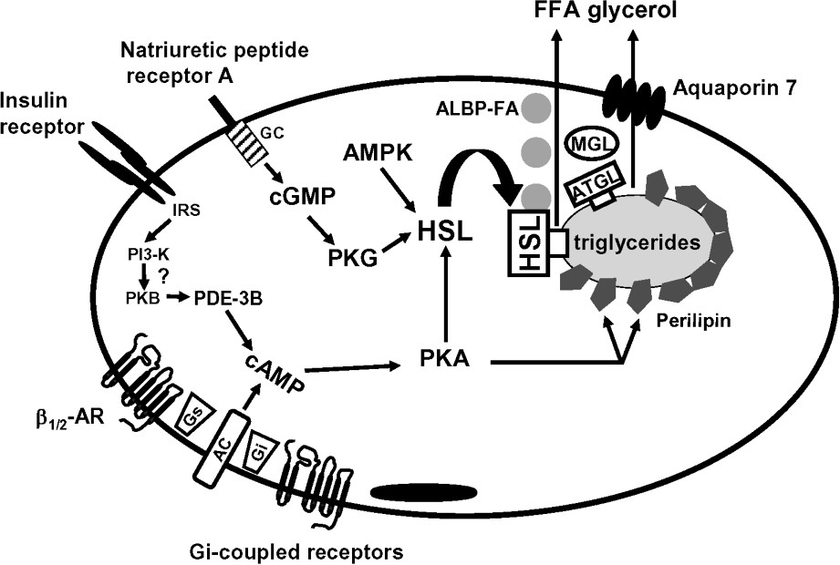
Schematic view of lipolysis in human adipose tissue. AC, adenylyl cyclase; ALBP, adipocyte lipid binding protein; AMPK, AMP-activated protein kinase; AR, adrenoceptor; ATGL, adipocyte triglyceride lipase; FFA, free fatty acid; GC, guanylyl cyclase; Gi, inhibitory GTP-binding protein; Gs, stimulatory GTP-binding protein; HSL, hormone-sensitive lipase; IRS, insulin receptor substrate; MGL, monoglyceride lipase; PDE-3B, phosphodiesterase 3B; PI3-K, phosphatidylinositol-3-phosphate kinase; PKA, protein kinase A; PKB, protein kinase B; PKG protein kinase G.

The reduced lipolysis commonly reported in subcutaneous WAT of obese subjects may involve defects at different steps of the transduction pathways. To date, the best characterized defect is the decreased expression of HSL. It is however still unclear whether altered lipolysis promotes the development and/or stabilization of obesity or is an early protective adaptation to limit excessive fatty acid release.

Acknowledgements

The author's work is supported by INSERM, the 6th framework Integrated Project Hepadip (Adipose tissue and liver in the metabolic syndrome), the French ‘Programme national de recherche en nutrition humaine’ and ‘Programme national de recherche sur le diabète’.


Bibliographie

[1] P. Mauriège; G.D. Pergola; M. Berlan; M. Lafontan Human fat cell beta-adrenergic receptors: beta agonist-dependent lipolytic responses and characterization of beta-adrenergic binding sites on human fat cell membranes with highly selective beta1-antagonists, J. Lipid Res., Volume 29 (1988), pp. 587-601

[2] P. Barbe; L. Millet; J. Galitzky; M. Lafontan; M. Berlan In situ assessment of the role of the β1-, β2- and β3-adrenoceptors in the control of lipolysis and nutritive blood flow in human subcutaneous adipose tissue, Br. J. Pharmacol., Volume 117 (1996), pp. 907-913

[3] B.B. Lowell; E.S. Bachman Beta-adrenergic receptors, diet-induced thermogenesis, and obesity, J. Biol. Chem., Volume 278 (2003), pp. 29385-29388

[4] J.R.S. Arch; S. Wilson Prospects for β3-adrenoceptor agonists in the treatment of obesity and diabetes, Int. J. Obesity, Volume 20 (1996), pp. 191-199

[5] G. Tavernier; P. Barbe; J. Galitzky; M. Berlan; D. Caput; M. Lafontan; D. Langin Expression of β3-adrenoceptors with low lipolytic action in human subcutaneous fat cells, J. Lipid Res., Volume 37 (1996), pp. 87-97

[6] D. Langin; M.-P. Portillo; J.-S. Saulnier-Blache; M. Lafontan Coexistence of three beta-adrenoceptor subtypes in white fat cells of various mammalian species, Eur. J. Pharmacol., Volume 199 (1991), pp. 291-301

[7] J. Galitzky; D. Langin; P. Verwaerde; J.-L. Montastruc; M. Lafontan; M. Berlan Lipolytic effects of conventional β3-adrenoceptor agonists and of CGP 12, 177 in rat and human fat cells: preliminary pharmacological evidence for a putative β4-adrenoceptor, Br. J. Pharmacol., Volume 122 (1997), pp. 1244-1250

[8] J. Galitzky; D. Langin; J.-L. Montastruc; M. Lafontan; M. Berlan On the presence of a putative fourth beta-adrenoceptor in human adipose tissue, Trends Pharmacol. Sci., Volume 19 (1998), pp. 164-166

[9] A. Konkar; Y. Zhai; J. Granneman β1-adrenergic receptors mediate β3-adrenergic-independent effects of CGP12177 in brown adipose tissue, Mol. Pharmacol., Volume 57 (2000), pp. 252-258

[10] C. Weyer; P.A. Tataranni; S. Snitker; E. Danforth; E. Ravussin Increase in insulin action and fat oxidation after treatment with CL 316, 243, a highly selective β3-adrenoceptor agonist in humans, Diabetes, Volume 47 (1998), pp. 1555-1561

[11] S.L.H. Schiffelers; E.E. Blaak; W.H.M. Saris; M.A. van Baak In vivo β3-adrenergic stimulation of human thermogenesis and lipid utilization, Clin. Pharmacol. Therap., Volume 67 (2000), pp. 558-566

[12] G. Tavernier; M. Gimenez; J.-P. Giacobino; N. Hulo; M. Lafontan; P. Muzzin; D. Langin Norepinephrine induces lipolysis in β1/β2/β3-adrenoceptor knockout mice, Mol. Pharmacol., Volume 68 (2005), pp. 793-799

[13] M.D. Jensen; M.W. Haymond; R.A. Rizza; P.E. Cryer; J.M. Miles Influence of body fat distribution on free fatty acid metabolism in obesity, J. Clin. Invest., Volume 83 (1989), pp. 1168-1173

[14] J.F. Horowitz; S. Klein Whole body and abdominal lipolytic sensitivity to epinephrine is suppressed in upper body obese women, Am. J. Physiol., Volume 278 (2000), p. E1144-E1152

[15] P. Bougnères; C.L. Stunff; C. Pecqueur; E. Pinglier; P. Adnot; D. Ricquier In vivo resistance of lipolysis to epinephrine. A new feature of childhood onset obesity, J. Clin. Invest., Volume 99 (1997), pp. 2568-2573

[16] S. Enoksson; M. Talbot; F. Rife; W.V. Tamborlane; R.S. Sherwin; S. Caprio Impaired in vivo stimulation of lipolysis in adipose tissue by selective β2-adrenergic agonist in obese adolescent girls, Diabetes, Volume 49 (2000), pp. 2149-2153

[17] S. Reynisdottir; K. Ellerfeldt; H. Wahrenberg; H. Lithell; P. Arner Multiple lipolysis defects in the insulin resistance (metabolic) syndrome, J. Clin. Invest., Volume 93 (1994), pp. 2590-2599

[18] M. Lafontan; M. Berlan Fat cell α2-adrenoceptors: the regulation of fat cell function and lipolysis, Endocrine Rev., Volume 16 (1995), pp. 716-738

[19] V. Stich; I. de Glisezinski; H. Suljkovicova; F. Crampes; J. Galitzky; D. Rivière; J. Hejnova; M. Lafontan; M. Berlan Activation of antilipolytic α2-adrenergic receptors by epinephrine during exercise in human adipose tissue, Am. J. Physiol., Volume 277 (1999), p. R1076-R1083

[20] V. Stich; I. de Glisezinski; F. Crampes; J. Hejnova; J.-M. Cottet-Emard; J. Galitzky; M. Lafontan; D. Rivière; M. Berlan Activation of α2-adrenergic receptors impairs exercise-induced lipolysis in SCAT of obese subjects, Am. J. Physiol., Volume 279 (2000), pp. 499-504

[21] P. Mauriège; J.-P. Després; D. Prud'homme; M.-C. Pouliot; M. Marcotte; A. Tremblay; C. Bouchard Regional variation in adipose tissue lipolysis in lean and obese men, J. Lipid Res., Volume 32 (1991), pp. 1625-1633

[22] C. Sengenès; M. Berlan; I. de Glisezinski; M. Lafontan; J. Galitzky Natriuretic peptides: a new lipolytic pathway in human adipocytes, FASEB J., Volume 14 (2000), pp. 1345-1351

[23] C. Sengenes; A. Zakaroff-Girard; A. Moulin; M. Berlan; A. Bouloumie; M. Lafontan; J. Galitzky Natriuretic peptide-dependent lipolysis in fat cells is a primate specificity, Am. J. Physiol. Regul. Integr. Comp. Physiol., Volume 283 (2002), p. R257-R265

[24] C. Sengenes; A. Bouloumie; H. Hauner; M. Berlan; R. Busse; M. Lafontan; J. Galitzky Involvement of a cGMP-dependent pathway in the natriuretic peptide-mediated hormone-sensitive lipase phosphorylation in human adipocytes, J. Biol. Chem., Volume 278 (2003), pp. 48617-48626

[25] J. Galitzky; C. Sengenes; C. Thalamas; M.-A. Marques; J.-M. Senard; M. Lafontan; M. Berlan The lipid-mobilizing effect of atrial natriuretic peptide is unrelated to sympathetic nervous system activation or obesity in young men, J. Lipid Res., Volume 42 (2001), pp. 536-544

[26] C. Moro; J. Galitzky; C. Sengenes; F. Crampes; M. Lafontan; M. Berlan Functional and pharmacological characterization of the natriuretic peptide-dependent lipolytic pathway in human fat cells, J. Pharmacol. Exp. Ther., Volume 308 (2004), pp. 984-992

[27] C. Moro; F. Crampes; C. Sengenes; I. de Glisezinski; J. Galitzky; C. Thalamas; M. Lafontan; M. Berlan Atrial natriuretic peptide contributes to physiological control of lipid mobilization in humans, FASEB J., Volume 18 (2004), pp. 908-910

[28] A.L. Birkenfeld; M. Boschmann; C. Moro; F. Adams; K. Heusser; G. Franke; M. Berlan; F.C. Luft; M. Lafontan; J. Jordan Lipid mobilization with physiological atrial natriuretic peptide concentrations in humans, J. Clin. Endocrinol. Metab., Volume 90 (2005), pp. 3622-3628

[29] M. Lafontan; C. Moro; C. Sengenes; J. Galitzky; F. Crampes; M. Berlan An unsuspected metabolic role for atrial natriuretic peptides: the control of lipolysis, lipid mobilization, and systemic nonesterified fatty acids levels in humans, Arterioscler. Thromb. Vasc. Biol., Volume 25 (2005), pp. 2032-2042

[30] C.H. Gravholt; O. Schmitz; L. Simonsen; J. Bulow; J.S. Christiansen; N. Moller Effects of a physiological GH pulse on interstitial glycerol in abdominal and femoral adipose tissue, Am. J. Physiol., Volume 277 (1999), p. E848-E854

[31] J.S. Samra; M.L. Clark; S.M. Humphreys; I.A. Macdonald; P.A. Bannister; D.R. Matthews; K.N. Frayn Suppression of the nocturnal rise in growth hormone reduces subsequent lipolysis in subcutaneous adipose tissue, Eur. J. Clin. Invest., Volume 29 (1999), pp. 1045-1052

[32] M.A. Heffernan; W.J. Jiang; A.W. Thorburn; F.M. Ng Effects of oral administration of a synthetic fragment of human growth hormone on lipid metabolism, Am. J. Physiol., Volume 279 (2000), p. E501-E507

[33] M.A. Heffernan; A.W. Thorburn; B. Fam; R. Summers; B. Conway-Campbell; M.J. Waters; F.M. Ng Increase of fat oxidation and weight loss in obese mice caused by chronic treatment with human growth hormone or a modified C-terminal fragment, Int. J. Obes. Relat. Metab. Disord., Volume 25 (2001), pp. 1442-1449

[34] H. Hauner; T. Petruschke; M. Russ; K. Röhrig; J. Eckel Effects of tumor necrosis factor alpha (TNFα) on glucose transport and lipid metabolism of newly differentiated human fat cells in culture, Diabetologia, Volume 38 (1995), pp. 764-771

[35] S.C. Souza; L.M. de Vargas; M.T. Yamamoto; P. Lien; M.D. Franciosa; L.G. Moss; A.S. Greenberg Overexpression of perilipin A and B blocks the ability of tumor necrosis factor-α to increase lipolysis in 3T3-L1 adipocytes, J. Biol. Chem., Volume 273 (1998), pp. 24665-24669

[36] S. Gasic; B. Tian; A. Green Tumor necrosis factor alpha stimulates lipolysis in adipocytes by decreasing Gi protein concentrations, J. Biol. Chem., Volume 274 (1999), pp. 6770-6775

[37] T. Rahn Landstrom; J. Mei; M. Karlsson; V. Manganiello; E. Degerman Down-regulation of cyclic-nucleotide phosphodiesterase 3B in 3T3-L1 adipocytes induced by tumour necrosis factor alpha and cAMP, Biochem. J., Volume 346 (2000), pp. 337-343

[38] J. Mei; L.S. Holst; T.R. Landstrom; C. Holm; D. Brindley; V. Manganiello; E. Degerman C2-ceramide influences the expression and insulin-mediated regulation of cyclic nucleotide phosphodiesterase 3B and lipolysis in 3T3-L1 adipocytes, Diabetes, Volume 51 (2002), pp. 631-637

[39] M. Ryden; A. Dicker; V.V. Harmelen; H. Hauner; M. Brunnberg; L. Perbeck; F. Lönnqvist; P. Arner Mapping of early signalling events in TNF-α-mediated lipolysis in human fat cells, J. Biol. Chem., Volume 277 (2002), pp. 1085-1091

[40] C. Marcus; H. Ehrén; P. Bolme; P. Arner Regulation of lipolysis during the neonatal period. Importance of thyrotropin, J. Clin. Invest., Volume 82 (1988), pp. 1793-1797

[41] T.K. Sinha; P. Thajchayapong; S.F. Queener; D.O. Allen; N.H. Bell On the lipolytic action of parathyroid hormone in man, Metabolism, Volume 25 (1976), pp. 251-260

[42] L. Akesson; B. Ahren; V.C. Manganiello; L.S. Holst; G. Edgren; E. Degerman Dual effects of pituitary adenylate cyclase-activating polypeptide and isoproterenol on lipid metabolism and signaling in primary rat adipocytes, Endocrinology, Volume 144 (2003), pp. 5293-5299

[43] L. Akesson; B. Ahren; G. Edgren; E. Degerman VPAC2-R mediates the lipolytic effects of pituitary adenylate cyclase-activating polypeptide/vasoactive intestinal polypeptide in primary rat adipocytes, Endocrinology, Volume 146 (2005), pp. 744-750

[44] C. Bing; Y. Bao; J. Jenkins; P. Sanders; M. Manieri; S. Cinti; M.J. Tisdale; P. Trayhurn Zinc-alpha2-glycoprotein, a lipid mobilizing factor, is expressed in adipocytes and is up-regulated in mice with cancer cachexia, Proc. Natl Acad. Sci. USA, Volume 101 (2004), pp. 2500-2505

[45] N. Gaudiot; A.-M. Jaubert; E. Charbonnier; D. Sabourault; D. Lacasa; Y. Giudicelli; C. Ribière Modulation of white adipose tissue lipolysis by nitric oxide, J. Biol. Chem., Volume 273 (1998), pp. 13475-13481

[46] K. Andersson; N. Gaudiot; C. Ribière; M. Elizalde; Y. Giudicelli; P. Arner A nitric oxide-mediated mechanism regulates lipolysis in human adipose tissue in vivo, Br. J. Pharmacol., Volume 126 (1999), pp. 1639-1645

[47] F. Karpe; K.N. Frayn The nicotinic acid receptor – a new mechanism for an old drug, Lancet, Volume 363 (2004), pp. 1892-1894

[48] S. Tunaru; J. Kero; A. Schaub; C. Wufka; A. Blaukat; K. Pfeffer; S. Offermanns PUMA-G and HM74 are receptors for nicotinic acid and mediate its anti-lipolytic effect, Nat. Med., Volume 9 (2003), pp. 352-355

[49] A. Wise; S.M. Foord; N.J. Fraser; A.A. Barnes; N. Elshourbagy; M. Eilert; D.M. Ignar; P.R. Murdock; K. Steplewski; A. Green; A.J. Brown; S.J. Dowell; P.G. Szekeres; D.G. Hassall; F.H. Marshall; S. Wilson; N.B. Pike Molecular identification of high- and low-affinity receptors for nicotinic acid, J. Biol. Chem., Volume 278 (2003), pp. 9869-9874

[50] Y. Zhang; R.J. Schmidt; P. Foxworthy; R. Emkey; J.K. Oler; T.H. Large; H. Wang; E.W. Su; M.K. Mosior; P.I. Eacho; G. Cao Niacin mediates lipolysis in adipose tissue through its G-protein coupled receptor HM74A, Biochem. Biophys. Res. Commun., Volume 334 (2005), pp. 729-732

[51] A.K. Taggart; J. Kero; X. Gan; T.Q. Cai; K. Cheng; M. Ippolito; N. Ren; R. Kaplan; K. Wu; T.J. Wu; L. Jin; C. Liaw; R. Chen; J. Richman; D. Connolly; S. Offermanns; S.D. Wright; M.G. Waters (d)-beta-Hydroxybutyrate inhibits adipocyte lipolysis via the nicotinic acid receptor PUMA-G, J. Biol. Chem., Volume 280 (2005), pp. 26649-26652

[52] K. Chatzipanteli; S. Rudolph; L. Axelrod Coordinate control of lipolysis by prostaglandin E2 and prostacyclin in rat adipose tissue, Diabetes, Volume 41 (1992), pp. 927-935

[53] J.D. Borglum; S.B. Pedersen; G. Ailhaud; R. Negrel; B. Richelsen Differential expression of prostaglandin receptor mRNAs during adipose cell differentiation, Prostaglandins Other Lipid Mediat., Volume 57 (1999), pp. 305-317

[54] H. Kather Purine accumulation in human fat cell suspensions, J. Biol. Chem., Volume 263 (1988), pp. 8803-8809

[55] H. Kather Pathways of purine metabolism in human adipocytes: Further evidence against a role of adenosine as an endogenous regulator of human fat cell function, J. Clin. Invest., Volume 265 (1990), pp. 96-102

[56] P. Lönnroth; P.-A. Jansson; B.B. Fredholm; U. Smith Microdialysis of intercellular adenosine concentration in subcutaneous adipose tissue in humans, Am. J. Physiol., Volume 256 (1989), p. E250-E255

[57] C. Schoelch; J. Kuhlmann; M. Gossel; G. Mueller; C. Neumann-Haefelin; U. Belz; J. Kalisch; G. Biemer-Daub; W. Kramer; H.P. Juretschke; A.W. Herling Characterization of adenosine-A1 receptor-mediated antilipolysis in rats by tissue microdialysis, 1H-spectroscopy, and glucose clamp studies, Diabetes, Volume 53 (2004), pp. 1920-1926

[58] P. Valet; M. Berlan; M. Beauville; F. Crampes; J.-L. Montastruc; M. Lafontan Neuropeptide Y and peptide YY inhibit lipolysis in human and dog fat cells through a pertussis toxin-sensitive G protein, J. Clin. Invest., Volume 85 (1990), pp. 291-295

[59] I. Castan; P. Valet; T. Voisin; N. Quideau; M. Laburthe; M. Lafontan Identification and functional studies of a specific peptide YY-preferring receptor in dog adipocytes, Endocrinology, Volume 131 (1992), pp. 1970-1976

[60] I. Castan; P. Valet; D. Larrouy; T. Voisin; A. Remaury; D. Daviaud; M. Laburthe; M. Lafontan Distribution of PYY receptors in human fat cells: an antilipolytic system alongside the alpha2-adrenergic one, Am. J. Physiol., Volume 265 (1993), p. E74-E80

[61] C. Serradeil-Le Gal; M. Lafontan; D. Raufaste; J. Marchand; B. Pou-zet; P. Casellas; M. Pascal; J.-P. Maffrand; G. Le Fur Characterization of NPY receptors controlling lipolysis and leptin secretion in human adipocytes, FEBS Lett., Volume 475 (2000), pp. 150-156

[62] L.C. Turtzo; R. Marx; M.D. Lane Cross-talk between sympathetic neurons and adipocytes in coculture, Proc. Natl Acad. Sci. USA, Volume 98 (2001), pp. 12385-12390

[63] K.N. Frayn Adipose tissue as a buffer for daily lipid flux, Diabetologia, Volume 45 (2002), pp. 1201-1210

[64] E. Degerman; T.R. Landstrom; J. Wijkander; L.S. Holst; F. Ahmad; P. Belfrage; V. Manganiello Phosphorylation and activation of hormone-sensitive adipocyte phosphodiesterase Type 3B, Methods, Volume 14 (1998), pp. 43-53

[65] J. Wijkander; T.R. Landstrom; V. Manganiello; P. Belfrage; E. Degerman Insulin-induced phosphorylation and activation of phosphodiesterase 3B in rat adipocytes: possible role for protein kinase B but not mitogen-activated protein kinase or p70 S6 kinase, Endocrinology, Volume 139 (1998), pp. 219-227

[66] R. Harmancey; J.-M. Senard; A. Pathak; F. Desmoulin; C. Claparols; P. Rouet; F. Smih The vasoactive peptide adrenomedullin is secreted by adipocytes and inhibits lipolysis through NO-mediated beta-adrenergic agonist oxidation, FASEB J., Volume 19 (2005), pp. 1045-1047

[67] J.E. Sullivan; K.J. Brocklehurst; A.E. Marley; F. Carey; D. Carling; R.K. Beri Inhibition of lipolysis and lipogenesis in isolated rat adipocytes with AICAR, a cell-permeable activator of AMP-activated protein kinase, FEBS Lett., Volume 353 (1994), pp. 33-36

[68] M. Daval; F. Diot-Dupuy; R. Bazin; I. Hainault; B. Viollet; S. Vaulont; E. Hajduch; P. Ferre; F. Foufelle Anti-lipolytic action of AMP-activated protein kinase in rodent adipocytes, J. Biol. Chem., Volume 280 (2005), pp. 25250-25257

[69] A.J. Garton; D.G. Campbell; D. Carling; D.G. Hardie; R.J. Colbran; S.J. Yeaman Phosphorylation of bovine hormone-sensitive lipase by the AMP-activated protein kinase. A possible antilipolytic mechanism, Eur. J. Biochem., Volume 179 (1989), pp. 249-254

[70] J. Osuga; S. Ishibashi; T. Oka; H. Yagyu; R. Tozawa; A. Fujimoto; F. Shionoiri; N. Yahagi; F.B. Kraemer; O. Tsutsumi; N. Yamada Targeted disruption of hormone-sensitive lipase results in male sterility and adipocyte hypertrophy, but not in obesity, Proc. Natl Acad. Sci. USA, Volume 97 (2000), pp. 787-792

[71] G. Haemmerle; R. Zimmermann; J.G. Strauss; D. Kratky; M. Riederer; G. Knipping; R. Zechner Hormone-sensitive lipase deficiency in mice changes the plasma lipid profile by affecting the tissue-specific expression pattern of lipoprotein lipase in adipose tissue and muscle, J. Biol. Chem., Volume 277 (2002), pp. 12946-12952

[72] H. Mulder; M. Sorhede-Winzell; J.A. Contreras; M. Fex; K. Strom; T. Ploug; H. Galbo; P. Arner; C. Lundberg; F. Sundler; B. Ahren; C. Holm Hormone-sensitive lipase null mice exhibit signs of impaired insulin sensitivity whereas insulin secretion is intact, J. Biol. Chem., Volume 278 (2003), pp. 36380-36388

[73] K. Harada; W.-J. Shen; S. Patel; V. Natu; J. Wang; J.-I. Osuga; S. Ishibashi; F.B. Kraemer Resistance to high-fat diet-induced obesity and altered expression of adipose-specific genes in HSL-deficient mice, Am. J. Physiol. Endocrinol. Metab., Volume 285 (2003), p. E1182-E1195

[74] R. Zimmermann; J.G. Strauss; G. Haemmerle; G. Schoiswohl; R. Birner-Gruenberger; M. Riederer; A. Lass; G. Neuberger; F. Eisenhaber; A. Hermetter; R. Zechner Fat mobilization in adipose tissue is promoted by adipose triglyceride lipase, Science, Volume 306 (2004), pp. 1383-1386

[75] C.M. Jenkins; D.J. Mancuso; W. Yan; H.F. Sims; B. Gibson; R.W. Gross Identification, cloning, expression, and purification of three novel human calcium-independent phospholipase A2 family members possessing triacylglycerol lipase and acylglycerol transacylase activities, J. Biol. Chem., Volume 279 (2004), pp. 48968-48975

[76] J.A. Villena; S. Roy; E. Sarkadi-Nagy; K.-H. Kim; H.S. Sul Desnutrin, an adipocyte gene encoding a novel patatin domain-containing protein, is induced by fasting and glucocorticoids: ectopic expression of desnutrin increases triglyceride hydrolysis, J. Biol. Chem., Volume 279 (2004), pp. 47066-47075

[77] D. Langin; A. Dicker; G. Tavernier; J. Hoffstedt; A. Mairal; M. Ryden; E. Arner; A. Sicard; C.M. Jenkins; N. Viguerie; V. Van Harmelen; R.W. Gross; C. Holm; P. Arner Adipocyte lipases and defect of lipolysis in human obesity, Diabetes, Volume 54 (2005), pp. 3190-3197

[78] G. Fredrikson; P. Strålfors; N.Ö. Nilsson; P. Belfrage Hormone-sensitive lipase of rat adipose tissue. Purification and some properties, J. Biol. Chem., Volume 256 (1981), pp. 6311-6320

[79] G. Haemmerle; R. Zimmermann; M. Hayn; C. Theussl; G. Waeg; E. Wagner; W. Sattler; T.M. Magin; E.F. Wagner; R. Zechner Hormone-sensitive lipase deficiency in mice causes diglyceride accumulation in adipose tissue, muscle, and testis, J. Biol. Chem., Volume 277 (2002), pp. 4806-4815

[80] G. Fredrikson; H. Tornqvist; P. Belfrage Hormone-sensitive lipase and monoacylglycerol lipase are both required for complete degradation of adipocyte triacylglycerol, Biochim. Biophys. Acta, Volume 876 (1986), pp. 288-293

[81] P. Belfrage; G. Fredrikson; P. Strålfors; H. Tornquist Adipose tissue lipases (B. Borgström; H. Brockman, eds.), Lipases, Elsevier, Amsterdam, Netherland, 1984, pp. 366-416

[82] D. Langin; S. Lucas; M. Lafontan Millenium fat cell lipolysis reveals unsuspected novel tracks, Horm. Metab. Res., Volume 32 (2000), pp. 443-452

[83] P. Strålfors; P. Björgell; P. Belfrage Hormonal regulation of hormone-sensitive lipase in intact adipocytes: identification of phosphorylated sites and effects on the phosphorylation by lipolytic hormones and insulin, Proc. Natl Acad. Sci. USA, Volume 81 (1984), pp. 3317-3321

[84] J.J. Egan; A.S. Greenberg; M.-K. Chang; S.A. Wek; J.M.C. Moos; C. Londos Mechanism of hormone-stimulated lipolysis in adipocytes: Translocation of hormone-sensitive lipase to the lipid storage droplet, Proc. Natl Acad. Sci. USA, Volume 89 (1992), pp. 8537-8541

[85] K.G. Soni; R. Lehner; P. Metalnikov; P. O'Donnell; M. Semache; W. Gao; K. Ashman; A.V. Pshezhetsky; G.A. Mitchell Carboxylesterase 3 (EC 3.1.1.1) is a major adipocyte lipase, J. Biol. Chem., Volume 279 (2004), pp. 40683-40689

[86] V. Large; S. Reynisdottir; D. Langin; K. Fredby; M. Klannemark; C. Holm; P. Arner Decreased expression and function of adipocyte hormone-sensitive lipase in subcutaneous fat cells of obese subjects, J. Lipid Res., Volume 40 (1999), pp. 2059-2066

[87] L. Hellström; D. Langin; S. Reynisdottir; M. Dauzats; P. Arner Adipocyte lipolysis in normal weight subjects with obesity among first-degree relatives, Diabetologia, Volume 39 (1996), pp. 921-928

[88] V. Large; P. Arner; S. Reynisdottir; J. Grober; V. van Harmelen; C. Holm; D. Langin Hormone-sensitive lipase expression and activity in relation to lipolysis in human fat cells, J. Lipid Res., Volume 39 (1998), pp. 1688-1695

[89] W.J. Shen; K. Sridhar; D.A. Bernlohr; F.B. Kraemer Interaction of rat hormone-sensitive lipase with adipocyte lipid-binding protein, Proc. Natl Acad. Sci. USA, Volume 96 (1999), pp. 5528-5532

[90] A.J. Smith; M.A. Sanders; B.R. Thompson; C. Londos; F.B. Kraemer; D.A. Bernlohr Physical association between the adipocyte fatty acid-binding protein and hormone-sensitive lipase: a fluorescence resonance energy transfer analysis, J. Biol. Chem., Volume 279 (2004), pp. 52399-52405

[91] N.R. Coe; M.A. Simpson; D.A. Bernlohr Targeted disruption of the adipocyte lipid-binding protein (aP2 protein) gene impairs fat cell lipolysis and increases cellular fatty acid levels, J. Lipid Res., Volume 40 (1999), pp. 967-972

[92] L. Scheja; L. Makowski; K.T. Uysal; S.M. Wiesbrock; D.R. Shimshek; D.S. Meyers; M. Morgan; R.A. Parker; G.S. Hotamisligil Altered insulin secretion associated with reduced lipolytic efficiency in aP2–/– mice, Diabetes, Volume 48 (1999), pp. 1987-1994

[93] R.A. Baar; C.S. Dingfelder; L.A. Smith; D.A. Bernlohr; C. Wu; A.J. Lange; E.J. Parks Investigation of in vivo fatty acid metabolism in AFABP/aP2(–/–) mice, Am. J. Physiol. Endocrinol. Metab., Volume 288 (2005), p. E187-E193

[94] C. Londos; C. Sztalryd; J.T. Tansey; A.R. Kimmel Role of PAT proteins in lipid metabolism, Biochimie, Volume 87 (2005), pp. 45-49

[95] J. Martinez-Botas; J.B. Anderson; D. Tessier; A. Lapillonne; B.H. Chang; M.J. Quast; D. Gorenstein; K.H. Chen; L. Chan Absence of perilipin results in leanness and reverses obesity in Leprdb/db mice, Nat. Genet., Volume 26 (2000), pp. 474-479

[96] J.T. Tansey; C. Sztalryd; J. Gruia-Gray; D.L. Roush; J.V. Zee; O. Gavrilova; M.L. Reitman; C.-X. Deng; C. Li; A.R. Kimmel; C. Londos Perilipin ablation results in a lean mouse with aberrant adipocyte lipolysis, enhanced leptin production, and resistance to diet-induced obesity, Proc. Natl Acad. Sci., Volume 98 (2001), pp. 6494-6499

[97] C. Sztalryd; G. Xu; H. Dorward; J.T. Tansey; J.A. Contreras; A.R. Kimmel; C. Londos Perilipin A is essential for the translocation of hormone-sensitive lipase during lipolytic activation, J. Cell Biol., Volume 161 (2003), pp. 1093-1103

[98] D.L. Brasaemle; G. Dolios; L. Shapiro; R. Wang Proteomicanalysis of proteins associated with lipid droplets of basal and lipolytically stimulated 3T3-L1 adipocytes, J. Biol. Chem., Volume 279 (2004), pp. 46835-46842

[99] J.A. Hamilton; F. Kamp How are free fatty acids transported in membranes? Is it by proteins or by free diffusion through the lipids?, Diabetes, Volume 48 (1999), pp. 2255-2269

[100] N. Maeda; T. Funahashi; T. Hibuse; A. Nagasawa; K. Kishida; H. Kuriyama; T. Nakamura; S. Kihara; I. Shimomura; Y. Matsuzawa Adaptation to fasting by glycerol transport through aquaporin 7 in adipose tissue, Proc. Natl Acad. Sci. USA, Volume 101 (2004), pp. 17801-17806

[101] M. Hara-Chikuma; E. Sohara; T. Rai; M. Ikawa; M. Okabe; S. Sasaki; S. Uchida; A.S. Verkman Progressive adipocyte hypertrophy in aquaporin-7-deficient mice: adipocyte glycerol permeability as a novel regulator of fat accumulation, J. Biol. Chem., Volume 280 (2005), pp. 15493-15496

[102] T. Hibuse; N. Maeda; T. Funahashi; K. Yamamoto; A. Nagasawa; W. Mizunoya; K. Kishida; K. Inoue; H. Kuriyama; T. Nakamura; T. Fushiki; S. Kihara; I. Shimomura Aquaporin 7 deficiency is associated with development of obesity through activation of adipose glycerol kinase, Proc. Natl Acad. Sci. USA, Volume 102 (2005), pp. 10993-10998


Commentaires - Politique


Ces articles pourraient vous intéresser

Molecular genetics of human obesity: A comprehensive review

Rajan Kumar Singh; Permendra Kumar; Kulandaivelu Mahalingam

C. R. Biol (2017)


Fundamental mechanisms of thermogenesis

Daniel Ricquier

C. R. Biol (2006)


Lipase reaction at interfaces as self-limiting processes

Pedro Reis; Krister Holmberg; Reinhard Miller; ...

C. R. Chim (2009)