


 CC-BY 4.0
1. Introduction
Rapid urbanization and population growth generate significant amounts of food waste. This biowaste can contaminate and degrade the environment and ecosystems. Among food biowaste, citrus peels represent 50% to 60% of the fruit mass [1]. Therefore, it is crucial to find tailored options for the sustainable management of this biowaste. Accordingly, several technologies have been investigated. They include composting, anaerobic digestion, thermochemical conversion, and the extraction of biomaterials such as pectin [2]. The last method promotes the circular economy concept and also improves the sustainability of food production systems [3].
Pectin is a naturally occurring biopolymer that it is abundantly found in various plant cell walls such as lemon, orange, sugar beet, apple, etc. It is a heteropolysaccharide compound, formed of linear chains of α-(1 → 4)-linked D-galacturonic acid residues and combined with small quantities of neutral sugars like L-rhamnose, D-galactose, L-arabinose, and D-xylose [4]. The degree of esterification (DE) of pectin refers to the percentage of carboxyl groups esterified with methanol and determines its gelling and functional properties [5]. Pectins with high DE usually form rigid gels, while those with low DE form softer gels or act as thickening agents [6]. Additionally, the presence of acetyl groups can also significantly influence the gelling properties of pectins [7].
Due to its unique physicochemical properties, pectin has become the subject of numerous researches in several application fields [8], notably in the food, pharmaceutical and medical industries, material engineering, and environmental sciences [9, 10, 11, 12, 13, 14]. In this context, several methods have been tested for pectin extraction from plant sources. They mainly include the use of enzymes [15], microwaves [16], ultrasound [17], subcritical water [18], pressurized carbon dioxide or deionized water [19], and acids [20, 21]. The last method has been widely tested. It consists in treating the plant material with an acid to break down the cell walls and release pectin [22]. Then, the resulting mixture is filtered and pectin is usually precipitated by adding alcohol. Pectin extraction yield is dependent on both the plant peel type and acid used. For instance, in a comparative study, Yu et al. [23] studied pectin extraction efficiency from potato pulp using five different acids: nitric (HNO3), hydrochloric (HCl), sulfuric (H2SO4), acetic (C2H4O2), and citric (C6H8O7). They found that the highest pectin yield was observed when using the citric acid. It is important to underline that in another study [24], pectin extraction yield from unripe Cavendish banana peels by citric acid was evaluated at 11.52% with a pH of 1.5. It has been shown that the yield of pectins extracted from banana and mango peels under optimal conditions using citric acid at pH 2.0 was the highest (12.98%). In contrast, a lower yield (5.28%) was found for extraction with hydrochloric acid (pH 1.5). The yield and characteristics of acid-extracted pectin also depend on the extraction parameters used, such as contact time and temperature [25].
Currently, the increasing number of foodborne pathogenic bacteria such as Pseudomonas aeruginosa, E. coli, and Staphylococcus aureus has become a major public health problem, since it can cause serious foodborne diseases [26]. Therefore, it is crucial to develop natural, biodegradable, efficient, and renewable alternatives. Gao et al. [27] showed a good antibacterial activity of pectin against the growth of E. coli and S. aureus at minimum inhibitory concentrations (MICs) of 25.0 and 50.0 g/L, respectively. In addition, Di Rong et al. [28] investigated citrus pectin oligosaccharides on anti-adhesion activity against E. coli and showed that they could be used as a reliable antibacterial agent in functional foods. In addition, Tripathi et al. [29] evaluated the antibacterial and antioxidant activity of pectin obtained from banana peels. They found a maximum antibacterial activity against Staphylococcus aureus (19.6 mm in the well-diffusion method). Furthermore, their antioxidant activity study showed that pectin, up to a concentration of 75 μg/mL, increases the free radical scavenging activity of 2-2-diphenyl-1-picrylhydrazil (DPPH). On the other hand, Santana et al. [30] showed that sulfated pectin extracted from Citrus sinensis has interesting anticoagulant activity. The current study provides a new contribution to a better understanding of the antibacterial, antioxidant, and anticoagulant behavior of pectin obtained by a simple extraction from an abundant and renewable raw material (orange peel waste). The main aim of this work is to convert Algerian citrus peel waste into value-added biomaterial through pectin extraction using a mild organic acid (citric acid) and a strong mineral acid (hydrochloric acid) to assess their antibacterial, antioxidant, and anticoagulant effects. This approach will clarify the trade-off between extraction efficiency and structural integrity, and it would also address inconsistencies in prior works. Practically, the specific objectives are: (i) to study the role of the acid used on pectin extraction yield, (ii) to fully characterize the extracted pectins by various analytical techniques, and (iii) to assess the potential biomedical application of these pectins through the assessment of their antibacterial, antioxidant, and anticoagulant activities.
2. Materials and methods
2.1. Materials collection and preparation
The citrus fruit used consists in sweet oranges (Citrus sinensis). They were gathered from an agricultural farm located in the Sidi Matmar region in the northwestern part of Algeria. The orange peels were thoroughly washed with distilled water, then cut into small pieces, and subsequently dried at 50 °C for 24 h. Finally, the dried pieces were manually crushed and sieved. The bacteria used in this work, Pseudomonas aeruginosa (ATCC:27853), Escherichia coli (ATCC:25922), and Staphylococcus epidermidis (ATCC:12228) were provided by the Laboratory of Microbiology, Hassani Abdelkader Hospital, Sidi Bel Abbes, Algeria. They were kept at +4 °C and renewed every 24 h on nutrient agar at 37 °C.
2.2. Pectin extraction
Pectin extraction from orange peels was carried out with two different acids, citric and hydrochloric, as follows.
2.2.1. Extraction by citric acid
Pectin extraction by citric acid was performed according to the following steps [31]: (i) agitation of 40 g of dried orange peel waste in 1 L of a 0.1 N citric acid solution at a constant pH of 2 and temperature of 70 °C for 40 min, (ii) storing this suspension for 24 h at room temperature, (iii) recovery of the liquid phase by centrifugation at 6000 rpm for 10 min, (iv) mixing (1:2 v/v) this solution with 95% ethanol at 25 °C for 24 h to allow the precipitation and flotation of pectin, (v) filtration of the precipitated pectin using a Büchner funnel and washing twice with ethanol (70%), and (vi) drying in an oven at 65 °C for 24 h. The resulting product was named PCT-1.
2.2.2. Extraction by hydrochloric acid
Pectin extraction by hydrochloric acid was carried out through the boiling of 40 g of dried orange peels in 0.8 L of a 0.1 N HCl solution in a reflux system at 90 °C for 45 min. After 6 min, the suspension was placed on ice to stop the hydrolysis reaction. Then, it was filtered and the filtrate precipitated in ethanol. This filtrate was washed with 60%, 80%, and 98% ethanol, then centrifuged at 10 000 rpm for 20 min and dried at 50 °C for 2 h, then crushed. The obtained product was called PCT-2 and used in the experimental study below.
For both methods, the pectin extraction yield was evaluated on the basis of three parallel experiments and the mean values are presented in this work. The following equation was used for the calculus of these yields:
| \begin {equation}\label {eq1} \mathrm {Yield}~(\%)=\frac {\mbox {Obtained product mass}} {\mbox {Initial orange peel waste mass}}\times 100 \end {equation} | (1) |
2.3. Pectin characterization
2.3.1. Solubility
The solubility of the pectins in different solvents was assessed in batch mode. The assays consisted in stirring 0.05 g of pectin for 3 h in 10 mL of ethanol, petroleum ether, ketone, cyclohexane, dichloromethane, cold water, or hot water.
2.3.2. Humidity
The humidity (H) of the pectins was evaluated through the drying of 1 g of pectin at 105 °C for 24 h in a porcelain capsule. The humidity value is determined as follows:
| \begin {equation}\label {eq2} \mathrm {H}(\%)=\frac {m_{1}-m_{2}}{m_{1}} \end {equation} | (2) |
2.3.3. Organic matter and ash contents
The organic matter (OM) and ash (Cd) contents were assessed according to the protocol given by Soliman et al. [32]. It consists in placing a porcelain capsule containing 4 g of pectin in a muffle furnace (Biobase, China) at 550 ± 15 °C for 5 h until a light gray or whitish color is obtained. Then, the capsule is cooled in a desiccator until a constant weight. The OM content is calculated as follows:
| \begin {equation}\label {eq3} \mathrm {OM}~(\%)=\frac {m_{3}-m_{4}}{m_{3}}\times 100 \end {equation} | (3) |
| \begin {equation}\label {eq4} \mathrm {Cd}~(\%)=100-\mathrm {OM}~(\% ) \end {equation} | (4) |
2.3.4. Degree of esterification
The degree of esterification (DE) of the extracted pectins was assessed according to the following experimental protocol [33]: first, 0.2 g of the dried pectin is moistened with ethanol and dissolved in 20 mL of distilled water, then three drops of phenolphthalein are added to the sample and titrated by a 0.1 N NaOH solution. The result is recorded as the initial titration volume once the pink color appears and the number of free carboxyl groups can be deduced. After that, 10 ml of a 0.1 N NaOH solution is added to neutralize the polygalacturonic acid. The sample was capped with a cork and shaken vigorously for 5 h, then kept at room temperature for 2 h to permit the deesterification of the pectin. After that, 10 mL of a 0.1 N HCl solution was added to neutralize the excess of NaOH and the sample was shaken until its pink color disappears. Then, three drops of phenolphthalein were added to the sample and titrated with a 0.1 N NaOH solution. The titration volume was recorded as the final titration volume once a pink color appears, and the number of esterified carboxyl groups can be deduced. The DE of pectins is defined as the ratio of the esterified galacturonic acid to the galacturonic acid groups and calculated as follows [26]:
| \begin {eqnarray} &&\mathrm {DE}~(\%)\nonumber =\textstyle \frac {\mathrm {Final~titration~volume~(mL)}} {\mathrm {Initial~titration~volume~(mL)}\,+\,\mathrm {Final~titration~volume~(mL)}}\nonumber \times \, 100\label {eq5} \end {eqnarray} | (5) |
All the above parameters were evaluated through triplicate assays and the mean and standard variation were calculated using Excel 2016 software.
2.3.5. Morphology and structure
The morphology of the extracted PCT-1 and PCT-2 was determined through scanning electronic microscopy (SEM, brand: Carl Zeiss, model: Sigma 300 VP) coupled with an energy dispersive X-ray (EDX) detector capable of elemental surface analysis. Moreover, in order to study their crystalline structure, the pectins were examined using an X-ray diffraction (XRD) apparatus (Bruker model D8 ADVANCE). The diffraction data was recorded in a 2𝜃 range of 5° to 40° at a scan rate of 1°/min. The X’Pert High score program was used for the analysis. The International Center for Diffraction Data (ICDD) database was used to identify the crystalline peaks.
2.3.6. Surface chemistry
The richness in functional groups of the extracted pectins was assessed using a Fourier-transform infrared (FTIR) spectrometer (Bruker alpha-P) equipped with an ATR (attenuated total reflectance) diamond crystal. The infrared spectra of the pectin samples were obtained over a 400–4000 cm−1 wavenumber range.
2.3.7. Thermal properties
Thermal analysis of the extracted pectins was carried out using a differential scanning calorimetry (DSC) device (DSC-NETZSCH DSC-214 polyma). During this analysis, 10 mg of the sample was placed in the crucible and heated for 45 min at a rate of 10 °C/min.
2.4. Antibacterial activity
The antibacterial action of the extracted pectins was studied using the disk diffusion method and the bacteria Pseudomonas aeruginosa, Escherichia coli, and Staphylococcus epidermidis were chosen for this study [34]. For this purpose, pure colonies were collected, isolated, and stored at 4 °C. This method is particularly suitable for studying the action of antibiotics on the growth of bacteria; it allows determining their antibiograms (data not shown), which reflect the specific sensitivity of different bacterial species to given antibiotics. Petri dishes containing an already solidified suitable agar medium were inoculated with the tested microbial strain [34]. The antibiotic discs were then placed on the surface of the agar; the antibiotics used in these tests were: fusidic acid, vancomycin, spiramycin, ampicillin, oxacillin, norfloxacin, cefotaxime, doxycycline, and cefixime. The bacterial strains were stored at 4 °C, and these strains were renewed every 24 h on nutrient agar at 37 °C. The most commonly used medium for antibacterial susceptibility testing is Mueller Hinton Agar (MHA) [35]. Using a sterilized (Bunsen burner) Pasteur pipette, a few pure colonies were collected and isolated in a test tube containing 9 mL of saline solution and then thoroughly vortexed. The resulting bacterial solution was inoculated over the entire surface of the culture medium using a swab in each Petri dish.
Furthermore, the antibacterial action of PCT-1 and PCT-2 was assessed at different concentrations (5, 2.5, 1.66, and 1.25 mg/mL) against the selected bacteria on MHA culture medium, then incubated at 37 °C for 24 h. Whatman paper discs of diameter 6 mm were prepared and sterilized in the autoclave at 120 °C for 15 min. The discs were removed using sterilized forceps, and then soaked with the PCT solutions of each concentration for 30 s. Using sterile forceps, five discs at different concentrations were placed in each Petri dish containing a bacterium to be tested and then incubated at 37 °C in the oven for 24 h. The reading was carried out by measuring the diameter of the inhibition zone around the two tested pectins PCT-1 and PCT-2 [28].
2.5. Antioxidant activity
Unlike most free radicals, DPPH∙ is stable in solution; it cannot dimerize, due to steric hindrance around the nitrogen atom carrying the free electron. It exhibits a characteristic absorbance in the 512–517 nm range. The purple color disappears rapidly after reduction of DPPH to diphenylpicrylhydrazine by a compound with anti-radical properties, resulting in discoloration. The color intensity is proportional to the proton-donating capacity of antioxidants present in the medium [29]. The antiradical scavenging activity of the extracted pectins was studied by measuring the retention power of the DPPH∙ radical according to the protocol described by Qian et al. [36]. However, this radical can be reduced by a hydrogen transfer from the various antioxidants found in the reaction medium. Pectin (0.5, 1, 2, and 4 mg/mL) was mixed with DPPH (0.2 mmol/L) in ethanol (0.5 mL) and incubated at 37 °C for 35 min. After 2 h, the optical density (OD) of the samples was determined by a UV spectrophotometer (Shimadzu UV-2401PC) at a wavelength of 517 nm. These results were compared to those from blank tests prepared by replacing the sample solution with anhydrous ethanol. The antiradical power is deduced as follows:
| \begin {equation}\label {eq6} Y_{1}~(\%)=\frac {A_{\mathrm {control}}-A_{\mathrm {sample}}}{A_{\mathrm {control}}} \times 100 \end {equation} | (6) |
| \begin {equation*} \text {DPPH}\bullet +(\mathrm {AH})_{n}\rightarrow \text {DPPH}-\mathrm {H}+(\mathrm {AH})_{n-1}\mathrm {A}\bullet \end {equation*} |
2.6. Anticoagulant activity
The anticoagulant activity of the pectin samples was evaluated in vitro through the activated partial thromboplastin time (APTT) and prothrombin time (PT), with a saline solution as negative control and heparin sodium as positive control, according to the experimental protocol given by Souza et al. [38]. The APTT and PT of the pectins were measured through the analysis of 100 μL of citrated normal human plasma mixed with 50 μL of PCT-1 or PCT-2 incubated at 37 °C for 15 min by a specific automated device.
It is worth mentioning that antibacterial, antioxidant, and anticoagulant activities were assessed in triplicate. The mean values and the standard variation of the experimental data are given in the corresponding figures.
3. Results and discussion
3.1. Pectin extraction yields
The pectin extraction yields with citric acid (PCT-1) and hydrochloric acid (PCT-2) were determined according to the experimental protocol given in Section 2.2.2 at 6.3% and 4.7%, respectively (Table 1).
Main physicochemical parameters of the extracted pectins
| Biomaterials | Extraction process | Yield (%) | H (%) | Cd (%) | DE (%) |
|---|---|---|---|---|---|
| PCT-1 | Citric acid | 6.3 ± 0.21 | 4.4 ± 0.26 | 3.5 ± 0.32 | 40.0 ± 2.10 |
| PCT-2 | Hydrochloric acid | 4.7 ± 0.20 | 3.6 ± 0.21 | 4.6 ± 0.28 | 57.0 ± 3.56 |
The higher extraction yield observed with citric acid is mainly due to its larger extraction time (overnight). Moreover, the cleavage action of hydrochloric acid on pectin’s glycoside and ester bonds leads to a reduction in pectin extraction yield. A similar trend was observed by Chan et al. [39] when investigating pectin extraction by these same acids from cocoa husks. Our results are also in agreement with those reported by Nateghi et al. [40] who reported that citric acid has a greater pectin extraction ability than hydrochloric acid and sulfuric acid due to the chelating characteristic of citric acid. On the other hand, Maran et al. [41] obtained a higher pectin extraction yield (9.0%) when using citric acid assisted with ultrasound for pectin extraction from an industrial waste (Musa balbisiana). It is worth mentioning that pectin extraction yields are very dependent on the citrus variety studied. Comparable yields were reported in previous published works [42].
3.2. Characterization of pectins
3.2.1. Solubility, humidity, organic matter, and ash contents
Solubility tests showed that both extracted pectins are perfectly soluble in hot and cold water. This finding may be attributed to pectin’s richness in hydrophilic groups (i.e., hydroxyl OH, carboxyl COOH). Moreover, ionization of the carboxyl groups into negatively charged carboxylate ions can enhance pectin solubility in water through electrostatic interaction with water molecules. A similar outcome was reported for pectins extracted from sweet oranges (Citrus sinensis L) by citric acid [43]. However, PCT-1 and PCT-2 were completely insoluble in all the organic solvents tested (ethanol, petroleum ether, ketone, cyclohexane, dichloromethane). This result is in agreement with those reported by Würfel et al. [44]. Pure white powdered pectin dissolves in water with a 1:20 (w/v) ratio to form a negatively charged viscous colloidal solution, but it is insoluble in organic solvents such as ether and acetone. Generally, the solubility of pectin in water is linked to its long chains of galacturonic acid and richness in hydrophilic carboxyl and hydroxyl groups, which easily form hydrogen bonds with water. The longer the polygalacturonic acid chain, the lower the pectin solubility in water. As shown in Table 1, PCT-1 had a higher humidity than PCT-2. This finding can be explained by the characteristics of the chelator present in citric acid [43]. Moreover, the ash contents of PCT-1 and PCT-2 were evaluated at 3.5 ± 0.32% and 4.6 ± 0.28%, respectively. Akhtar et al. [45] investigated the extraction of pectin from orange and lime peels using hydrochloric, sulfuric, nitric, and citric acids. The pectin extracted from orange and lime peels had ash contents of 4.26% ± 0.12% and 2.41% ± 0.07%, respectively, which are consistent with our results. Mamiru et al. [46] found higher ash contents for pectins extracted from watermelon peels and suggested that ash content variation is primarily affected by both the feedstock type and the extraction conditions. Comparable values were reported for pectin extracted from orange peels using the acid extraction and a Box-Behnken design [47]. It is important to underline that the ash content of pectins is usually dependent on the mineral composition of the raw material. For instance, due to the high content in mineral salts and heavy metals in dragon fruit peels (Hylocereus polyrhizus), the ash contents of the related extracted pectins under three different extraction conditions were higher than our measured value (7% to 11%) [48].
3.2.2. Degree of esterification
The degree of esterification (DE) of pectin has significant implications for its functional properties in various applications, such as food and pharmaceuticals [49]. Pectins with a DE above 50% are designated high-methoxyl pectins (HMPs), while those with a DE under 50% are designated low methoxyl pectins (LMPs). Such a difference in the DE value is due to the distribution of methyl ester groups along the polygalacturonic acid chain, the molecular weight, and the type and amount of neutral sugars bound to the pectin molecules [50]. The DE of the extracted pectins is highly dependent on the extracting acid used. Indeed, the DE of PCT-2 extracted with hydrochloric acid was evaluated at 57%, which makes PCT-2 a HMP (Table 1). This value was obtained at an acidic pH of 3.3 with a short extraction time (45 min), conditions favorable to the preparation of sugar-rich products [51]. However, the DE of PCT-1 extracted using citric acid is much lower (40%) and indicates a LMP (Table 1). A similar trend was reported by Mansor et al. [52] who show that the DE of pectins depends on both the type of extracting acid and its interactions with the citrus matrix.
On the other hand, the reduction in carboxylate functions is responsible for the diminishing repulsive forces of the polysaccharides, which favors pectin gelation and results in more precipitated pectin at lower pH values. This finding is in agreement with those reported for pectins extracted from apple pomace and sugar beet pulp [49, 53]. It is important to mention that the DE highly influences the functional properties of pectins. Indeed, high DEs usually result in more rigid gels [54], whereas lower DEs lead to softer gels or even to pectin dissolution. This property is crucial in various food applications, such as in the production of jams, jellies, and fruit-based desserts, where the desired texture and stability depend on the DE of the pectin used [55]. Additionally, the DE affects the interactions of pectin with other molecules (e.g., sugars, proteins) which significantly impact the overall texture and stability of food products [54].
3.2.3. Morphology and structure
Figure 1 shows the SEM images of both PCT-1 and PCT-2. These images indicate that the small pectin particles exhibit the same rough and irregular morphological characteristics with no coherent porous surface. However, differences were observed when changing the image acquisition and resolution, as shown in Figure 1. On the other hand, these particles are able to disperse in solutions thus improving the solubility and intermolecular interactions through H-bonds and dipole formation [56].
SEM micrographs of PCT-1 (left) and PCT-2 (right).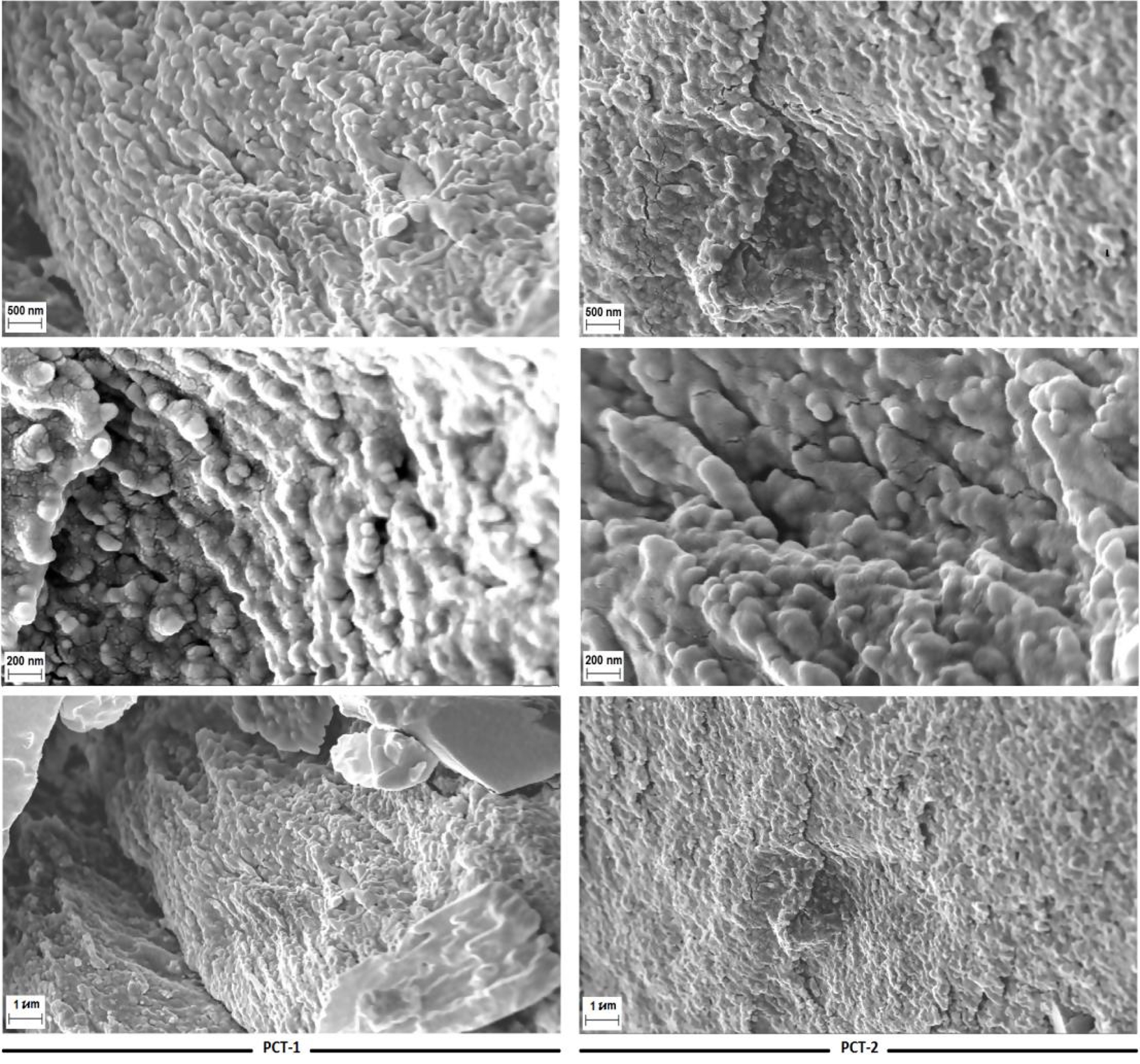
The XRD analyses show that PCT-1 and PCT-2 exhibit only a weak characteristic diffraction peak at 2𝜃 around 21° and another small peak at 2𝜃 of 6° for PCT-2, as shown in Figure 2. This result shows that both pectins have a semi-crystalline nature. This finding is in agreement with that reported for pectins extracted from apple waste [57].
XRD patterns of PCT-1 and PCT-2.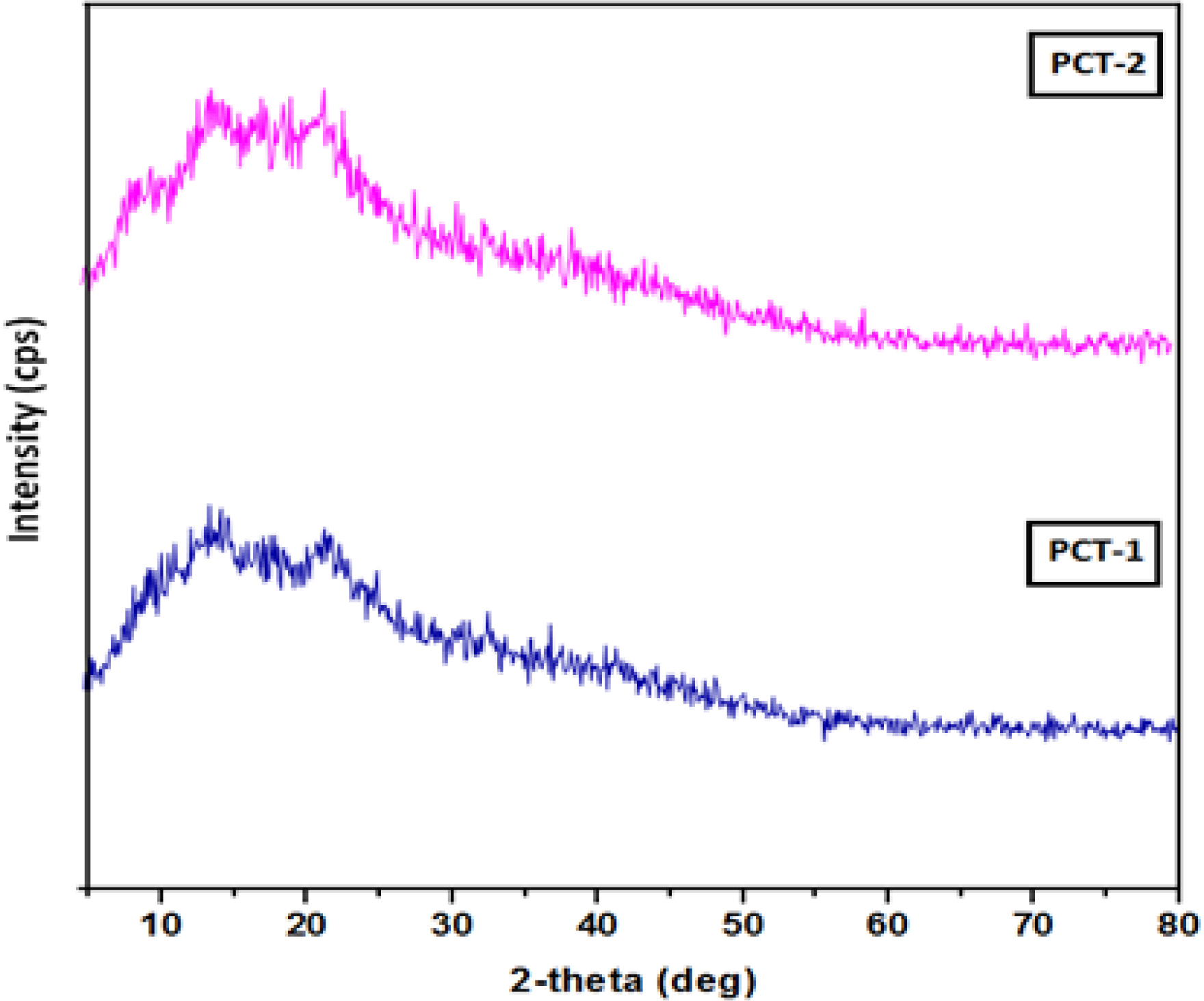
This can be attributed to the variation in their molecular weights due to the use of different extraction conditions. Similar observations have been reported by previous studies on pectin extracted from black carrot pomace, fruit peels, citrus fruits [56, 57, 58], sunflowers [59], and sweet lemon [60].
3.2.4. Surface chemistry
The FTIR spectra of both extracted pectins are shown in Figure 3. The wave number range of 850 and 1250 cm−1 corresponds to the “fingerprint” region of carbohydrates [61]. In this spectral region, and for both PCT-1 and PCT-2, the characteristic peaks observed at 920, 1016, 1090, and 1130 cm−1 confirm the typical profile of polygalacturonic acid [62].
FTIR spectra of PCT-1 and PCT-2.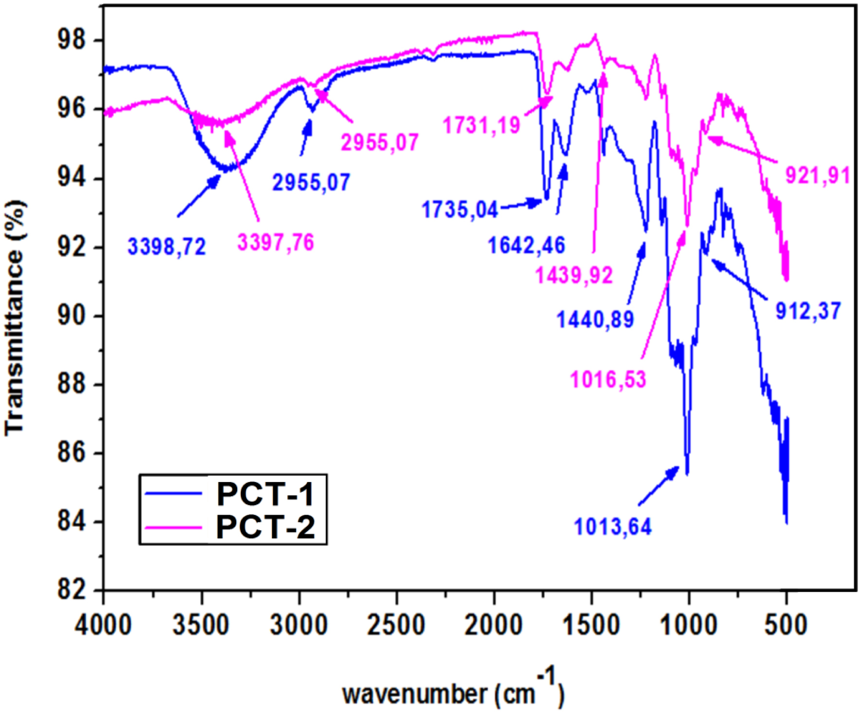
Moreover, other specific functional groups are present in both extracted pectins. They include a broad peak at 3700–3000 cm−1 corresponding to O–H stretching due to hydrogen bonds in galacturonic acid [62]. Peaks at 2954 and 2941 cm−1 for PCT-1 and PCT-2, respectively, can be attributed to C–H bond stretching [62]. Peaks at 1730 and 1620 cm−1 correspond to esterified and free carboxyls, respectively. Bands at 1100–1020 cm−1 can be imputed to C–O–C stretching vibrations and confirm the presence of pyranoses in the structure of both pectins. Peaks around 1000–1030 cm−1 correspond to the C–O stretching vibrations of ether or ester groups.
The asymmetric stretching observed around 1643–1626 cm−1 can be imputed to carbohydrate functions, and the bands in the 1300–800 cm−1 region correspond to the main carbohydrate chemical groups in galacturonic acid [62]. Finally, the peaks observed at 920–820 cm−1 refer to the absorption of D-glucopyranosyl and α-D-mannopyranose, respectively.
3.2.5. Thermal behavior
The thermograms (Figure 4) show endothermic peaks at 89.0 and 77.7 °C for PCT-1 and PCT-2, respectively. These peaks correspond to residual water retention due to existing hydrogen bonds between the galacturonic acid units [58]. Moreover, exothermic peaks are recorded at 260 and 253 °C for PCT-1 and PCT-2, respectively; they are related to the thermal degradation of polymers and pectin [58]. A close value (240 °C) was reported by Aldemir et al. [50] for pectin extracted from crab apple peels.
Differential scanning calorimetry thermograms of PCT-1 (left) and PCT-2 (right).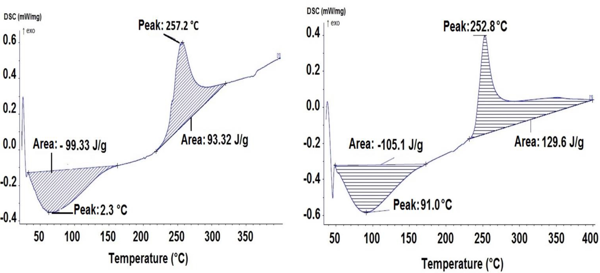
In addition, PCT-1 has a higher degradation temperature than PCT-2. This can be explained by the different operating conditions used during the two extraction processes. Moreover, PCT-1 exhibits greater changes than PCT-2 during the heating process (Figure 4). This finding indicates that PCT-1 has a higher thermal stability than PCT-2. Similar trends were observed by Ezzati et al. [59] and Rahmani et al. [60] when studying pectin extraction from sunflowers and sweet lemon, respectively.
3.2.6. Antibacterial action
The antibacterial action of PCT-1 and PCT-2 on Pseudomonas aeruginosa, E. coli, and Staphylococcus epidermidis has been assessed in vitro according to the experimental protocol given in Section 2.4. Results (data not shown) indicate that neither PCT-1 nor PCT-2 has an antibacterial effect on Pseudomonas aeruginosa or E. coli. Both pectins exhibit significant antibacterial potential against Staphylococcus epidermis. Indeed, for a content of 1 mg/mL, the zone of inhibition was evaluated at 11 and 9.5 mm for PCT-2 and PCT-1, respectively. This result highlights the effect of the acid extraction method used and of the pectin’s properties (humidity H, ash content Cd, and degree of esterification DE). The high resistance level of these Gram-negative bacteria to the extracted pectins can be attributed to the complexity of their cell envelope, which contains double membranes [63]. It is important to underline that antibacterial activity is determined not only by the number of free OH groups, but also by the overall structure of the molecule; molecular mass and monosaccharide composition can influence its ability to reach and interact with the bacterial membrane. In general, a lower DE, or a higher number of free hydroxyl groups, is associated with stronger antibacterial activity. This is because free OH groups can interact more easily with bacterial membranes, disrupting their structure and function, leading to increased antibacterial activity. They are more reactive and can interact with bacterial cellular components. Moreover, free OH groups can form hydrogen bonds with phospholipids and proteins in the bacterial cell membrane, increasing its permeability and leading to cell death [63, 64]. In contrast to that, a significant antibacterial effect was observed on the Staphylococcus epidermidis strain (Figure 5a). Indeed, PCT-1 and PCT-2 show a concentration-dependent inhibitory effect against Staphylococcus epidermidis strain (Figure 5a). Pectin suspensions of 1 mg/mL showed a significant reduction in the growth curve of Staphylococcus epidermidis compared to the lowest concentration group of 1/6 mg/mL. In contrast, neither PCT showed an antibacterial effect at 1/8 mg/mL. Likewise, Abdelgawad et al. [63] observed a concentration-dependent inhibitory effect of pectin against two bacterial strains (E. faecalis and F. nucleatum). This significant antibacterial activity can be explained by a higher sensitivity of Staphylococcus epidermidis, as a Gram-positive bacteria with a simple membrane structure, to the action of the pectins. Pectins easily act on their cell wall leading to a significant change in cell permeability [64].
(a) Illustration of the antibacterial action of PCT-1 and PCT-2 against Staphylococcus epidermidis, (b) DPPH radical scavenging activity of PCT-1 and PCT-2, (c) Anticoagulant activity of PCT-1 and PCT-2 fractions.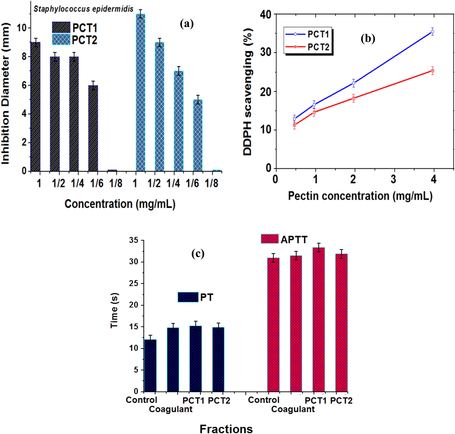
3.2.7. Antioxidant activity
The antioxidant activity of PCT-1 and PCT-2 was evaluated based on the experimental protocol given in Section 2.5. Results (Figure 5b) show that both pectins exhibit a significant DPPH∙ scavenging capacity. Moreover, this effect is highly dependent on the pectin concentration used (Figure 5b). Indeed, for an initial concentration of 0.5 mg/mL, the DPPH scavenging capacity was evaluated at 13.8% and 12.0% for PCT-1 and PCT-2, respectively. Increasing pectin concentration to 4 mg/L allowed this capacity to reach more than 36.3% and 26.0% for PCT-1 and PCT-2, respectively. This result is imputed to the presence of more free hydroxyl groups in the polysaccharide structure [64]. Moreover, PCT-1 has a better antioxidant activity than PCT-2 (Figure 5b). This may be linked to its lower DE (see Table 1). Indeed, polysaccharides with low DE values have more terminal reducing hydroxyl groups and more effectiveness in scavenging free radicals. Relatively high antioxidant capacities were also reported for pectins extracted using citric acid [65, 66]. It is worth mentioning that the antioxidant capacity of polysaccharides is mainly affected by electron- or hydrogen-donating abilities. The higher DPPH∙ scavenging capacity of PCT-1 compared to PCT-2 may be attributed to its higher total sugar content, making it the better hydrogen donor [65].
3.2.8. Anticoagulant activity
The anticoagulant activity of PCT-1 and PCT-2 was evaluated as described in Section 2.6. Experimental results (Figure 5c) show the PT values for PCT-1 (15.3 s) and PCT-2 (14.9 s) as well as the APTT values for PCT-1 (33.4 s) and PCT-2 (31.9 s).
These values are comparable to those of heparin (a sulfated polysaccharide of animal origin) indicating an interesting potential use for our pectins. Likewise, Chaouch et al. [78] developed two sulfated pectins. They showed that their extracted pectins exhibited a strong anticoagulant activity and could be considered as potential alternatives of heparin. Furthermore, Bae et al. assessed the anticoagulant activity of commercial pectin and found PTs and APTTs of 22–38 s and 10–14 s, respectively [79]. Further research is crucial to determine whether our pectin can be used safely and effectively as a substitute for heparin, particularly in the context of PT/APTT testing in normal plasma.
Table 2 gives a comparison of the antibacterial, antioxidant, and anticoagulant effects of our pectins with those of common polysaccharides from plants, animals, and marine algae.
Antibacterial, antioxidant, and anticoagulant activities of different polysaccharides compared with our pectins
| Origin | Antibacterial activity | Antioxidant activity | Anticoagulant effect | ||||||||
|---|---|---|---|---|---|---|---|---|---|---|---|
| Polysaccharides | Strain | Inhibition zone | Notes | Reference | DPPH∙ scavenging capacity | Notes | Reference | Effect | Notes | Reference | |
| Plant polysaccharides | Cellulose composites | S. epidermis | Variable | Often combined with chitosan oils | [67] | Very low | Increases only with added antioxidant compounds | [67] | None (native) | Modified cellulose sulfate shows antioxidant effect | [68] |
| Pectin | S. epidermis | Variable | Moderate activity alone | [67] | Moderate to higher depending on form | Increases with concentration | [69] | Weak | Slight prolongation PT/ATPP | [70] | |
| Animal polysaccharides | Chitosan composites | S. epidermis | Variable (often measurable) | Depends on formulation | [71] | High to medium | Higher when modified with phenolic compounds | [67] | Weak | Sulfated chitosan | [72] |
| Marine Algae | Alginate | S. epidermis | Low/None | Often combined | [67] | Low to moderate | Weak activity | [73] | Minimal | Sulfated alginate extracts | [74] |
| Agar | S. epidermis | No effect | No activity alone | [75] | Low | Enhanced in blends with antioxidants | [76] | Very limited | Not pure agar | [77] | |
| Algerian Citrus sinensis Peels | Pectin extracted | S. epidermis | Significant | Moderate | This study | Significant | Dependent on concentration | This study | Interesting | Comparable to a sulfated animal polysaccharide | This study |
4. Conclusion
The aim of this work is to turn abundant agricultural waste (local Algerian orange peels) into an effective biomaterial. Two pectins were extracted using citric (PCT-1) and hydrochloric acid (PCT-2). Their thorough characterization shows that both pectins have interesting thermal and physicochemical properties. PCT-1 is more stable. It showed a better antibacterial activity with a concentration-dependent inhibitory effect against Staphylococcus epidermidis but had no antibacterial effect on Pseudomonas aeruginosa or E. coli. PCT-1 also exhibits greater antioxidant activity than PCT-2. The prothrombin and activated partial thromboplastin time values of PCT-1 (15.3 and 33.4 s) and PCT-2 (14.9 and 31.9 s) demonstrated that the extracted pectins could be considered as a potential alternative to heparin. Future work will be undertaken to optimize the extraction process using other methods (i.e., microwave, ultrasound), to evaluate the antimicrobial capacity of the extracted pectins on other pathogenic bacteria, and the antioxidant and anticoagulant activities under a wider range of experimental conditions.
Acknowledgments
The authors would like to express their gratitude to the Directorate General of Scientific Research and Technological Development (DGRSDT), Laboratory of Microbiology, Hassani Abdelkader Hospital, Sidi Bel Abbes and the Ministry of Higher Education and Scientific Research (MESRS), Algeria, for their support to this research work.
Declaration of interests
The authors do not work for, advise, own shares in, or receive funds from any organization that could benefit from this article, and have declared no affiliation other than their research organizations.