


 CC-BY 4.0
1. Introduction
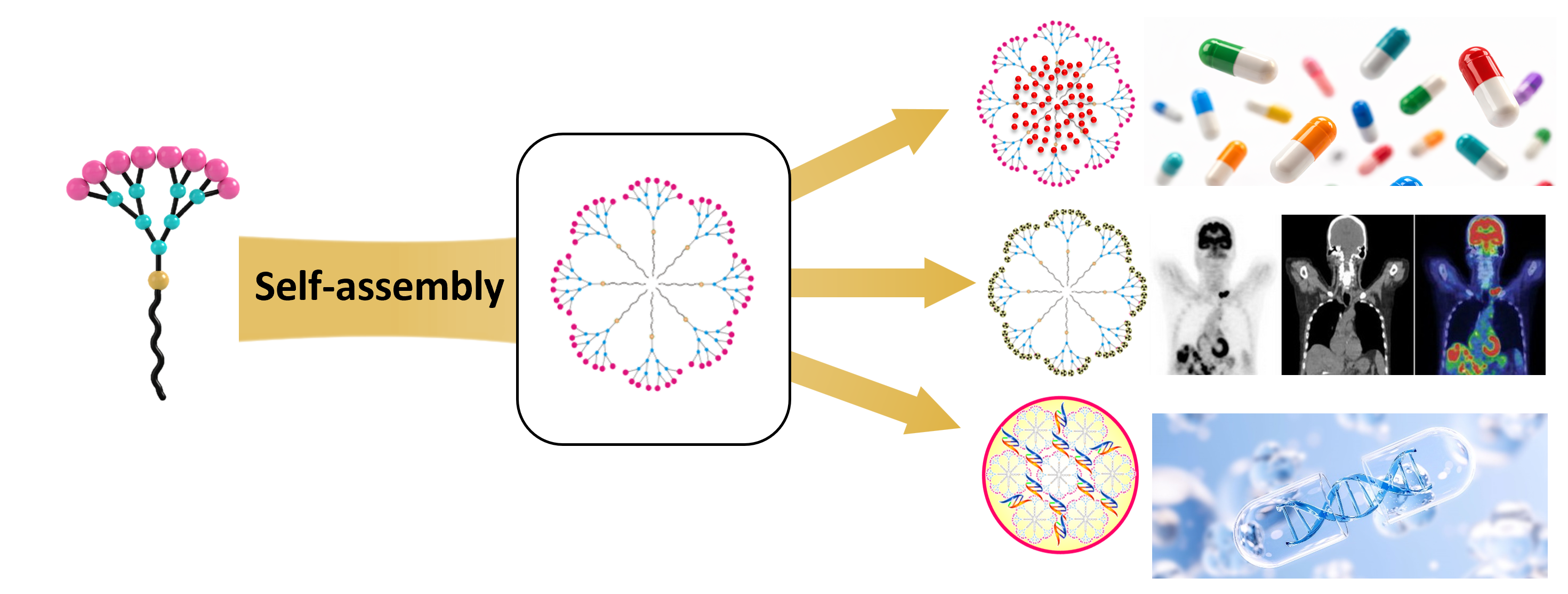
Dendrimers are a unique class of structurally well-defined synthetic molecules with precisely controlled architectures [1, 2, 3, 4, 5]. They consist of three distinct structural components: (1) a central core from which the dendrimer emanates; (2) repetitive branching units that enable growth into geometrically organized radial layers, referred to as generations; and (3) numerous terminal functional groups on the periphery (Figure 1) [6]. The dendritic architecture, together with their precisely controlled structures, distinguishes dendrimers from other synthetic macromolecules and polymers. As a result, dendrimers exhibit unique multivalent cooperativity confined within a small and well-defined three-dimensional space, leading to distinct functional properties. Consequently, dendrimers are attractive precision materials for a wide range of applications, including sensors, devices, catalysis, diagnostics, therapeutics, and delivery systems, spanning areas from environmental protection and energy production to human health [3, 4, 7, 8].
Schematic illustration of a conventional covalent dendrimer, which is composed of three structural components: core (orange), branching units (blue) and terminals (pink). Starting from the core, increasing branching generates dendrimers with increasing generations G1, G2, G3, etc. Adapted with permission from [6] (copyright 2018 Science China Press and Springer Nature).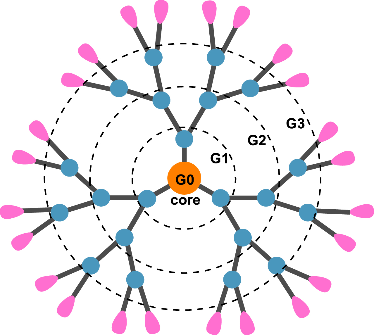
Dendrimer synthesis is inherently unique and technically demanding. Usually, dendrimers are constructed through stepwise synthesis, which can be achieved using either the divergent strategy from a central core to the periphery, or the convergent strategy from the periphery toward a central core, or the combination of both strategies, a hybrid divergent–convergent approach, also known as the double-stage convergent strategy [1].
Recently, we have pioneered the concept of supramolecular dendrimer construction via the self-assembly of small amphiphilic dendritic building blocks through non-covalent interactions (Figure 2) [9, 10, 11]. This approach relies on the spontaneous organization of small amphiphilic dendritic building blocks into large, non-covalent supramolecular dendrimers. In contrast to conventional covalent dendrimer synthesis, self-assembly into supramolecular dendrimers is readily achieved through reversible, yet highly specific and controllable interactions, requiring significantly reduced synthetic effort. This strategy enables a simplified and cost-effective route to dendrimers with entirely new and tunable properties [10, 11].
Schematic illustration of a self-assembling supramolecular dendrimer constructed using a small amphiphilic dendrimer. Adapted with permission from [9] (copyright 2016 Wiley-VCH Verlag GmbH & Co. KGaA, Weinheim).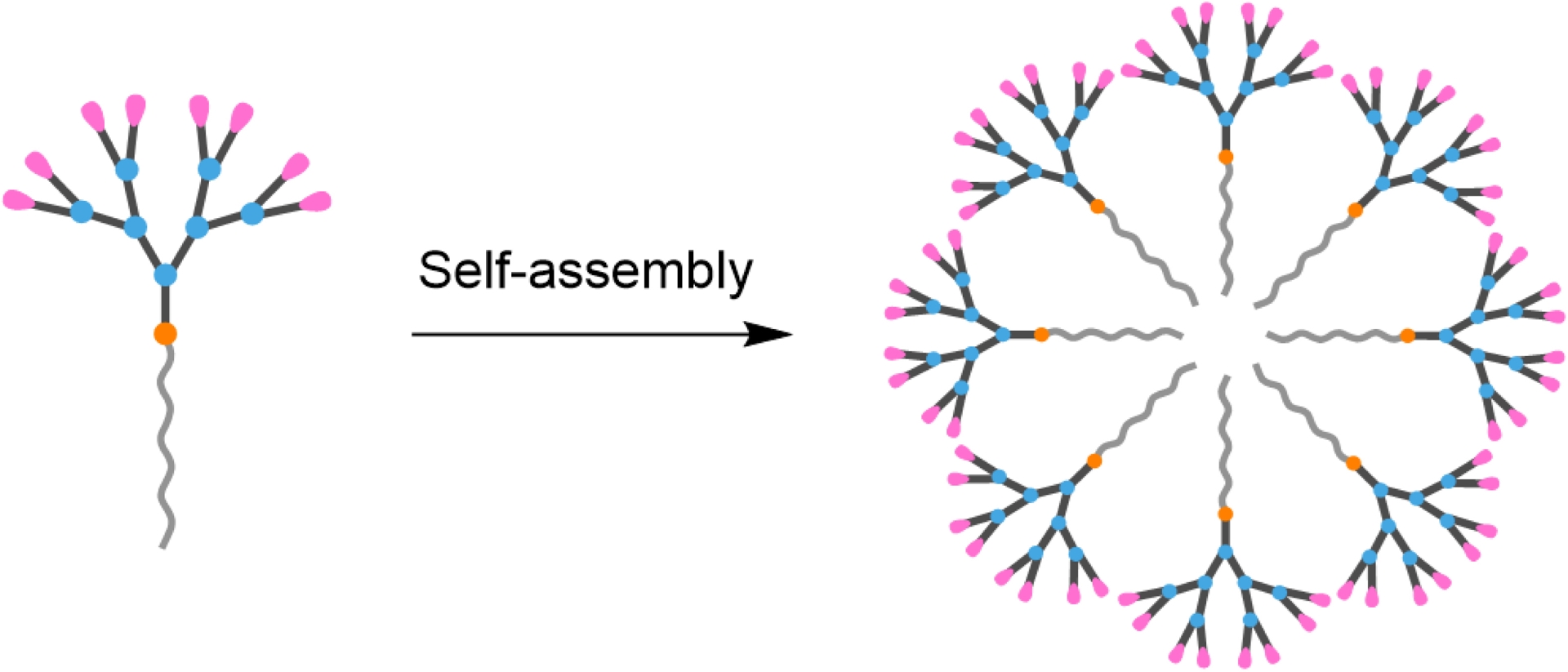
Importantly, the self-assembling supramolecular dendrimers can mimic covalent dendrimers not only in terms of their radial dendritic architecture emanating from a central core, but also in their suitability for biomedical applications [11]. In particular, the self-assembled supramolecular dendrimers are modular and adaptive for drug delivery (Figure 3). Notably, the dendritic structure of the amphiphilic building units enables the formation of large internal void spaces within the assembled supramolecular dendrimers for physical encapsulation of pharmaceutical agents, while the abundant surface functionalities allow both physical and chemical conjugation of biologically active entities for diverse biomedical applications.
Schematic illustration of self-assembling supramolecular dendrimers as delivery vehicles for pharmaceutical agents in biomedical applications. Reproduced from [11], licensed under CC BY-NC-ND 4.0.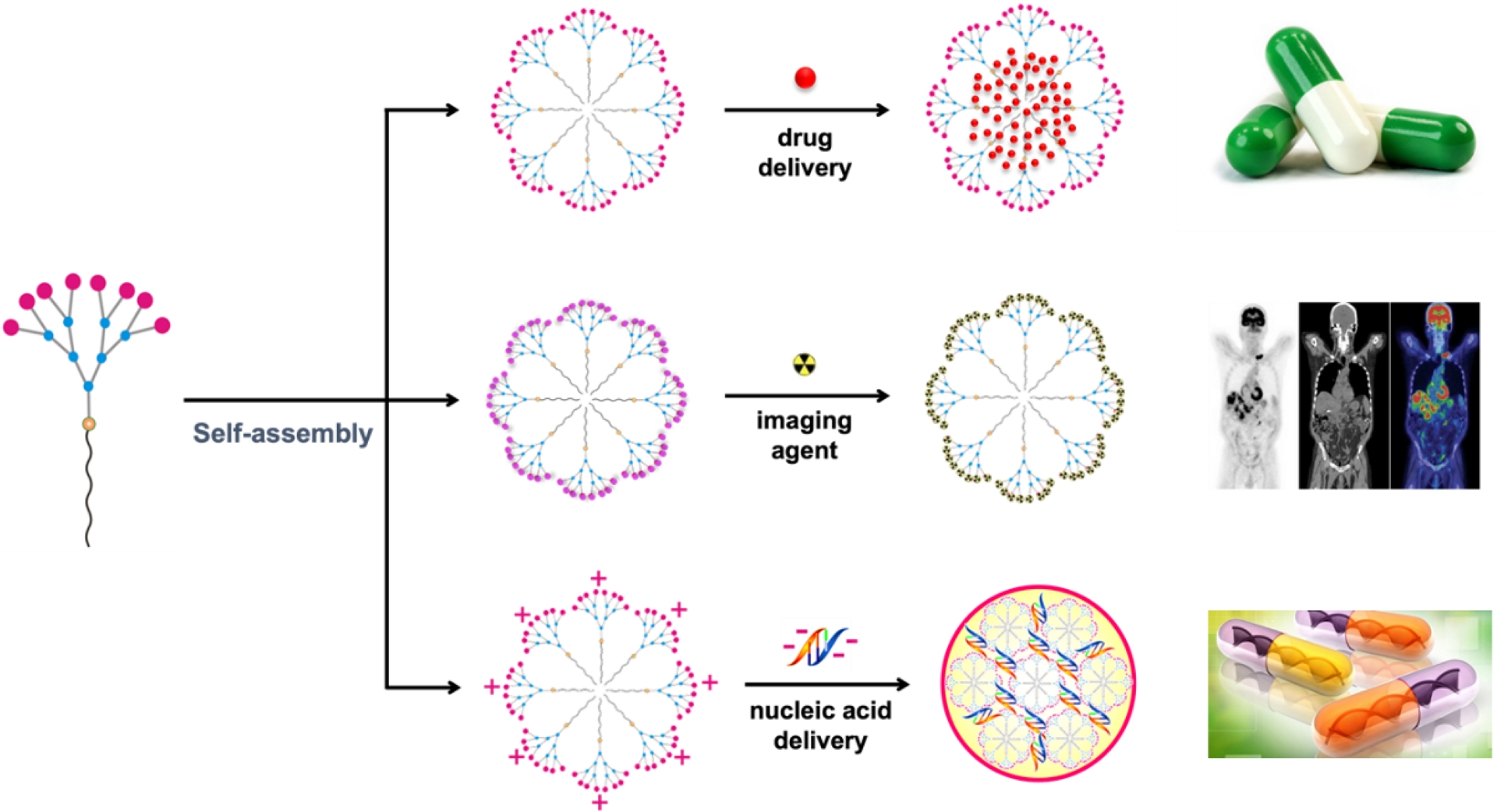
In this short Account, we provide a brief overview of our efforts over the past decade to develop self-assembling supramolecular dendrimers for biomedical applications in drug delivery, bioimaging and gene therapy (Figure 3) by highlighting representative studies. We will then outline future perspectives in this emerging field.
2. Drug delivery
Drug delivery aims to deliver the therapeutic agent to the desired site, at the correct dose, and for an optimal duration, thereby maximizing therapeutic efficacy while minimizing adverse effects [12, 13]. Supramolecular dendrimers formed through the self-assembly of small amphiphilic dendritic building blocks offer distinct advantages for this purpose. In particular, these supramolecular systems possess large internal void spaces within their cores, hence enable efficient encapsulation of drug molecules at high loading capacities, outperforming lipid- or polymer-based drug delivery systems [11].
In our very first study, we explored the use of amphiphilic dendrimer 1 (Figure 4A) to construct a supramolecular dendrimer nanosystem for the delivery of the anticancer drug doxorubicin (DOX) [14]. The spacious internal cavities of the resulting supramolecular dendrimers enabled stable DOX encapsulation (Figure 4B) with high drug-loading efficiency of up to 40%. The drug-loaded dendrimer nanomicelles DOX/1@, though small in size (12 nm), were bigger than the empty dendrimer nanomicelles 1@ (Figure 4B). This size expansion upon drug encapsulation can be ascribed to the accommodation of drug molecules within the micellar core. Following systemic administration, DOX-loaded nanomicelles preferentially accumulated in tumors (Figure 4C) via the enhanced permeability and retention (EPR) effect and were internalized by cancer cells predominantly through macropinocytosis (Figure 4D). This uptake pathway enhanced intracellular drug enrichment while circumventing efflux-mediated drug resistance (Figure 4D). Consequently, the supramolecular dendrimer formulation significantly improved the therapeutic efficacy of DOX, overcame doxorubicin resistance in multiple cancer models—including drug-resistant breast tumors—and substantially reduced the systemic toxicity associated with free DOX. This work pioneered supramolecular dendrimers as a promising platform for anticancer drug delivery.
(A) Chemical structure of amphiphilic dendrimer 1. (B) Anticancer drug doxorubicin (DOX) encapsulated within the nanomicelles formed by 1, and (C) accumulated in tumor. (D) Schematic illustration of cellular uptake of the DOX/1@ nanoparticles via macropinocytosis, overcoming efflux pump-mediated drug resistance. Adapted with from [14], licensed under CC BY 4.0.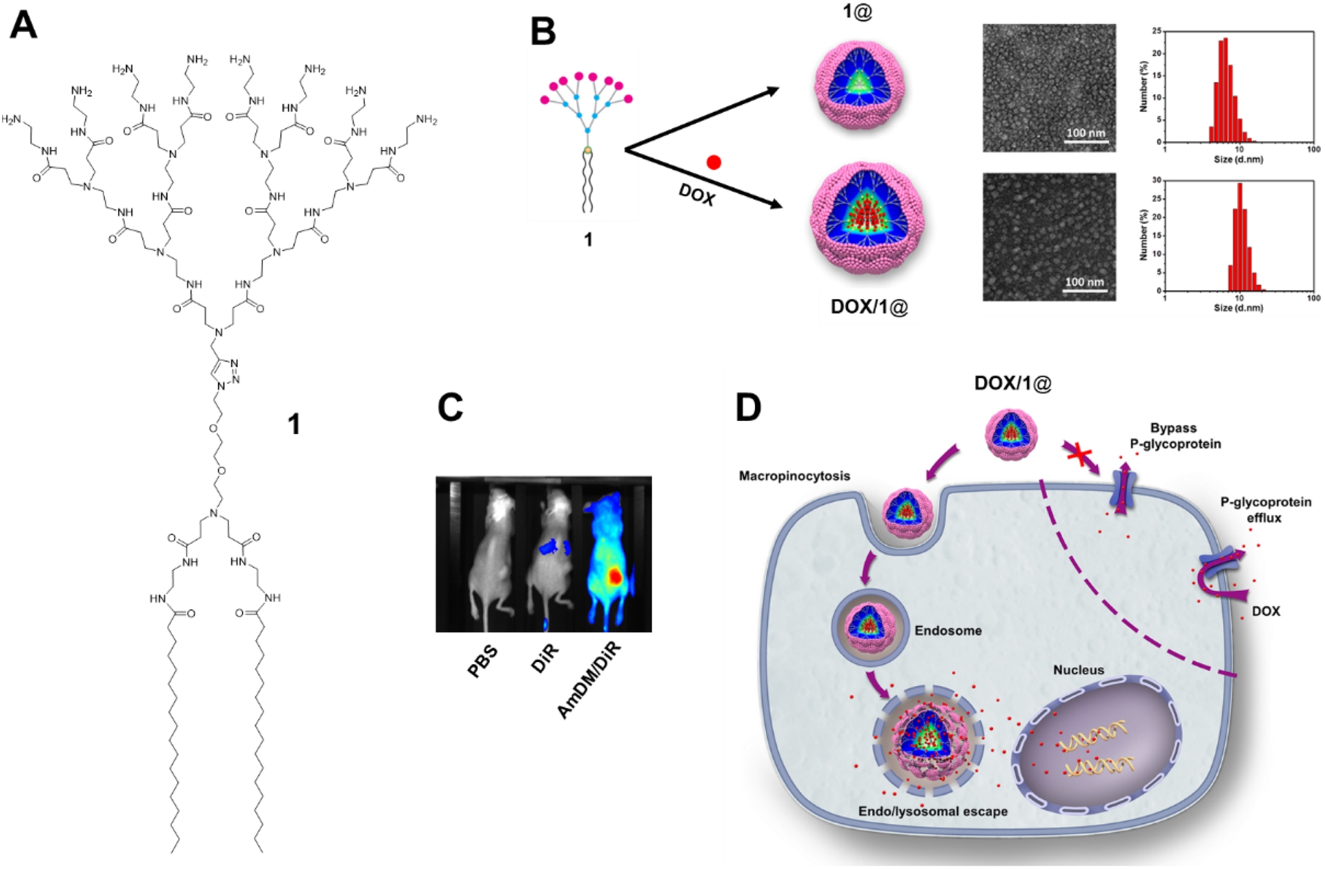
Despite the high drug-loading capacity, the nanomicelles formed by dendrimer 1 exhibited relatively slow drug release, with only approximately 40% of the encapsulated DOX released after 72 h [14]. This behavior can be ascribed to the presence of two long alkyl chains in dendrimer 1, which promote the formation of highly stable and compact nanomicelles that impede effective drug release. To address this limitation, we studied amphiphilic dendrimer 2 (Figure 5A), which contains a single alkyl chain and was expected to form less compact and stable assemblies based on its hydrophilicity–hydrophobicity balance [15]. Indeed, nanomicelles derived from 2 displayed substantially improved release profiles, with nearly 80% drug release after 72 h [15], while maintaining high drug-loading efficiencies (29–39%) and excellent encapsulation efficiencies (85–99%) for multiple anticancer agents, including doxorubicin, paclitaxel, and rapamycin [16]. Most importantly, we discovered that nanomicelles formed by 2 were able to hijack tumor-secreted extracellular vesicles (EVs) in vivo, enabling intercellular drug delivery and deep tumor penetration (Figure 5B) [16]. EVs are endogenous vesicles secreted by cells and play key roles in intercellular communication while retaining the characteristics of their parental cells [17]. In tumors, EVs are often overproduced [18]. By exploiting this intrinsic and dynamic transport network, dendrimer-based nanomicelles achieved efficient penetration into deep tumor tissue, overcame tumor heterogeneity, and enhanced anticancer effect [16].
(A) Chemical structure of amphiphilic dendrimer 2. (B) Schematic illustration of the anticancer drug encapsulated within the nanomicelle formed by 2, administrated via intravenous injection to tumor-xenograft mouse, accumulated within tumor via EPR effect, and delivered intercellularly via hijacking of extracellular vesicles (EVs) generated in situ by cancer cells for deep penetration into tumor tissue, achieving effective treatment while overcoming tumor heterogeneity and dynamic evolution. Reproduced from [16], licensed under CC BY-NC-ND 4.0.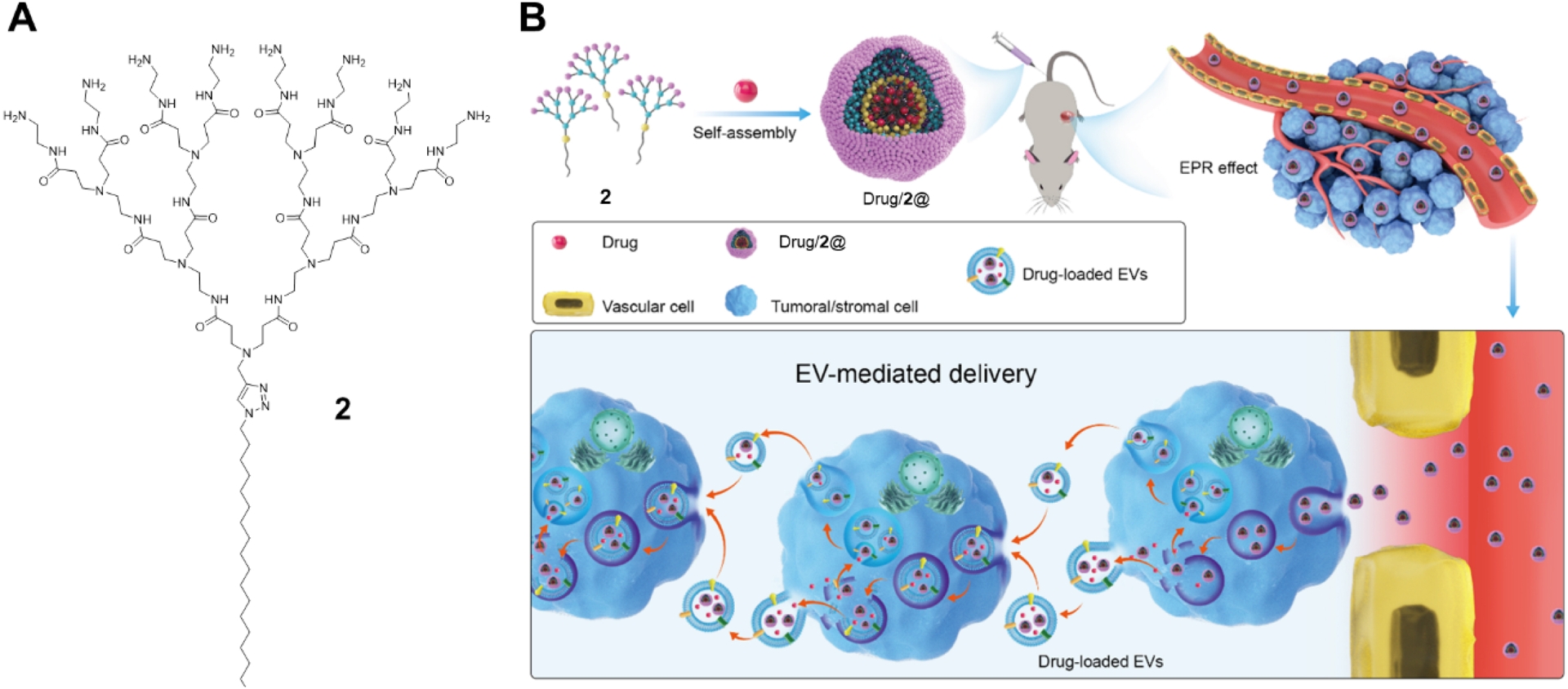
Further investigation revealed a drawback of 2-based nanodrug formulation: although it exhibited favorable drug release under acidic conditions (pH 5.0), mimicking the tumor microenvironment and endosomes, it also showed appreciable drug leakage (∼30%) at physiological pH (7.4), raising concerns about premature release during systemic circulation [15]. To mitigate this issue, we designed fluorinated dendrimer 3 (Figure 6A), which preserved high drug-loading capacity and acid-triggered release while reducing drug leakage at pH 7.4 to below 10% (Figure 6B) [15]. In vivo studies demonstrated that DOX-loaded nanomicelles derived from 3 were markedly more effective at suppressing tumor growth in patient-derived xenograft models [15]. These findings underscore the importance of molecular-level dendrimer engineering in tuning drug encapsulation and release profiles.
(A) Chemical structure of fluorinated amphiphilic dendrimer 3. (B) Time- and pH-dependent drug release profile of the anticancer drug doxorubicin (DOX) from the nanodrug formed by 3 (DOX/3@) at pH 7.4 and pH 5.0, respectively. Adapted from [15], licensed under CC BY 4.0.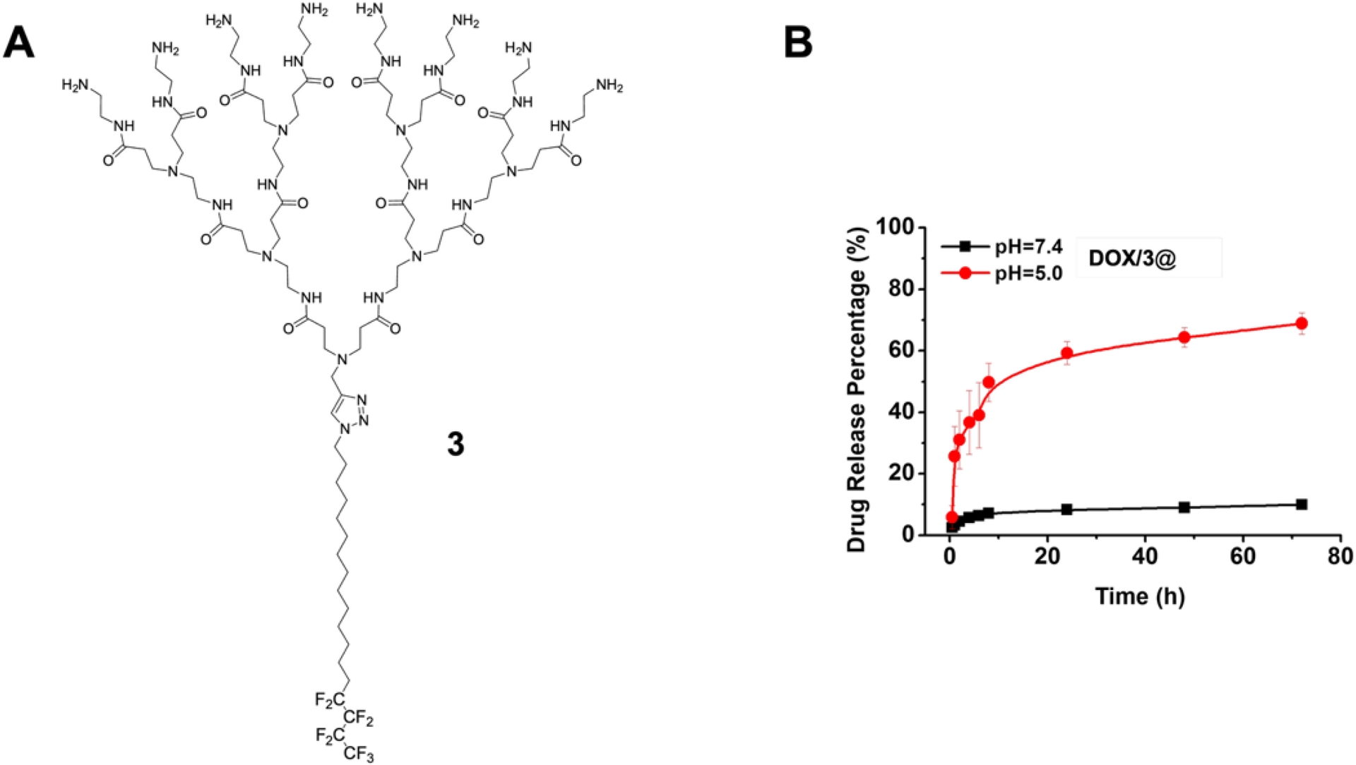
We also extended supramolecular dendrimers for drug delivery to address cardiotoxicity associated with hERG channel binding, a major cause of drug attrition and market withdrawal [19, 20]. In a proof-of-concept study, self-assembling dendrimer nanosystems formed from dendrimers 2, 4, and 5 (Figure 7) were used to formulate three structurally distinct drugs: the anticancer drug doxorubicin (DOX), the antimalarial drug chloroquine (CQ), and the NUPR1 inhibitor ZZW115, respectively [21]. Encapsulation within dendrimer nanomicelles reduced hERG binding affinities by three- to eight-fold relative to the free drugs (Figure 7B), translating into the elimination of the related toxicity in animal models. Additionally, these formulations prolonged systemic circulation, enhanced accumulation at disease sites, and improved therapeutic efficacy against both malaria infection and tumor burden [21]. This study illustrates the potential of dendrimer-based nanodrug formulation to mitigate hERG-related cardiotoxicity while simultaneously enhancing drug performance.
(A) Chemical structures of amphiphilic dendrimers 4 and 5. (B) Dendrimer nanodrug formulation reduced hERG binding affinities compared to the free drugs. Adapted from [21], licensed under CC BY-NC 4.0.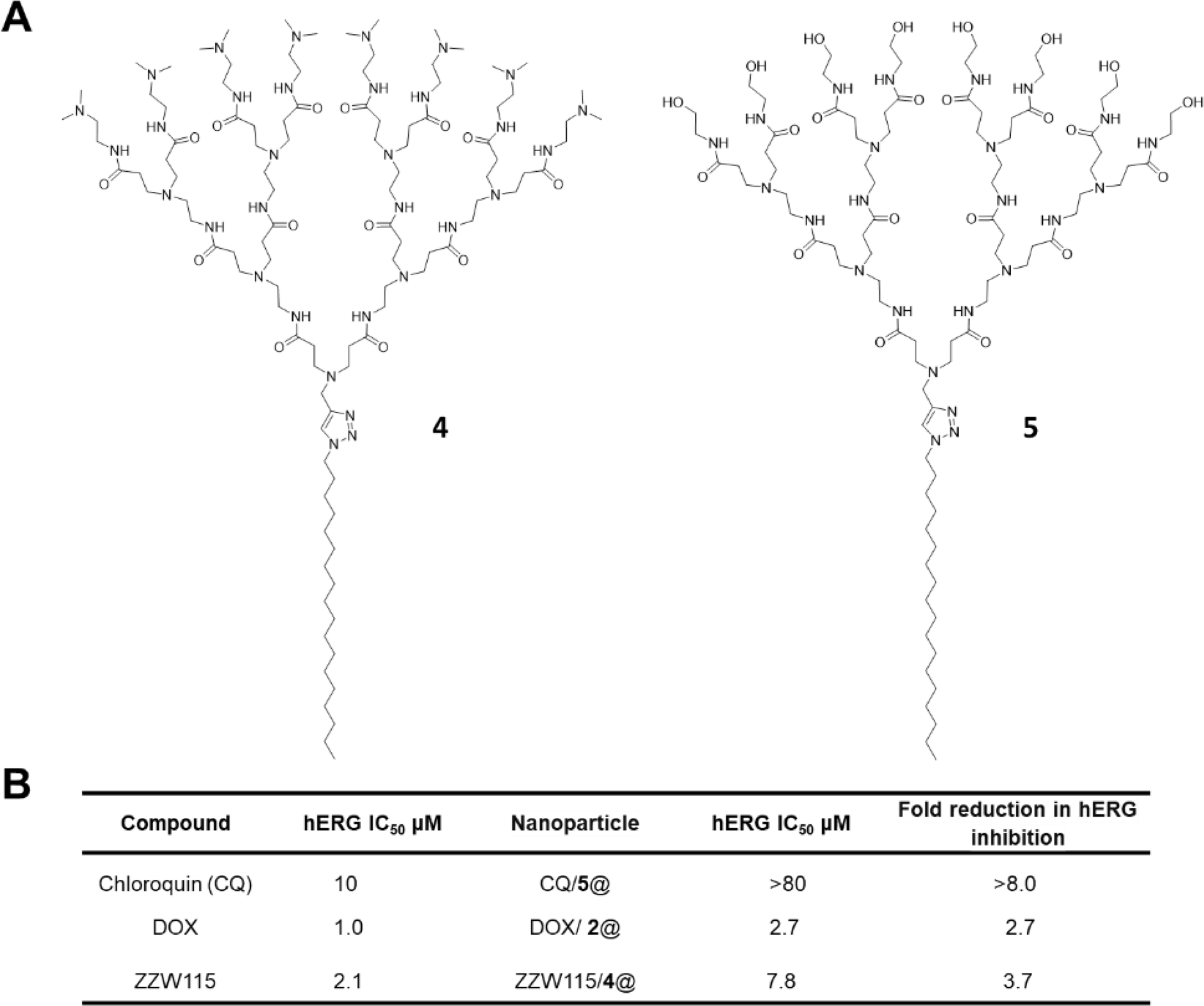
We further extended our design framework to bipolar(bola)-amphiphilic dendrimer 6 (Figure 8) capable of ROS (reactive oxygen species)–responsive delivery because of the presence of the thioacetal functionality [22]. Dendrimer 6 was employed to encapsulate the anticancer drug imatinib (IM) to target CD117 (c-Kit) [23], a key mediator of ovarian cancer stem cells (CSCs), for the treatment of metastatic ovarian cancer (Figure 8). The resulting formulation achieved significantly enhanced CSC targeting compared to free IM, leading to robust inhibition of cancer cell survival, stemness, and metastatic potential in metastatic and drug-resistant ovarian cancer models. Moreover, encapsulated IM exhibited synergistic anticancer activity when combined with the first-line chemotherapeutic agent cisplatin in patient-derived xenograft models, without observable adverse effects. Collectively, these findings established IM delivered by 6 as a versatile platform that potentiates imatinib for targeted and effective treatment of metastatic ovarian cancer, representing a meaningful advance toward addressing this unmet clinical need.
(A) Chemical structure of bola-amphiphilic dendrimer 6. (B) Nanodrug formulation formed by imatinib (IM) with 6 (IM/6@) for targeting cancer stem cells in metastatic and drug-resistant ovarian cancer models. Reproduced from [23], licensed under CC BY-NC-ND 4.0.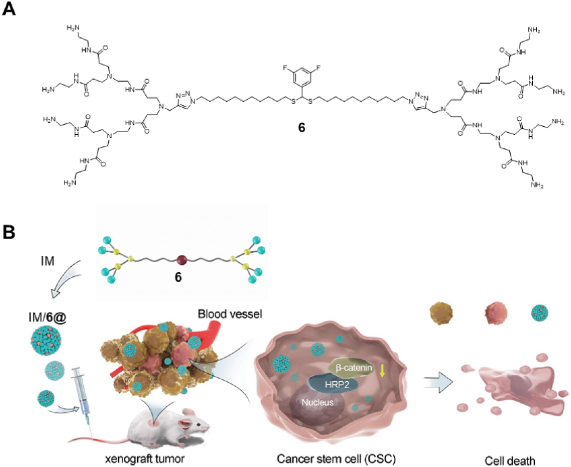
Beyond physical encapsulation, chemical conjugation of drug molecules to amphiphilic dendrimer terminals offers an alternative strategy for controlled drug delivery. In a pilot study, a prodrug of gemcitabine was covalently conjugated to the amphiphilic dendrimer surface (7 and 8, Figure 9) via click chemistry to enhance metabolic stability of gemcitabine while achieving high drug loading (40%) [24]. Although the higher-generation dendrimer 8 was insoluble in water, the lower-generation dendrimer 7 self-assembled into nanomicelles that were readily internalized by cells via endocytosis, bypassing nucleoside transporter–mediated uptake and thereby overcoming gemcitabine resistance. In addition, these nanomicelles exhibited pH- and enzyme-responsive drug release, resulting in enhanced anticancer efficacy and reduced systemic toxicity [24].
Chemical structures of amphiphilic dendrimers 7 and 8.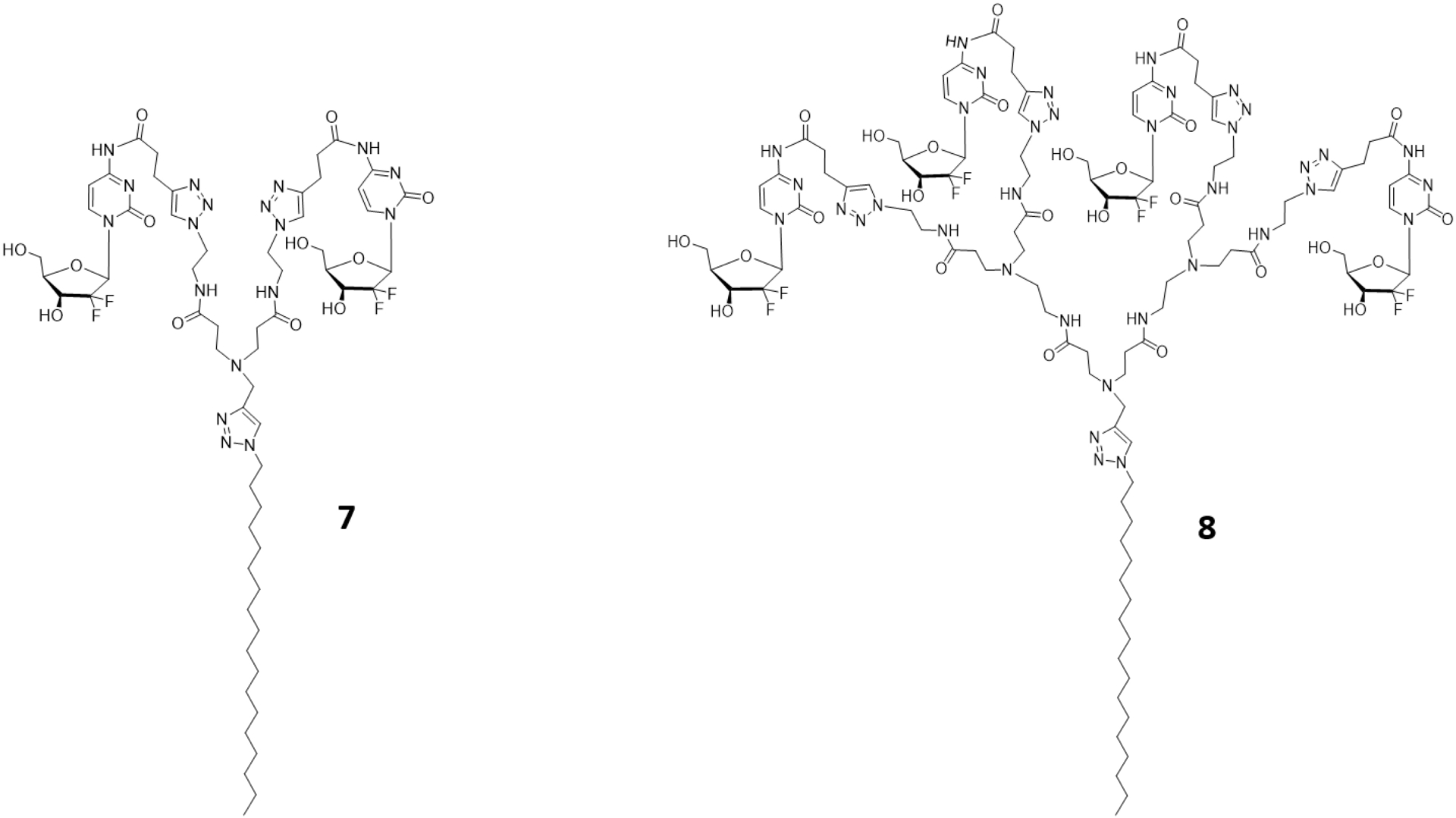
In another study, a nucleoside anticancer candidate was appended to dendrimer terminals through biodegradable ester linkages (9 and 10, Figure 10) to enable esterase-triggered drug release [25]. Notably, higher-generation dendrimer 10 released the active molecule significantly more slowly than lower-generation dendrimer 9, reflecting increased steric hindrance that limited enzymatic access to the ester bonds for effective action. Although such “negative” dendritic effects may appear disadvantageous, they can be strategically exploited to achieve generation-dependent and temporally controlled drug release.
Chemical structures of amphiphilic dendrimers 9 and 10.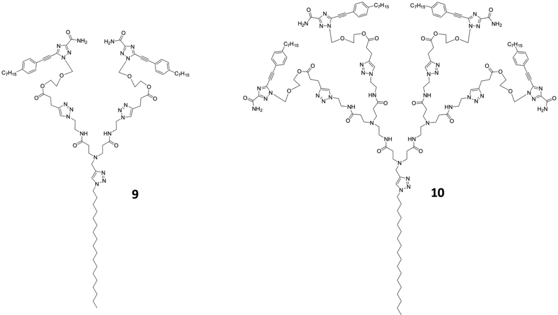
We further developed bola-amphiphilic dendrimers 11 and 12 (Figure 11), bearing glucose and mannose terminals, respectively [26, 27]. They were designed to target astrocytes and microglia—two major glial cell populations essential for the maintenance of brain homeostasis. Dysfunction of these glial cells is implicated in a wide range of central nervous system (CNS) disorders [28, 29]. Although targeting astrocytes and microglia, or modulating their activity, represents an attractive therapeutic strategy, it remains challenging because of the restrictive nature of the blood–brain barrier (BBB) and the scarcity of cell-specific targeting platforms. We demonstrated that both 11 and 12 efficiently reached the brain following intranasal administration, which bypassed the BBB [26]. More importantly, the glucose-functionalized dendrimer 11 selectively targeted astrocytes, whereas the mannose-functionalized dendrimer 12 preferentially accumulated in microglia [26]. Collectively, these findings underscore the potential of glycodendrimers as precision targeting platforms in the brain and highlight their promise for the treatment of CNS diseases.
Chemical structures of bola-amphiphilic dendrimers 11 and 12.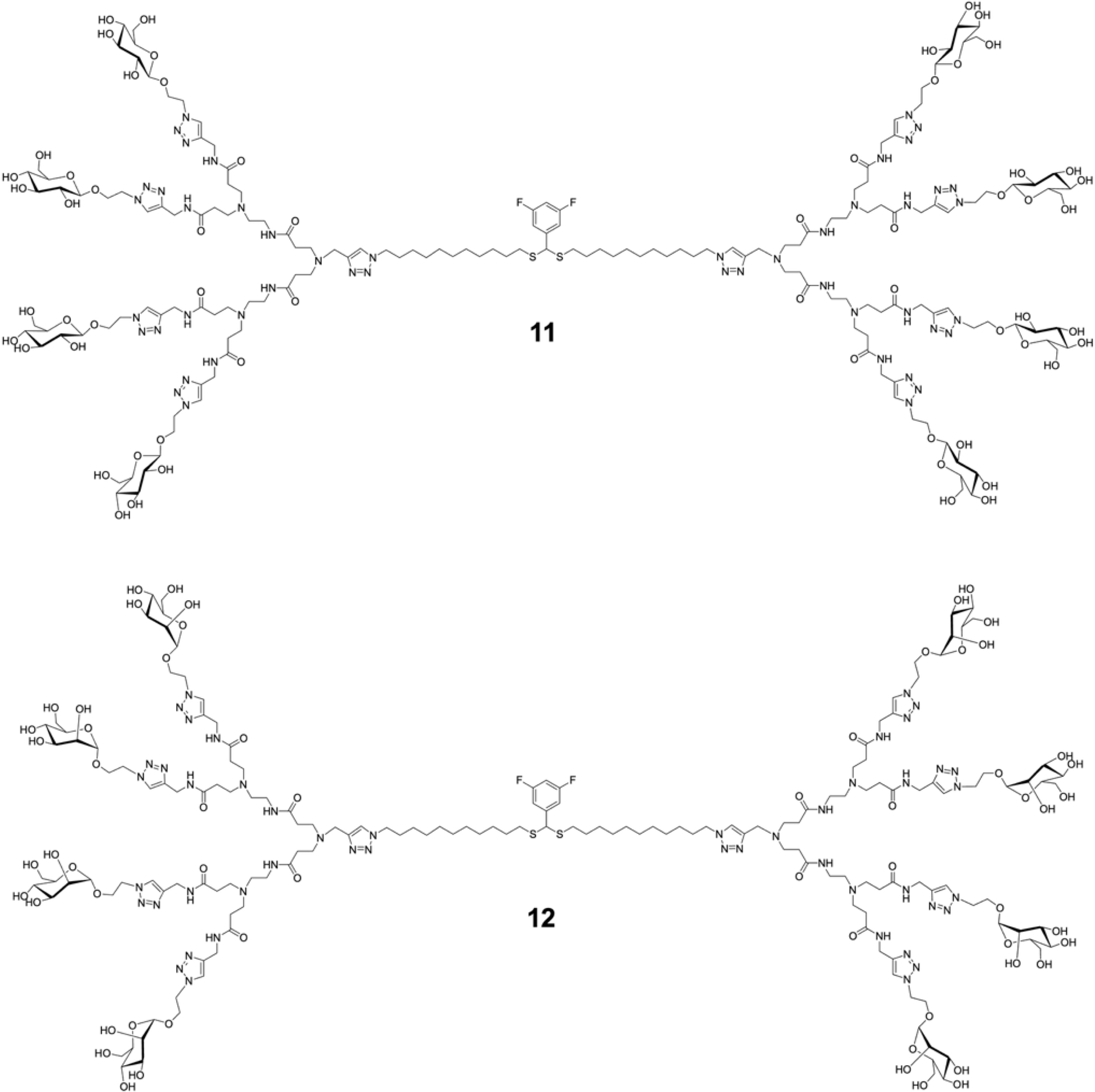
Together, these studies not only establish supramolecular dendrimers as tunable drug delivery platforms, but also highlight a set of design rules for engineering effective self-assembling dendrimer-based drug delivery systems. First, amphiphilicity-driven self-assembly of dendritic entities creates internal voids that enable high drug-loading capacity. Second, the number and chemistry of hydrophobic chains dictate stability of the nanoassemblies and therefore control drug release kinetics, where overly stable assemblies hinder release and less compact structures promote payload liberation. Third, subtle structural modifications—such as fluorination or generation tuning—provide powerful means to suppress premature drug leakage under physiological conditions while preserving responsive release in diseased microenvironments. Fourth, supramolecular adaptability enables biological “hijacking” mechanisms, such as extracellular vesicle–mediated intercellular transport, to enhance deep tissue penetration and overcome tumor heterogeneity. Finally, dendrimer generation and linkage chemistry can be exploited to achieve enzyme- and pH-responsive, generation-dependent drug release, offering controlled and tunable release. Collectively, these studies highlight self-assembling dendrimers as programmable nanocarriers enabling therapeutic performance via engineering and interplay between molecular design, self-assembly, and biological context.
3. Biomedical imaging and theranostics
Biomedical imaging plays an important role in disease diagnosis, monitoring, and treatment planning [30, 31]. Advances in nanotechnology have created new opportunities to improve imaging performance by enabling high local concentrations of imaging agents, integration of multiple different imaging reporters within a single probe for multimodal imaging, and the development of theranostic systems that unify diagnosis and therapy [32, 33]. In addition, nanoscale imaging agents can exploit the enhanced permeability and retention (EPR) effect to achieve passive targeting, thereby further improving imaging specificity and clinical relevance [34, 35].
Among various noninvasive imaging modalities, single-photon emission computed tomography (SPECT) is the most widely used in the clinic, because of its relatively low cost, femtomolar sensitivity, quantitative capability, and unlimited tissue penetration depth [30]. We designed amphiphilic dendrimer 13 (Figure 12A) to construct self-assembling dendrimer nanomicelles as the nanoprobe for SPECT imaging [36]. Dendrimer 13 bears multiple SPECT-reporting units at its periphery, based on the chelation of [111In]In3+ by 1,4,7,10-tetraazacyclododecane-1,4,7,10-tetraacetic acid (DOTA), a gold-standard chelator for indium and other clinically relevant radionuclides. This dendrimer spontaneously assembled into uniform nanomicelles that accumulated efficiently in tumors via the EPR effect, enabling effective SPECT imaging (Figure 12B).
(A) Chemical structure of amphiphilic dendrimer 13. (B) Schematic illustration of the self-assembling dendrimer 13 as the nanoprobe (13@) for tumor detection using SPECT imaging. Arrow indicates tumor location. Adapted from [36], licensed under CC BY 3.0.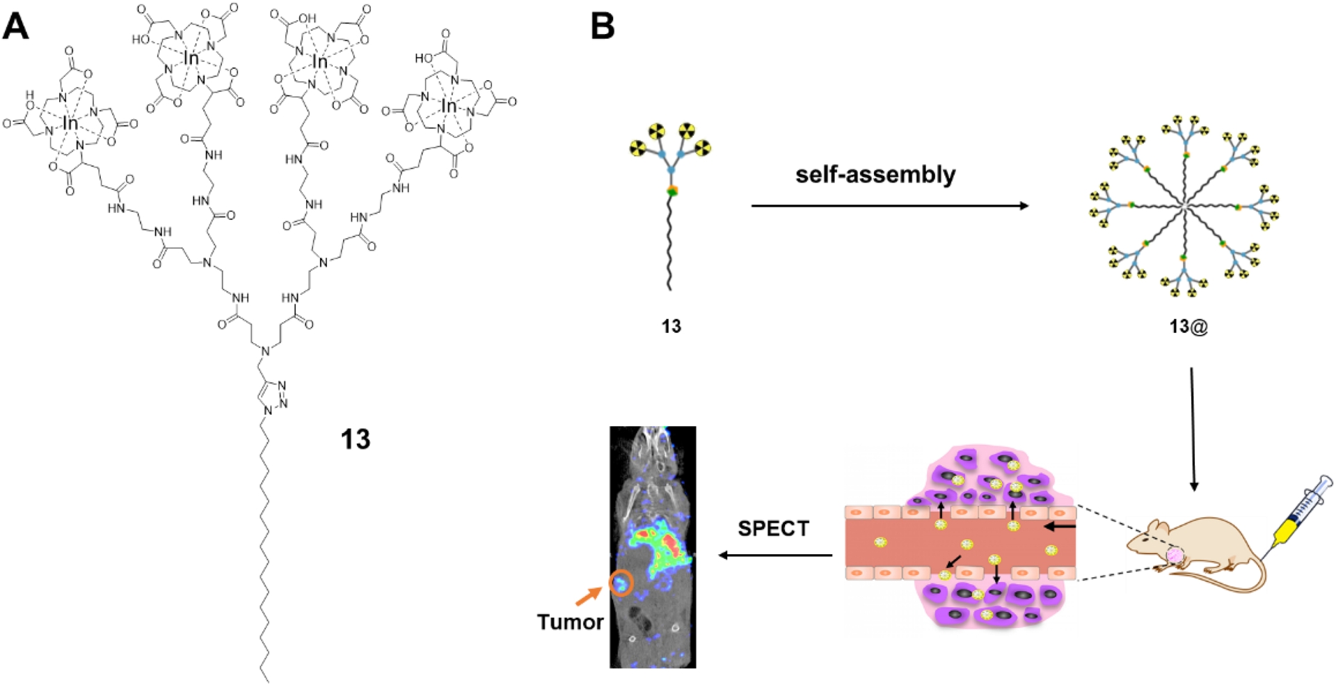
It should be mentioned that notable liver uptake was also observed when using 13 for SPECT imaging (Figure 12B) [36]. To address this concern, we developed amphiphilic dendrimer 14 (Figure 13A), in which DOTA was replaced by the smaller macrocyclic chelator 1,4,7-triazacyclononane-1,4,7-triacetic acid (NOTA) for [111In]In3+ chelation [37]. NOTA forms neutral and kinetically more stable complexes with small metal ions such as In3+ or Ga3+, owing to its reduced ring size and improved geometric match [38, 39]. Importantly, 14 formed supramolecular nanostructures with size and morphology comparable to those of 13, but exhibited a markedly different surface charge and considerably improved biodistribution profile, characterized by substantially reduced hepatic uptake and enhanced tumor-to-background contrast (Figure 13B) [37]. These results demonstrate how subtle molecular-level modifications can profoundly influence the in vivo biodistribution, highlighting the importance of chelator and surface chemistry in governing biological fate of imaging nanoprobes.
(A) Chemical structure of amphiphilic dendrimer 14. (B) Comparison of the SPECT/CT images of tumors obtained with the imaging probes 13@ and 14@ formed by the self-assembling of 13 and 14, respectively. Arrows indicate tumor locations. Adapted with permission from [37] (copyright 2020 Wiley-VCH GmbH).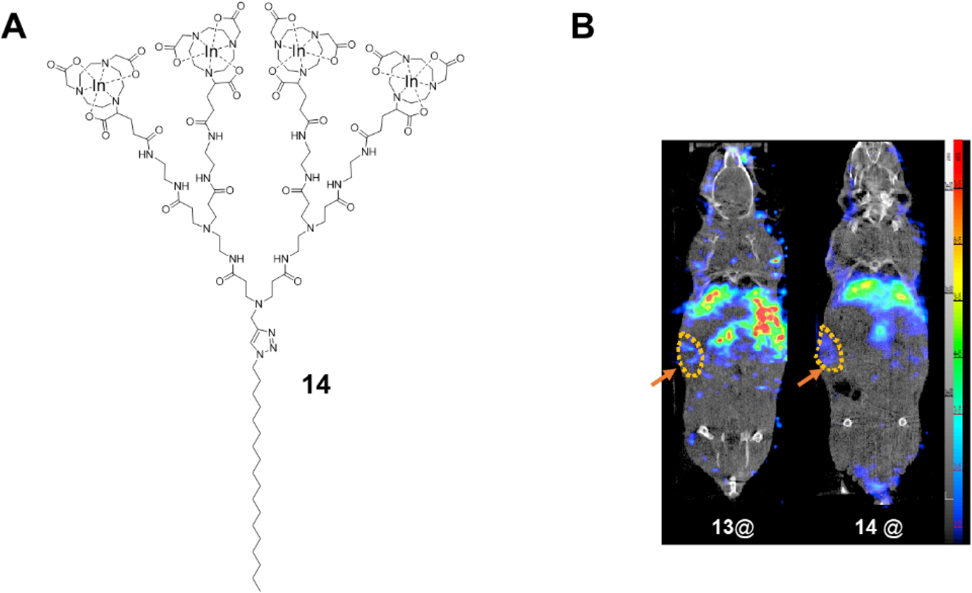
We also explored the self-assembling dendrimer nanoprobes for positron emission tomography (PET), which offers higher sensitivity and spatial resolution than SPECT [30]. Amphiphilic dendrimer 15 (Figure 14A) was established as a PET nanoprobe using [68Ga]Ga3+ as the radionuclide and NOTA as the chelator [40]. The excellent thermodynamic stability and kinetic inertness of the Ga3+–NOTA complex, combined with dendrimer multivalency and EPR-mediated tumor accumulation, enabled PET imaging with sensitivity and specificity that surpassed the clinical gold reference [18F]FDG (2-deoxy-2-[fluorine-18]fluoro-D-glucose) for tumor imaging (Figure 14B). Notably, this dendrimer nanoprobe allowed detection of tumors that were not discernible using [18F]FDG (Figure 14B), underscoring the advantages of this dendrimer nanoprobe for oncological imaging.
(A) Chemical structure of amphiphilic dendrimer 15. (B) Comparison of the PET images of tumors obtained with the clinical reference [18F]FDG and the nanoprobe 15@ formed by self-assembling of 15. Arrows indicate tumor locations. Reproduced from [40], licensed under CC BY-NC-ND 4.0.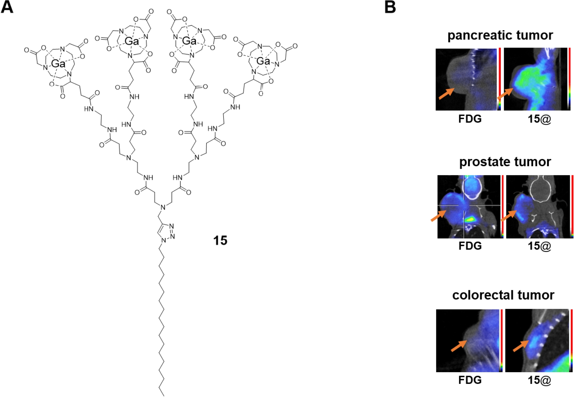
We further engineered self-assembling dendrimer nanosystems for multimodal imaging, which can combine complementary strengths of different imaging modalities while overcoming their limitation. Magnetic resonance imaging (MRI) provides excellent spatial resolution but suffers from relatively low sensitivity [41, 42], while near-infrared fluorescence (NIRF) imaging offers high sensitivity and resolution but limited tissue penetration [43, 44]. Integrating these modalities within a single nanosystem can provide synergistic benefits. To this end, we developed amphiphilic dendrimer 16 as a multimodal platform that combines 1H-MRI and NIRF imaging (Figure 15) [45]. Dendrimer 16 bears multiple Gd3+-based MRI contrast units at its terminals (Figure 15A) and self-assembles into uniform nanomicelles with high relaxivity and a favorable safety profile. Moreover, these nanomicelles were efficiently cleared from the liver within 24 h while selectively accumulating in tumors via the EPR effect, resulting in high tumor specificity and minimal off-target accumulation. In vivo MRI studies revealed up to a two-fold enhancement in tumor contrast relative to surrounding muscle tissue. Further encapsulation of the NIRF dye DiR within the hydrophobic core yielded a bimodal MR/NIRF imaging agent that achieved effective MRI at Gd3+ doses as low as one-tenth of the standard clinical dose, while simultaneously providing strong fluorescence enhancement. This dose-sparing capability highlights the potential of dendrimer-based multimodal probes for safer and more precise imaging.
(A) Chemical structure of amphiphilic dendrimer 16. (B) Schematic illustration of the self-assembling dendrimer nanosystems based on 16 as probes for 1H-MRI and 1H-MRI/NIRF multimodal imaging. (C) 1H-MRI and (D) NIRF imaging of tumors. Circles indicate tumor locations. Adapted from [45], licensed under CC BY 4.0.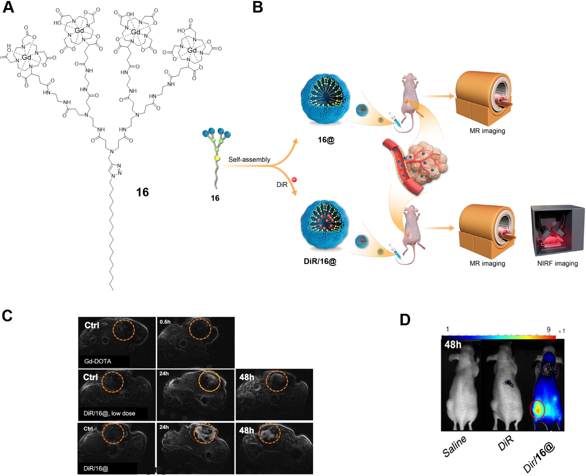
Although 1H-MRI is widely used clinically, its contrast is often limited by high background signals from endogenous water. To overcome this intrinsic limitation, fluorine magnetic resonance imaging (19F-MRI) is a promising technique, which offers background-free imaging thanks to the absence of fluorine in biological tissues [46, 47]. We therefore designed a fluorinated amphiphilic dendrimer 17 (Figure 16A) in which charged groups were strategically positioned near the fluorinated moieties at the dendrimer periphery [48]. This design prevented fluorine aggregation while maintaining high fluorine content, optimal molecular mobility, and excellent aqueous solubility. The resulting supramolecular dendrimer nanoassemblies exhibited favorable relaxation properties and enabled sensitive and specific 19F-MRI. Importantly, the modular architecture allowed pharmaceutical agents to be encapsulated within the nanomicellar core without compromising the 19F-MRI signal generated by the fluorinate entities at the periphery. Leveraging this feature, we co-encapsulated the NIRF dye DiR and the anticancer drug paclitaxel (PTX) to create a multimodal 19F-MRI/NIRF theranostic system (Figure 16B). This platform enabled simultaneous tumor visualization using multiple imaging modalities (1H-MRI, 19F-MRI, and NIRF) and effective therapy in a pancreatic cancer patient-derived xenograft model.
(A) Chemical structure of amphiphilic dendrimer 17. (B) Schematic illustration of the self-assembling dendrimer nanosytems based on 17 as probes for 19F-MRI and 19F-MRI/NIRF multimodal imaging as well as for 19F-MRI/NIRF/paclitaxel as theranostics. (C) 19F-MRI (red) superimposed on 1H-MRI (white) of tumor. Circles indicate tumor locations. (D) NIRF imaging (red) of tumor. (E) Inhibition of tumor growth upon treatment of the theranostics (PTX/16@). Reproduced from [48], licensed under CC BY-NC-ND 4.0.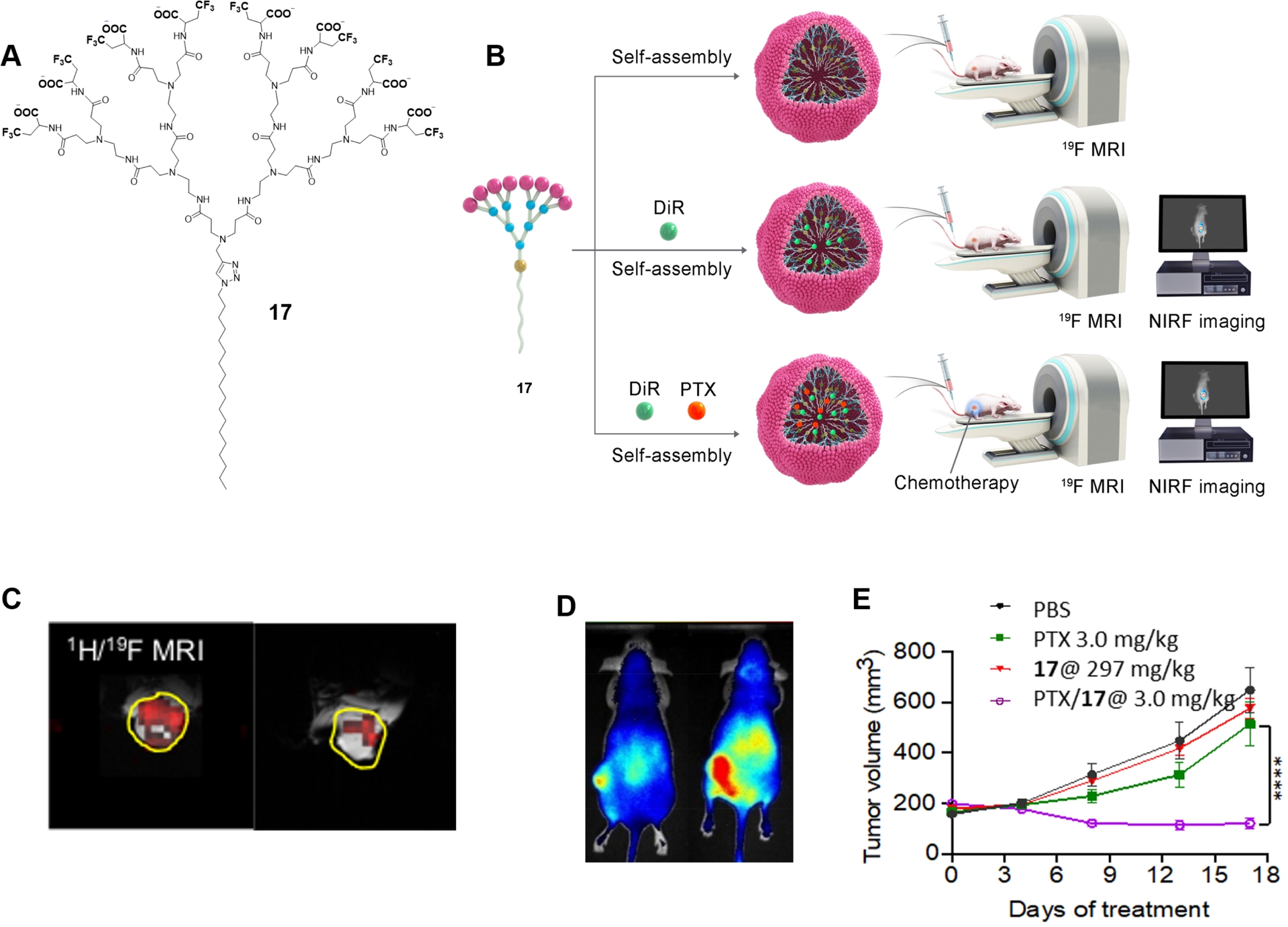
Altogether, our studies demonstrate how self-assembling supramolecular dendrimers can be rationally engineered as versatile platforms for advanced biomedical imaging. By integrating multiple imaging reporters within amphiphilic dendrimers, these dendrimer-based nanoprobes achieve high sensitivity, and enhanced tumor specificity through the EPR effect, enabling high-performance PET imaging that outperformed the clinical standard [18F]FDG. Systematic molecular design—particularly chelator selection and surface chemistry—proved critical in governing in vivo behavior, as exemplified by the marked improvement in biodistribution achieved by replacing DOTA with NOTA. Beyond single-modality imaging, the self-assembling dendrimers enabled multimodal MRI/NIRF- and MRI-based theranostic systems that combine anatomical resolution, sensitivity, and therapeutic capability within a single nanosystem. Collectively, these studies establish supramolecular dendrimers as adaptable and high-performance platforms for bioimaging and theranostics.
4. Nucleic acid delivery
Nucleic acid therapeutics have emerged as a powerful drug modality that treats disease by directly modulating gene expression [49, 50]. They offer unique access to address conventionally “undruggable” targets, rapid responses to emerging pathogens, and new opportunities for precision medicine. However, nucleic acid therapeutics suffer from poor bioavailability and low stability arising from their intrinsic polyanionic character and labile ester linkages. Effective delivery systems are therefore essential to protect nucleic acids from degradation, promote cellular uptake, facilitate endosomal escape, and achieve targeted and safe therapeutic action.
Cationic and ionizable dendrimers have attracted considerable attention for nucleic acid delivery because of their well-defined architectures and capacity to electrostatically complex polyanionic nucleic acids [3, 51, 52]. We previously developed a family of structurally flexible PAMAM dendrimers 18 (Figure 17) featuring an extended triethanolamine (TEA) core, which proved effective for the delivery of diverse nucleic acid cargos—including small interfering RNA (siRNA), small activating RNA (saRNA), microRNA (miRNA), and DNA—in both in vitro and in vivo settings [53, 54, 55, 56, 57, 58, 59, 60, 61, 62, 63, 64]. Notably, a fifth-generation dendrimer enabled efficient delivery of saRNA targeting C/EBPα for the treatment of liver cancer [56], leading to the first clinical trial of saRNA-based gene therapy for advanced hepatocellular carcinoma and other malignancies [65, 66].
Chemical structure of the TEA-core PAMAM dendrimer 18. For the sake of clarity, only the dendrimer of generation 3 is presented.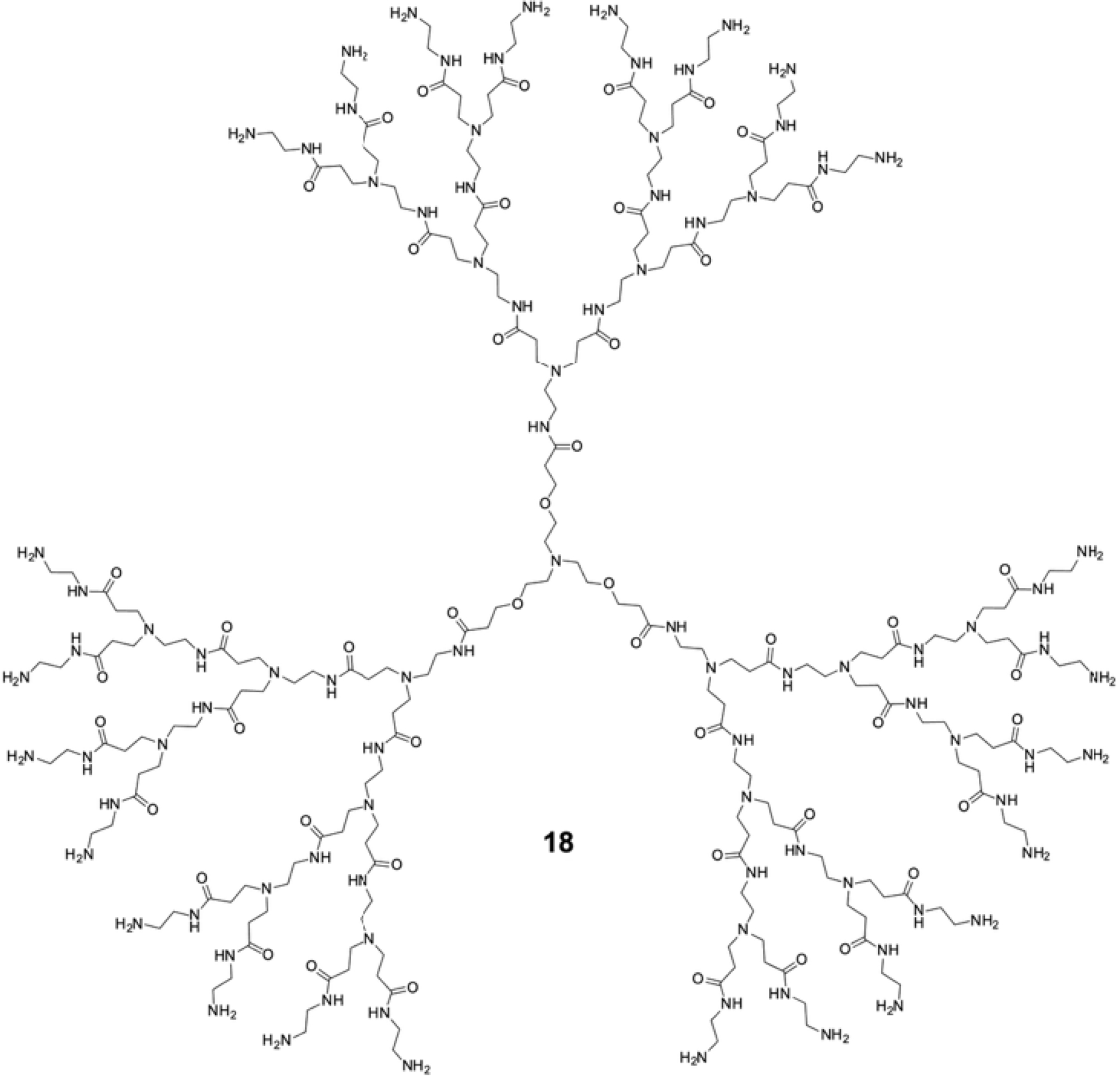
Building on this foundation, amphiphilic dendrimer 2 (Figure 5) was the very first amphiphilic dendrimer that we investigated for its self-assembly with the aim of nucleic acid delivery (Figure 18A). This dendrimer spontaneously formed supramolecular nanomicelles in water that efficiently complexed with siRNA and protected it from enzymatic degradation [9]. This enabled effective siRNA delivery and robust gene silencing by integrating the key advantages of lipid and polymer vectors, namely membrane fusion–mediated cellular uptake and endosomal escape associated with lipid vectors, together with the proton sponge–mediated endosomal release characteristic of polymer vectors (Figure 18B) [51]. In contrast, the corresponding hydrophilic dendron 19 (Figure 18C) or hydrophobic alkyl chain components alone were inactive; neither was the dendrimer 20 (Figure 18C) bearing a hydrophilic chain without amphiphilicity. These findings established amphiphilicity and self-assembly as key for amphiphilic dendrimers in nucleic acid delivery. Systematic modulation of the hydrophobic chain length in dendrimer 2 revealed a narrow structural window for delivery activity: a shorter alkyl chain prevented the formation of stable assemblies required for effective delivery, whereas a longer chain produced overly stable assemblies with strong siRNA binding, thereby hindering siRNA release and abolishing delivery capacity [9]. This highlights the critical role of hydrophilic–hydrophobic balance in controlling supramolecular assembly of amphiphilic dendrimers in nucleic acid delivery. In addition, this dendrimer not only integrated favorable features of lipid and polymer vectors, but also incorporated EPR-based passive tumor-targeting properties.
(A) Schematic illustration of self-assembling dendrimer vector for siRNA delivery and gene silencing. (B) Schematic illustration of amphiphilic dendrimer for siRNA delivery via combination of the delivery feature of lipid and polymer vectors. (C) Chemical structures of dendrimers 19 and 20. Adapted with permission from [9] (copyright 2016 Wiley-VCH GmbH) and [51] (copyright 2022 ShanghaiTech University and American Chemical Society).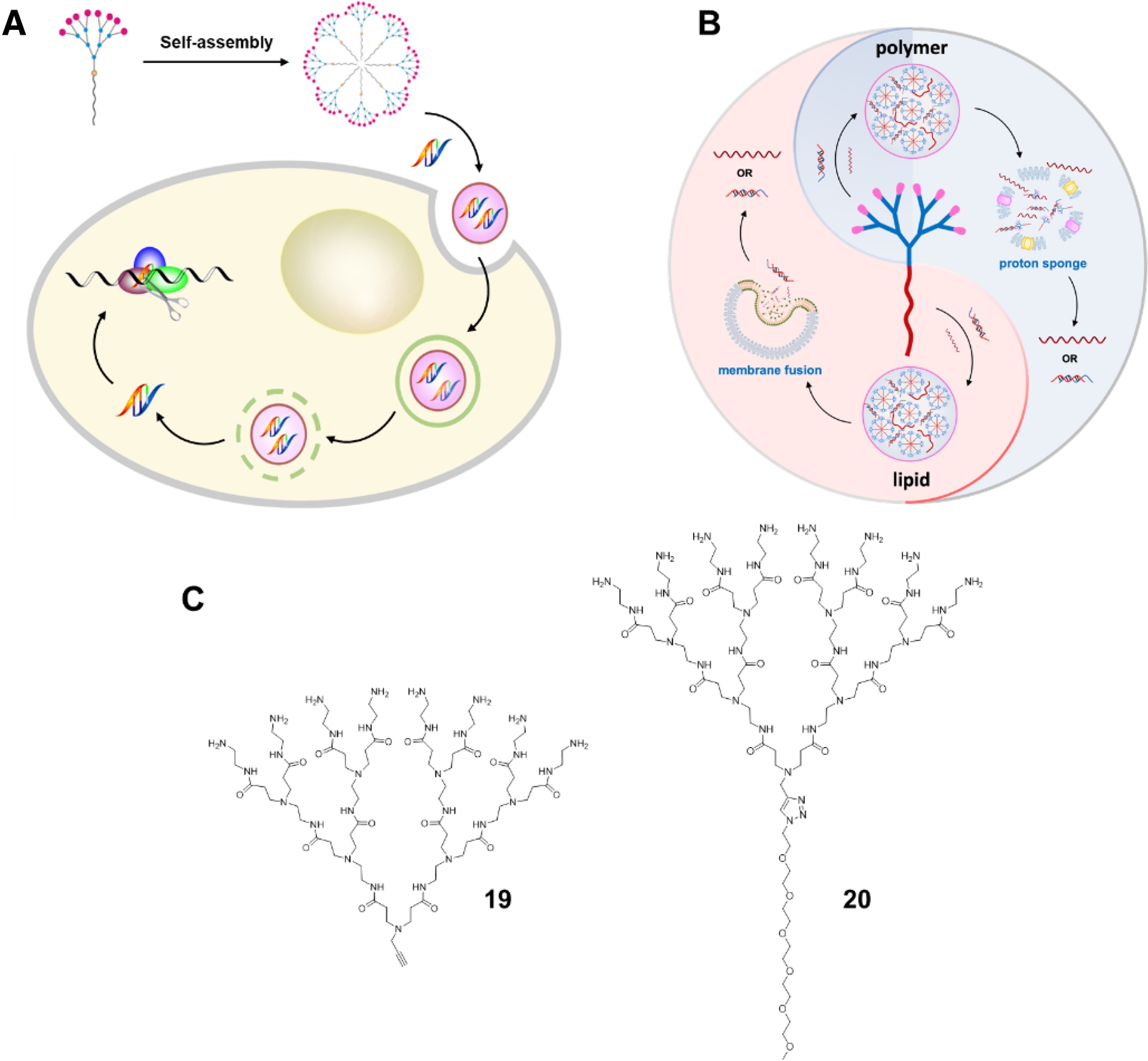
Further structural refinements were introduced to improve biocompatibility and delivery efficiency of dendrimer 2. Substitution of the terminal primary amines in dendrimer 2 with tertiary amines afforded dendrimer 4 (Figure 4), which retained siRNA delivery efficacy while substantially reducing cytotoxicity [67]. Interestingly, both dendrimer 2 and dendrimer 4 also acted as permeabilizers of the bacterial envelope and assisted in the internalization of oligonucleotide mimics for antibacterial activity, yet without any notable toxicity [68]. This finding demonstrated that amphiphilic dendrimers effectively assisted oligonucleotide mimics in killing bacteria, highlighting their potential as a novel strategy against antimicrobial-resistant pathogens.
Inspired by arginine-rich cell-penetrating peptides (CPPs) [69], we elaborated dendrimer 21 (Figure 19A) bearing arginine residues at the terminals with the view to mimicking the cell penetration feature of CPPs for enhanced cellular uptake [70]. Consistent with the design strategy, dendrimer 21 outperformed dendrimer 2 in cellular uptake of siRNA (Figure 19B/C) as well as in siRNA delivery for gene silencing (Figure 19D), underscoring the importance of terminal functionality in governing biological interactions and nucleic acid delivery efficiency.
(A) Chemical structure of amphiphilic dendrimer 21. (B) Confocal image and (C) quantification of cellular uptake of the Cy3-labeled siRNA delivered by dendrimer 21 in comparison to that by 2. Adapted with permission from [70] (copyright 2015 Royal Society of Chemistry).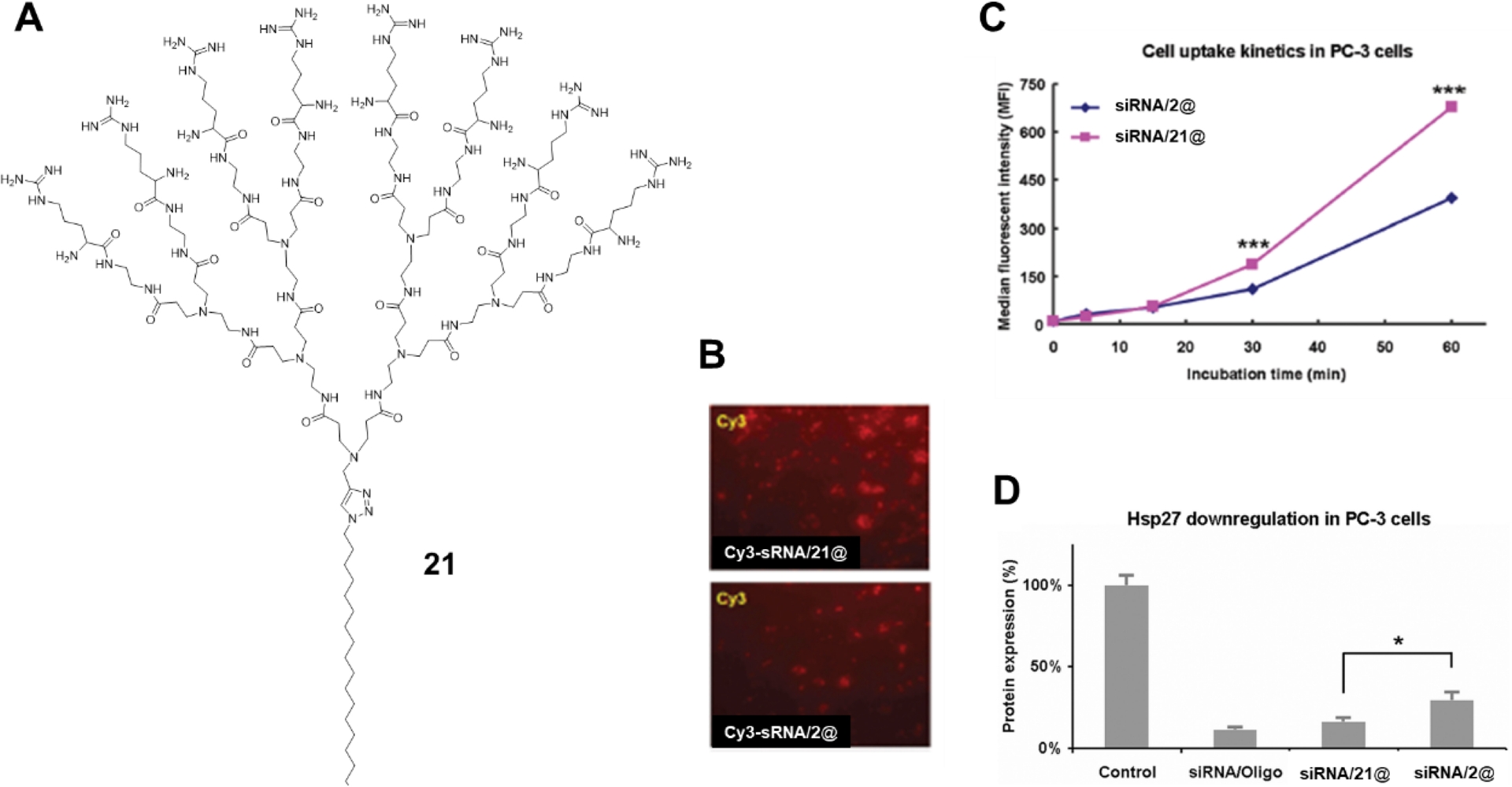
We also developed poly(aminoester) (PAE) dendrimers [71, 72, 73] as biodegradable platforms for nucleic acid delivery. These dendrimers integrate hydrolysable ester linkages with ionizable amine functionalities, enabling efficient complexation of nucleic acids through electrostatic interactions at physiological pH, while facilitating cargo release via dendrimer degradation in acidic/basic or enzyme-rich environment. Amphiphilic PAE dendrimer 22 (Figure 20), consisting of a second-generation PAE dendron appended with a long alkyl chain, exhibited effective siRNA delivery, whereas its PAMAM analogue 23 showed negligible activity [74]. These results highlight that biodegradable amphiphilic dendrimers can further improve delivery efficiency. Additional studies revealed a pronounced generation-dependent hydrolytic degradation behavior: the higher-generation dendrimer 25 (Figure 20) underwent rapid degradation, whereas the lower-generation analogue 24 (Figure 20) degraded substantially more slowly [75]. This trend can be attributed to the dendritic architecture, in which increasing generation generates steric congestion that destabilizes the ester linkages and enhances their susceptibility to hydrolysis. This “dendritic effect” and generation-dependent degradation can be exploited as a tunable strategy for generation-dependent delivery.
Chemical structures of amphiphilic dendrimers 22, 23, 24 and 25.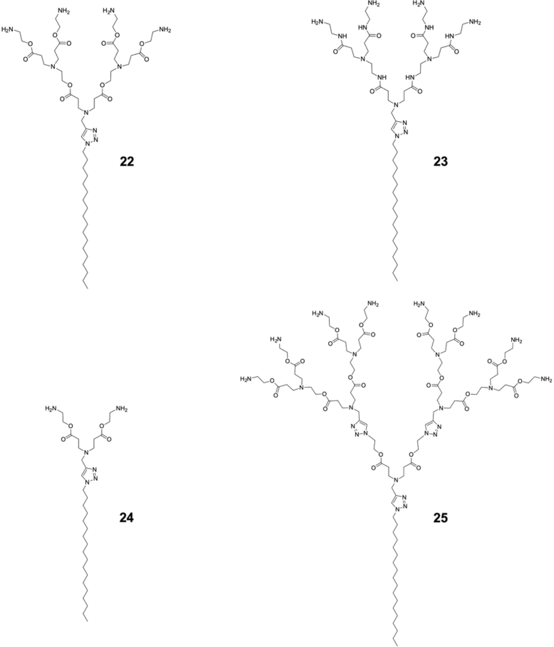
Subsequent investigations on amphiphilic dendrimer 1 revealed its excellent delivery performance not only in siRNA-mediated gene silencing but also in saRNA-induced gene activation (Figure 21A), surpassing commercially available vectors in both in vitro and in vivo models [76, 77, 78, 79, 80]. A recent study further demonstrated its capacity to deliver both single-stranded and double-stranded oligonucleotides to mitochondria, which are particularly challenging due to the presence of dual mitochondrial membranes [81]. Most strikingly, dendrimer 1 also enabled efficient siRNA delivery to primary immune cells such as primary T cells, primary natural killer cells, and primary macrophages and microglia (Figure 21B–I) [76, 78, 79]. It should be mentioned that primary immune cells are typically refractory to nonviral transfection agents and require electroporation for nucleic acid delivery. Therefore, dendrimer 1 constitutes a promising vector for nucleic acid delivery either in functional investigations on immune cells or in translational studies on immune therapies.
(A) Schematic illustration of the amphiphilic dendrimer 1 for the delivery of siRNA to cytosol for gene silencing and saRNA to nucleus for gene activation. Effective 1-mediated siRNA delivery and gene silencing achieved in (B,C) primary T cells, (D,E) primary natural killer cells, (F,G) primary macrophages, and (H,I) primary microglia. Adapted with permission from [76] (copyright 2021 Springer Nature).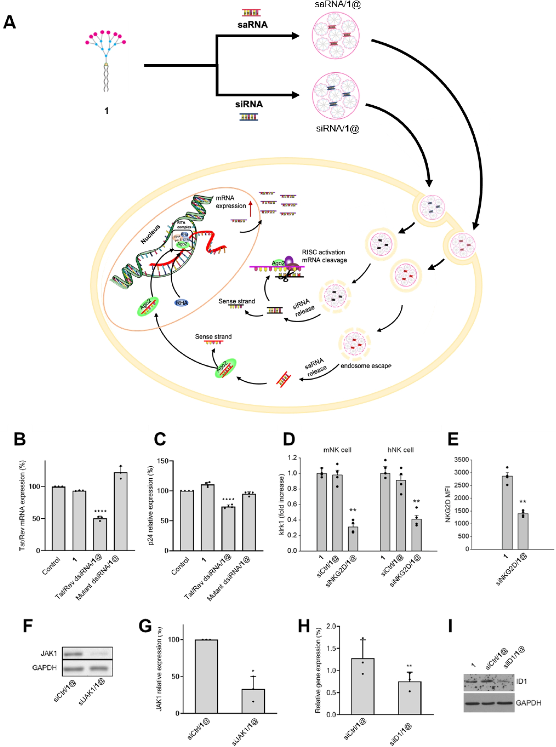
Moreover, dendrimer 1 functioned as a delivery platform for targeted siRNA transport, which could be realized through either physical or chemical conjugation with targeting moieties (Figure 22). In one representative approach, the positively charged siRNA/1@ complexes were decorated with a negatively charged peptide bearing targeting warheads via electrostatic interactions (Figure 22A) [82]. This strategy enabled selective siRNA delivery to cancer cells, resulting in enhanced therapeutic efficacy (Figure 22B). Alternatively, chemical conjugation of the siRNA/1@ complexes with an anti–PD-L1 antibody (Figure 22C) not only conferred cancer cell–specific delivery but also simultaneously blocked the PD-1/PD-L1 immune checkpoint interaction [83]. This dual functionality triggered immune activation in conjunction with siRNA-mediated anticancer activity, thereby affording synergistic therapeutic effects (Figure 22D). Collectively, these straightforward and modular strategies markedly improved delivery specificity and therapeutic outcomes while enabling dose reduction and minimizing associated off-target toxicity [82, 83, 84].
(A) Schematic illustration of targeted siRNA delivery via electrostatic decoration of positively charged siRNA/1@ complexes with negatively charged, warhead-bearing targeting peptides. (B) Resulting enhancement of anticancer activity relative to the nontargeted counterpart. (C) Schematic illustration of antibody-mediated targeting achieved through chemical conjugation of siRNA/1@ complexes with an anti–PD-L1 antibody. (D) Corresponding enhancement of anticancer activity. Adapted from [82], licensed under CC BY-NC 4.0, and [83], licensed under CC BY 4.0.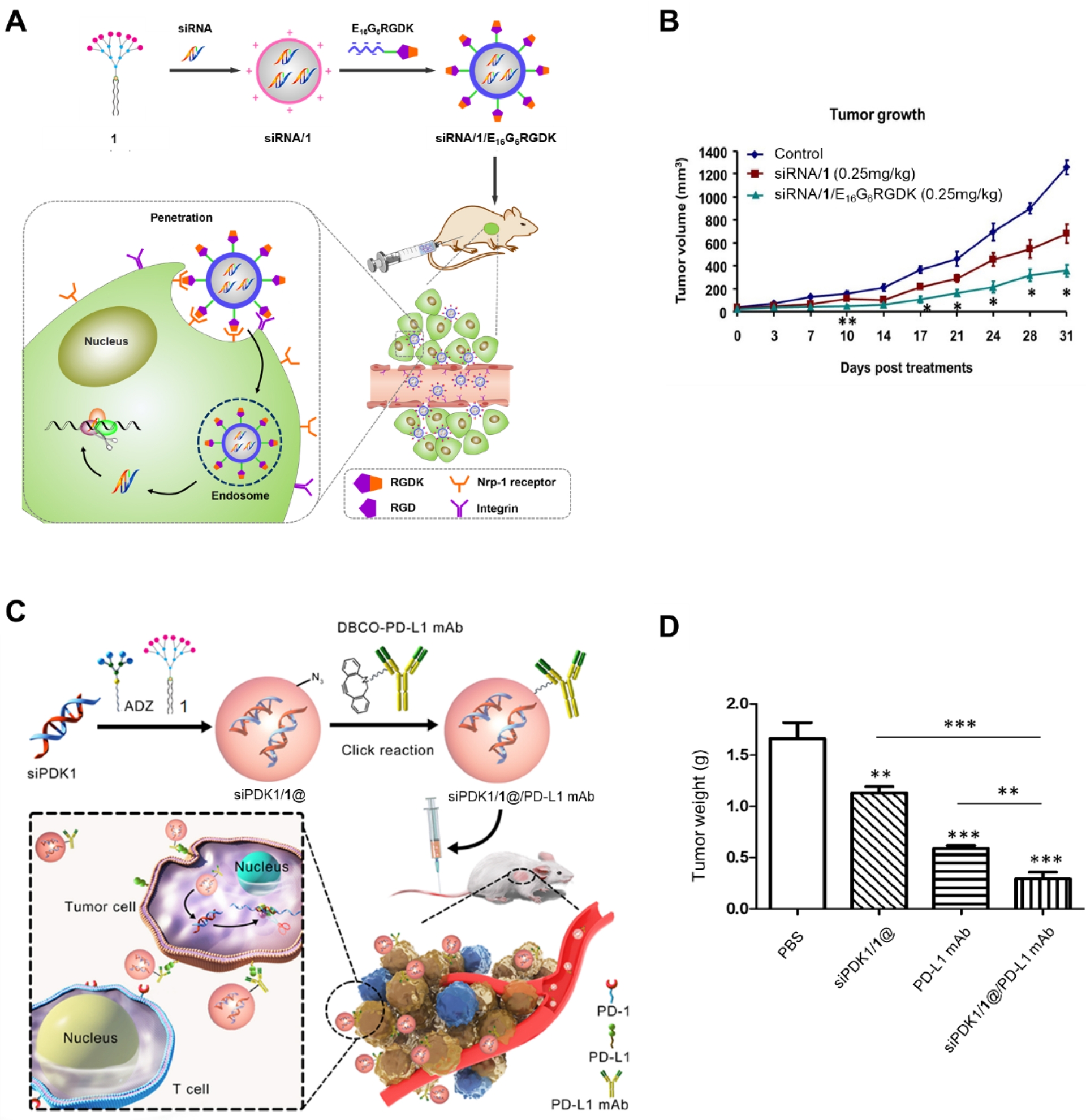
Remarkably the bola-amphiphilic dendrimers 6 and 27 are capable of selective and ROS-responsive delivery of DNA and siRNA (Figure 23) [22, 85]. Specifically, the second-generation dendrimer 6 preferentially delivered siRNA, whereas the third-generation analogue 27 excelled in DNA delivery. Systematic studies of cargo binding, cellular uptake, endosomal escape, and in vivo behavior revealed that size matching between dendrimer and nucleic acid cargo governs both nucleic acid loading and release via cooperative multivalent interactions, enabling adaptive and cargo-selective delivery [85]. In addition, these bola-amphiphilic dendrimers incorporated the thioacetal functionality for redox-responsive cargo release. As a result, they achieved tumor- and cancer cell–specific delivery of siRNA and DNA therapeutics via combined EPR effect and ROS-responsive delivery, leading to effective treatment in diverse cancer models, including aggressive and metastatic tumors, and outperforming existing delivery systems. The insights gained from this study provide a foundation for the rational design of tailor-made dendrimer platforms for cargo-selective delivery, advancing the development of nucleic acid therapeutics in precision medicine.
(A) Chemical structures of bola-amphiphilic dendrimers 26, 6, and 27. (B) Schematic illustration of cargo-selective delivery via matching nucleic acid size and dendrimer generation. Reproduced from [85], licensed under CC BY-NC-ND 4.0.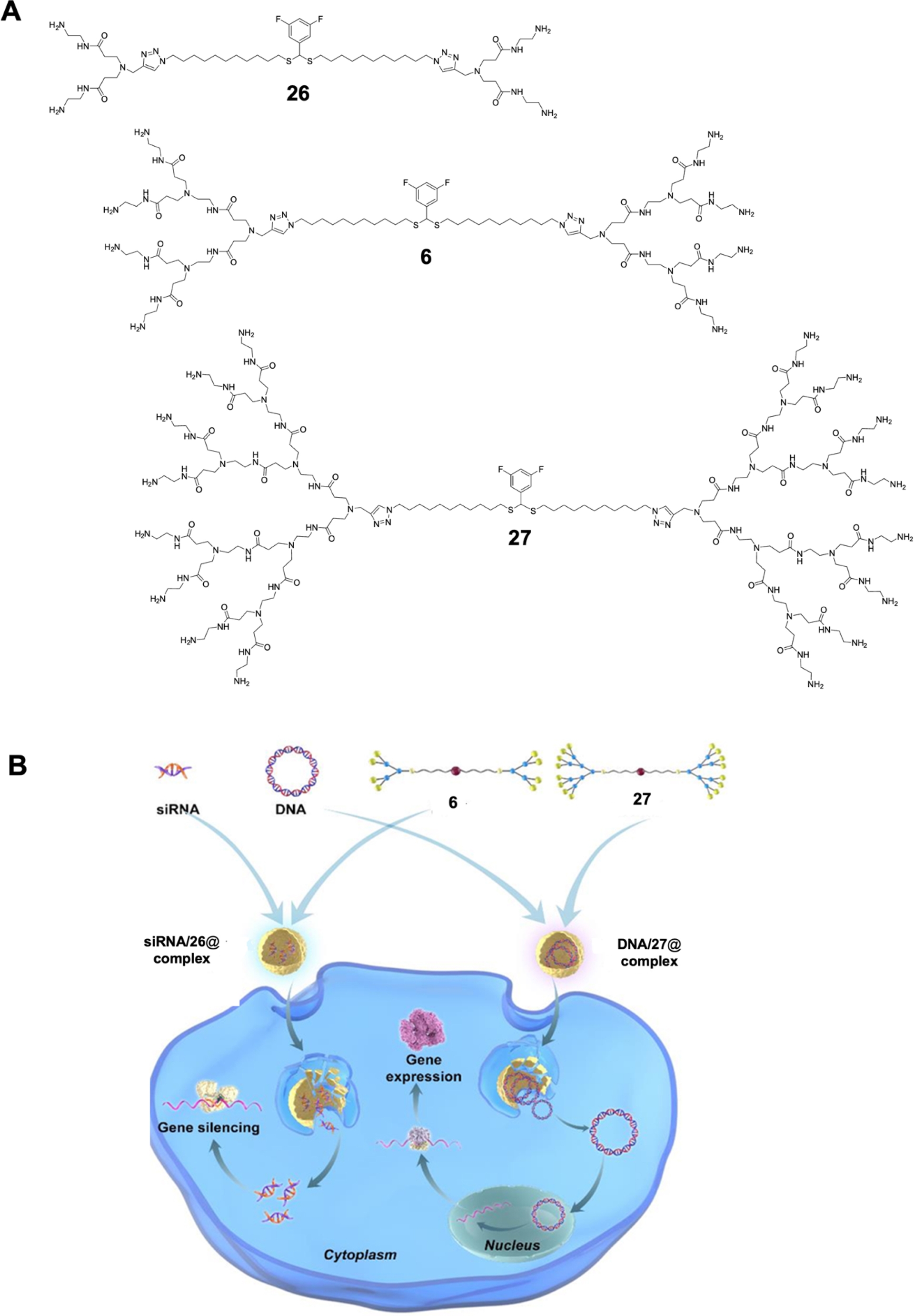
Interestingly, dendrimer 6 was also able to co-deliver nucleic acid therapeutics and anticancer drug for synergistic action in cancer chemo-immunotherapy [86]. Specifically, a combined anticancer drug DOX and siRNA formulation targeting CD47—an antiphagocytic “don’t eat me” signal—was elaborated. The co-delivery system was constructed by encapsulating DOX within the hydrophobic interior of the dendrimer nanoassemblies, while electrostatically complexing the negatively charged siRNA onto the positively charged dendrimer surface. This dual-cargo platform effectively downregulated CD47 expression, thereby attenuating antiphagocytic signaling, while simultaneously inducing DOX-mediated immunogenic cell death and activating antitumor immune responses. As a result, the system exhibited potent antitumor efficacy arising from the coordinated actions of DOX and siCD47, highlighting its promise as a strategy for synergistic cancer chemo-immunotherapy.
All these studies have established amphiphilic dendrimers as a versatile and programmable class of nonviral vectors for nucleic acid delivery. By systematically tuning dendrimer amphiphilicity, terminal functionality, generation, and supramolecular self-assembly, we investigated the factors governing nucleic acid complexation, cellular uptake, cargo release, and biological activity. Amphiphilicity-driven self-assembly was shown to be essential for effective siRNA delivery, with an optimal hydrophilic–hydrophobic balance required to form assemblies that are sufficiently stable for transport yet labile enough to enable intracellular cargo release. Terminal group engineering further improved biocompatibility and cellular internalization. Extension to bipolar-amphiphilic dendrimers revealed how dendrimer generation and size matching with nucleic acid cargos can be exploited to achieve adaptive and cargo-selective delivery of siRNA versus DNA, resulting in effective and tumor-specific therapeutic outcomes. Recently, Percec and coworkers have elaborated ionizable amphiphilic Janus dendrimer (IAJD) systems for mRNA delivery, which exhibited high delivery performance and organ-selective delivery [87, 88, 89, 90, 91, 92, 93], hence opening new avenues for more versatile and translatable mRNA therapeutics. Collectively, these findings define a rational engineering framework for amphiphilic dendrimer–mediated nucleic acid delivery.
5. Conclusion and perspectives
This short Account highlights our decade-long efforts in establishing self-assembling supramolecular dendrimers as modular and adaptable nanoplatforms for biomedical applications, including drug and nucleic acid delivery, advanced biomedical imaging and theranostics, etc. Amphiphilicity-driven self-assembly enables the formation of dendritic nanoassemblies with high drug loading capacity, controllable release kinetics, and tunable biological interactions. Through systematic modulation of dendrimer amphiphilicity, generation, surface chemistry, and degradable linkage, these systems were shown to overcome key barriers such as drug resistance, off-target toxicity, suboptimal biodistribution, and inefficient intracellular delivery. Importantly, subtle molecular-level modifications—such as hydrophobic chain engineering, fluorination, chelator selection, or terminal group functionalization—were found to exert profound effects on drug loading and release, cell uptake and endosome escape, in vivo biodistribution, toxicity and therapeutic efficacy, underscoring the sensitivity of supramolecular dendrimers to rational design. Taken together, the ability of self-assembling supramolecular dendrimers to precisely regulate cargo loading and release, biological interactions, and in vivo fate underpins their broad utility across drug and nucleic acid delivery alongside biomedical imaging and theranostics.
Beyond functioning as drug delivery vehicles, self-assembling supramolecular dendrimers can themselves serve as active therapeutic agents [94, 95, 96, 97]. For example, dendrimers 2 and 4 displayed pronounced antibacterial activity [95, 96, 97]. Dendrimer 2 exhibited broad-spectrum efficacy against both Gram-negative and Gram-positive bacteria, as well as drug-resistant strains and bacterial biofilms [95, 97]. In contrast, dendrimer 4 demonstrated selective yet potent activity toward multiple Gram-negative bacteria via targeting the bacterial membrane [95, 96]. Meanwhile, dendrimers bearing guanidinium or carboxylate terminal groups did not exhibit any appreciable antibacterial activity [95], underscoring the critical role of dendrimer structure and surface functionality in determining biological performance. This provides valuable design principles for the development of innovative antibacterial candidates.
In addition to amphiphilic PAMAM dendrimers developed in our group, numerous studies based on amphiphilic polyether or glycerol dendrimers [98, 99, 100, 101, 102, 103, 104, 105], phosphorus dendrimers [106, 107], carbosilane dendrimers [108, 109], or glycodendrimers [110, 111, 112], have been reported by other groups for creating self-assembling dendrimer nanosystems. All these studies have supported the concept of synthesizing supramolecular dendrimers via self-assembling as modular and adaptive nanosystems in biomedicine.
Looking forward, self-assembling dendrimers offer a powerful and flexible platform for next-generation precision medicine. Future developments will benefit from deeper integration of molecular design with biological insight, enabling dendrimers that respond to disease-specific microenvironments, biological transport pathways, and intracellular triggers. The convergence of therapeutic delivery, imaging, and targeting within a single supramolecular scaffold offers opportunities for truly integrated theranostic systems with improved efficacy and safety for precision medicine. As understanding of supramolecular chemistry continues to advance, supramolecular dendrimers are expected to play an increasingly important role in the rational design of multifunctional nanomedicines.
Acknowledgements
We thank all former and current group members as well as collaborators for their outstanding contributions to the studies presented in this Account. We acknowledge financial support from the Ligue Nationale Contre le Cancer, the French National Research Agency for funding of the Era-Net EURONANOMED projects “DENANORNA”, “Target4Cancer”, “NANOGLIO”, “TARBRAINFEC”, “NAN-4-TUM”, “NanoGUN”, “antineuropatho”, the European Union’s Horizon 2020 and Horizon Europe research and innovation projects “SAFE-N-MEDTECH” (no. 814607) and “HIT-GLIO” (no. 101136835), the EU MSCA ITN projects “OLIGOMED” (no. 956070) and “ON-TRACT” (no. 101227456), CNRS and Aix-Marseille Université.
Declaration of interests
The authors do not work for, advise, own shares in, or receive funds from any organization that could benefit from this article, and have declared no affiliations other than their research organization.